. 2001 Aug;10(8):1563-71.
doi: 10.1110/ps.7201.

Modeling of the bacterial luciferase-flavin mononucleotide complex combining flexible docking with structure-activity data

Affiliations

Modeling of the bacterial luciferase-flavin mononucleotide complex combining flexible docking with structure-activity data

L Y Lin et al. Protein Sci. 2001 Aug.

Abstract

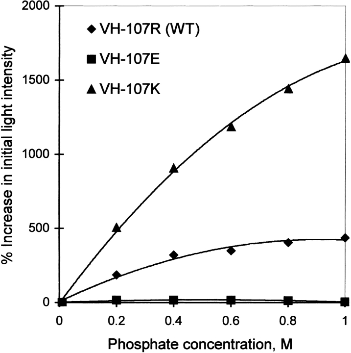
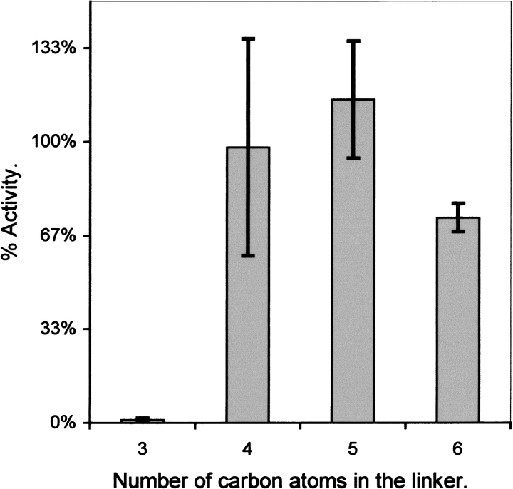
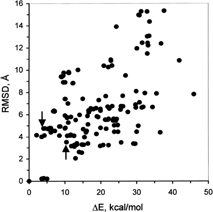
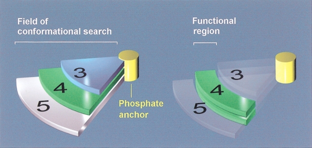
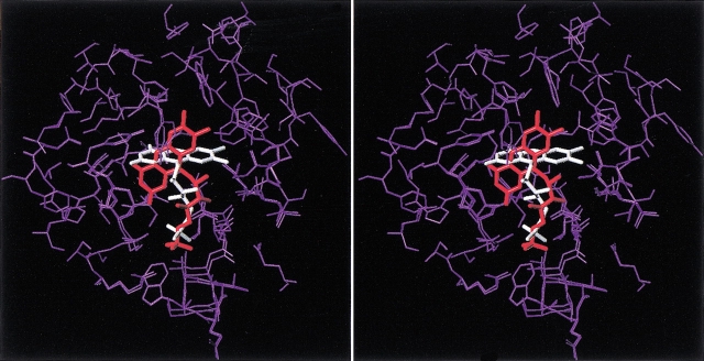
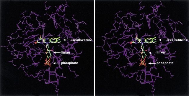
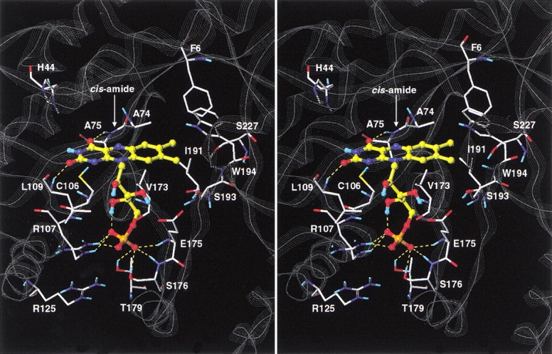
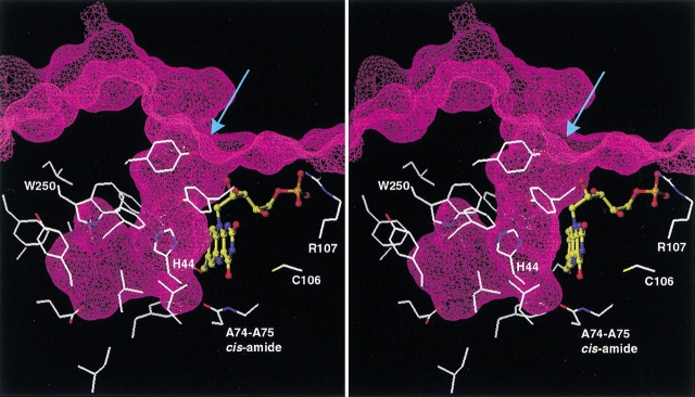
Although the crystal structure of Vibrio harveyi luciferase has been elucidated, the binding sites for the flavin mononucleotide and fatty aldehyde substrates are still unknown. The determined location of the phosphate-binding site close to Arg 107 on the alpha subunit of luciferase is supported here by point mutagenesis. This information, together with previous structure-activity data for the length of the linker connecting the phosphate group to the isoalloxazine ring represent important characteristics of the luciferase-bound conformation of the flavin mononucleotide. A model of the luciferase-flavin complex is developed here using flexible docking supplemented by these structural constraints. The location of the phosphate moiety was used as the anchor in a flexible docking procedure performed by conformation search by using the Monte Carlo minimization approach. The resulting databases of energy-ranked feasible conformations of the luciferase complexes with flavin mononucleotide, omega-phosphopentylflavin, omega-phosphobutylflavin, and omega-phosphopropylflavin were filtered according to the structure-activity profile of these analogs. A unique model was sought not only on energetic criteria but also on the geometric requirement that the isoalloxazine ring of the active flavin analogs must assume a common orientation in the luciferase-binding site, an orientation that is also inaccessible to the inactive flavin analog. The resulting model of the bacterial luciferase-flavin mononucleotide complex is consistent with the experimental data available in the literature. Specifically, the isoalloxazine ring of the flavin mononucleotide interacts with the Ala 74-Ala 75 cis-peptide bond as well as with the Cys 106 side chain in the alpha subunit of luciferase. The model of the binary complex reveals a distinct cavity suitable for aldehyde binding adjacent to the isoalloxazine ring and flanked by other key residues (His 44 and Trp 250) implicated in the active site.


Figures

Fig. 1.
Fig. 1.
Increase in bioluminescence activity with riboflavin as a function of phosphate concentration with luciferase mutants. Activities were measured using the dithionite assays described in Materials and Methods on injection of decanal into the assay mixture containing the indicated phosphate concentration and 50 μM riboflavin.
Fig. 2.
Fig. 2.
Relative bioluminescence activities of ω-phosphoalkylflavins with different numbers of carbon atoms linking the phosphate moiety to the isoalloxazine ring. The percentage of the initial light intensity of V. harveyi luciferase with FMN and different fatty aldehydes for each ω-phosphoalkylflavin has been taken from earlier data with the same aldehyde (Meighen and MacKenzie 1973), averaged (plus or minus standard deviation), and then plotted versus the number of carbon atoms in the alkyl chain.
Fig. 3.
Fig. 3.
Energetic discrimination of the FMN-binding modes. The root-mean-square deviation of the isoalloxazine ring nonhydrogen atoms (RMSD) is plotted against the relative internal energy (ΔE) of the corresponding luciferase–FMN complexes. The global minimum-energy conformation is taken as reference. (arrows) Two binding modes that are common to the active flavin analogs FMN, ω-phosphobutylflavin, and ω-phosphopentylflavin but inaccessible to the inactive flavin analog ω-phosphopropylflavin. (RMSD) root-mean-square deviation.
Fig. 4.
Fig. 4.
Geometrical constraints on the bound conformation of the isoalloxazine ring in luciferase. The length of the linker in the flavin analogs restricts the regions of conformational space accessible to the ring. (left) The accessible regions corresponding to each linker length are shown schematically by the three pie-shaped sectors extending from the phosphate anchor. The number labels correspond to the linker length. (right) The active bound conformation of the isoalloxazine ring will be in a region (green) accessible to both the four- and five-carbon linker flavin analogs but inaccessible to the three-carbon linker one.
Fig. 5.
Fig. 5.
Stereoview of the two binding modes of FMN to luciferase that are common to ω-phosphobutylflavin and ω-phosphopentylflavin, but inaccessible to the inactive ω-phosphopropylflavin. FMN is shown with capped-sticks, with the lower energy conformer colored in white and the higher energy conformer colored in red. The protein atoms that were allowed to move during flexible docking are shown in purple for both complexes. The superposition is based on the remaining part of the protein that was kept rigid during flexible docking (not shown). Hydrogen atoms are omitted for clarity.
Fig. 6.
Fig. 6.
Stereoview of the lowest energy common binding mode of FMN, ω-phosphobutylflavin, and ω-phosphopentylflavin to the α subunit of bacterial luciferase. The ligand molecules are represented as capped-sticks, with the carbon atoms colored in white for FMN, yellow for ω-phosphobutylflavin, and green for ω-phosphopentylflavin. The protein residues that were allowed to move during flexible docking are shown in purple for all complexes. The alignment is based on the remaining part of the protein that was kept rigid during flexible docking (not shown). Hydrogen atoms are omitted for clarity.
Fig. 7.
Fig. 7.
Stereoview of the modeled complex of FMN with the α subunit of bacterial luciferase. The FMN ligand is shown in ball-and-stick representation with the carbon atoms colored in yellow. Selected protein residues lining the binding site and discussed in the text are shown as capped-sticks and labeled. Inter- and intramolecular hydrogen bonds formed by the FMN molecule are shown as yellow dashed lines. Nonpolar hydrogen atoms are omitted for clarity. The backbone of the protein is displayed as a gray line-ribbon.
Fig. 8.
Fig. 8.
Stereoview of the proposed binding site of the fatty aldehyde substrate onto luciferase–FMN binary complex. The Connolly surface of the luciferase–FMN complex is shown as a mesh and Z-sliced to reveal the cavity protruding inside the protein. Side chains (including Cα atoms) of the luciferase residues lining this cavity are displayed. The arrow indicates the entrance into the cavity. The FMN ligand is shown in ball-and-stick representation with the carbon atoms colored in yellow. A few other key residues discussed in the text also are displayed and labeled for guidance, and hydrogen atoms are omitted for clarity.

References

    1. AbouKhair, N.K., Ziegler, M.M., and Baldwin, T.O. 1985. Bacterial luciferase: Demonstration of a catalytically competent altered conformational state following a single turnover. Biochemistry 24 3942–3947. - PubMed
    1. Abu-Soud, H.M., Clark, A.C., Francisco, W.A., Baldwin, T.O., and Raushel, F.M. 1993. Kinetic destabilization of the hydroperoxy flavin intermediate by site-directed modification of the reactive thiol in bacterial luciferase. J. Biol. Chem. 268 7699–7706. - PubMed
    1. Cornell, W.D., Cieplak, P., Bayly, C.I., Gould, I.R., Merz, Jr. K.M., Ferguson, D.M., Spellmeyer, D.C., Fox, T., Caldwell, J.W., and Kollman, P.A. 1995. A second generation force field for the simulation of protein, nucleic acids, and organic molecules. J. Am. Chem. Soc. 117 5179–5197.
    1. Chen, L.H. and Baldwin, T.O. 1989. Random and site-directed mutagenesis of bacterial luciferase: Investigation of the aldehyde binding site. Biochemistry 28 2684–2689. - PubMed
    1. Cline, T.W. and Hastings, J.W. 1972. Mutationally altered bacterial luciferase. Implications for subunit functions. Biochemistry 11 3359–3370. - PubMed

Publication types

LinkOut - more resources