. 2001 Aug;10(8):1635-44.
doi: 10.1110/ps.8101.

Reversible formation of on-pathway macroscopic aggregates during the folding of maltose binding protein

Affiliations

Reversible formation of on-pathway macroscopic aggregates during the folding of maltose binding protein

C Ganesh et al. Protein Sci. 2001 Aug.

Abstract

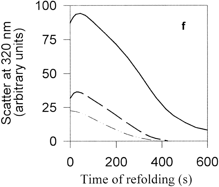
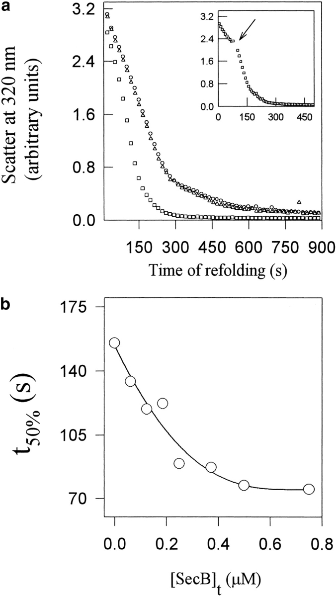
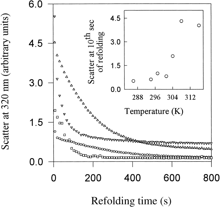
Maltose binding protein (MBP) is widely used as a model for protein folding and export studies. We show here that macroscopic aggregates form transiently during the refolding of MBP at micromolar protein concentrations. Disaggregation occurs spontaneously without any aid, and the refolded material has structure and activity identical to those of the native, nondenatured protein. A considerable fraction of protein undergoing folding partitions into the aggregate phase and can be manually separated from the soluble phase by centrifugation. The separated MBP precipitate can be resolubilized and yields active, refolded protein. This demonstrates that both the soluble and aggregate phases contribute to the final yield of refolded protein. SecB, the cognate Escherichia coli cytosolic chaperone in vivo for MBP, reduces but does not entirely prevent aggregation, whereas GroEL and a variety of other control proteins have no effect. Kinetic studies using a variety of spectroscopic probes show that aggregation occurs through a collapsed intermediate with some secondary structure. The aggregate formed during refolding can convert directly to a near native state without going through the unfolded state. Further, optical and electron microscopic studies indicate that the MBP precipitate is not an amyloid.


Figures

Fig. 1.
Fig. 1.
Kinetics of refolding of MBP at 25°C in the absence of aggregation at low protein concentrations. Refolding was monitored by the changes in intrinsic tryptophan (Trp) fluorescence (a) and changes in circular dichroism (CD) mean residue ellipticity (M.R.E.) at 222 nm (b) that occur with time after dilution of denaturant. The fluorescence measurements in (a) were made in a stopped-flow machine after rapid mixing (deadtime = 1.4 msec) at a protein concentration of 0.5 μM. Each curve is an average of at least a dozen independent traces. Curves in (a) were referenced with respect to the unfolded state signal which is set to zero. The CD measurements in (b) were made following manual mixing of solutions. The MBP concentration was 0.7 μM. The broken line in (b) represents the residual ellipticity in unfolded MBP. In both panels, dots represent the data points and the solid lines represent exponential fits to the data.
Fig. 2.
Fig. 2.
Precipitation and spontaneous resolubilization of MBP while refolding in GdnHCl at high protein concentrations. (a–d) zero time (before addition of MBP), and after 30 sec, 2 min, and 12 hr equilibration of refolding 25 μM MBP in 0.1 M GdnHCl. Sample was mixed intermittently by inverting the cuvette. (e and inset) Aggregation kinetics of 28 μM MBP following rapid mixing in a stopped-flow device, of unfolded MBP with refolding buffer containing 0.4 M GdnHCl. (f) Aggregation kinetics of 2 (dashed/dotted line), 4 (dashed line) and 8 (solid line) μM MBP in 0.2 M GdnHCl following manual mixing. (g) Near-UV CD spectra of native MBP (dashed line) and that of the precipitated and spontaneously redissolved, refolded protein (dotted line). All measurements were made at pH 7.3 and 24°C.
Fig. 2.
Fig. 2.
Precipitation and spontaneous resolubilization of MBP while refolding in GdnHCl at high protein concentrations. (a–d) zero time (before addition of MBP), and after 30 sec, 2 min, and 12 hr equilibration of refolding 25 μM MBP in 0.1 M GdnHCl. Sample was mixed intermittently by inverting the cuvette. (e and inset) Aggregation kinetics of 28 μM MBP following rapid mixing in a stopped-flow device, of unfolded MBP with refolding buffer containing 0.4 M GdnHCl. (f) Aggregation kinetics of 2 (dashed/dotted line), 4 (dashed line) and 8 (solid line) μM MBP in 0.2 M GdnHCl following manual mixing. (g) Near-UV CD spectra of native MBP (dashed line) and that of the precipitated and spontaneously redissolved, refolded protein (dotted line). All measurements were made at pH 7.3 and 24°C.
Fig. 3.
Fig. 3.
Kinetics of refolding of aggregated and nonaggregated protein. (a) Refolding of unaggregated protein present in supernatant (top trace) and protein from the resolubilized pellet (bottom trace), after centrifugation, were measured at 25°C as described in Materials and Methods. A single exponential fit of the data obtained from the supernatant yields an observed rate constant of 0.01 sec−1, while the Trp fluorescence in the resolubilized pellet does not change appreciably with time. (b) Refolding of unaggregated protein present in the supernatant upon refolding at 4°C, using three different protein concentrations: 2 μM (solid line), 10 μM (inverted triangles) and 20 μM (circles). In the latter two cases, aggregated protein was removed by a brief centrifugation prior to measurement. The centrifugation step was omitted for the 2 μM MBP sample because no aggregation was detected for that concentration at the temperature of measurement. In each case, the MBP was refolded in 0.26 M GdnHCl for 10 sec at 4°C prior to removal of aggregate by centrifugation. Refolding was monitored by measurement of tryptophan fluorescence as a function of time of folding. In each case, fluorescence values at any time of folding were normalized to a value of 1 for the value of fluorescence after 500 sec of folding.
Fig. 4.
Fig. 4.
Specific effect of SecB on MBP aggregation. (a) Disaggregation of 2 μM MBP refolding in the absence (circles) and presence of 1 μM SecB (squares) or 1 μM GroEL (triangles). The inset shows the effect of pulsed addition of 1 μM SecB at the indicated time arrow). (b) Effect of SecB concentration on the disaggregation kinetics of 1.4 μM MBP. In all cases, the temperature was 30°C and the samples were stirred magnetically.
Fig. 5.
Fig. 5.
MBP aggregation as a function of temperature. Aggregation kinetics were monitored by optical scatter at 320 nm, during the refolding of 2 μM MBP in refolding buffer at (○) 24°C, (□) 31°C, (▵) 35°C and (▽) 43°C. The inset shows the extent of aggregation after 10 sec of refolding at various temperatures.
Fig. 6.
Fig. 6.
Electron micrographs of (a) insulin amyloid fibrils and (b) the transient MBP precipitate obtained during refolding. Scale bar, 200 nm.

Similar articles

Cited by

References

    1. Agashe, V.R., Shastry, M.C., and Udgaonkar, J.B. 1995. Initial hydrophobic collapse in the folding of barstar. Nature 377 754–757. - PubMed
    1. Anfinsen, C.B. 1973. Principles that govern the folding of protein chains. Science 181 223–230. - PubMed
    1. Betton, J. and Hofnung, M. 1996. Folding of a mutant maltose-binding protein of Escherichia coli which forms inclusion bodies. J. Biol. Chem. 271 8046–8052. - PubMed
    1. Betton, J.M., Sassoon, N., Hofnung, M., and Laurent, M. 1998. Degradation versus aggregation of misfolded maltose-binding protein in the periplasm of Escherichia coli. J. Biol. Chem. 273 8897–8902. - PubMed
    1. Brems, D.N. 1990. Deciphering the second half of the genetic code. In Protein folding (eds. L.M. Gierasch and J. King), pp. 129–135. American Association For The Advancement of Science.

Publication types

MeSH terms