. 2001 Sep;69(3):516-27.
doi: 10.1086/323043. Epub 2001 Jul 20.

Molecular characterization and gene content of breakpoint boundaries in patients with neurofibromatosis type 1 with 17q11.2 microdeletions

Affiliations

Molecular characterization and gene content of breakpoint boundaries in patients with neurofibromatosis type 1 with 17q11.2 microdeletions

D E Jenne et al. Am J Hum Genet. 2001 Sep.

Abstract

Homologous recombination between poorly characterized regions flanking the NF1 locus causes the constitutional loss of approximately 1.5 Mb from 17q11.2 covering > or =11 genes in 5%-20% of patients with neurofibromatosis type 1 (NF1). To elucidate the extent of microheterogeneity at the deletion boundaries, we used single-copy DNA fragments from the extreme ends of the deleted segment to perform FISH on metaphase chromosomes from eight patients with NF1 who had large deletions. In six patients, these probes were deleted, suggesting that breakage and fusions occurred within the adjacent highly homologous sequences. Reexamination of the deleted region revealed two novel functional genes FLJ12735 (AK022797) and KIAA0653-related (WI-12393 and AJ314647), the latter of which is located closest to the distal boundary and is partially duplicated. We defined the complete reading frames for these genes and two expressed-sequence tag (EST) clusters that were reported elsewhere and are associated with the markers SHGC-2390 and WI-9521. Hybrid cell lines carrying only the deleted chromosome 17 were generated from two patients and used to identify the fusion sequences by junction-specific PCRs. The proximal breakpoints were found between positions 125279 and 125479 in one patient and within 4 kb of position 143000 on BAC R-271K11 (AC005562) in three patients, and the distal breakpoints were found at the precise homologous position on R-640N20 (AC023278). The interstitial 17q11.2 microdeletion arises from unequal crossover between two highly homologous WI-12393-derived 60-kb duplicons separated by approximately 1.5 Mb. Since patients with the NF1 large-deletion syndrome have a significantly increased risk of neurofibroma development and mental retardation, hemizygosity for genes from the deleted region around the neurofibromin locus (CYTOR4, FLJ12735, FLJ22729, HSA272195 (centaurin-alpha2), NF1, OMGP, EVI2A, EVI2B, WI-9521, HSA272196, HCA66, KIAA0160, and WI-12393) may contribute to the severe phenotype of these patients.


Figures
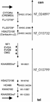
Figure  1
Figure 1
Contigs in 17q11.2 around the neurofibromin locus, order, and transcriptional orientation of functional genes (vertical arrows) and known markers (center) associated with these genes (left). Horizontal arrows (top and bottom) indicate the boundaries of the microdeleted segment that is shown in centromere (top) to telomere (bottom) orientation. Publicly available assemblies of draft segments are shown schematically (right), with indications of the direction (vertical arrows) of numbering. Conflicts between these tentative sequence paths and experimental data are reported in the present study. Asterisks indicate the four genes (AJ314645 [FLJ22729 and SHGC-2390], AJ314646 [KIAA0665rel and WI-9521], AJ314647 [KIAA0563rel and 1I-12393], and AJ314648 [FLJ12735]) whose cDNA sequences have been completed and submitted to GenBank.
Figure  2
Figure 2
Structure and gene content of 17q11.2 breakpoint regions in patients with NF1 who have microdeletions. A, the recombination event between highly homologous sequences located 400 kb upstream (top) and 700 kb downstream of the NF1 gene. BAC clones characterized and used as FISH probes in this study are shown above and below the lines that represent the proximal and distal breakpoint regions. The exons of both functional genes (KIAA0160 and WI-12393), as well as the nine exons of a pseudogene fragment derived from KIAA0160 and several pseudogene exons derived from WI-12393 by duplications and local rearrangements, are shown (black vertical bars with numbers). The vertical long arrows indicate the fusion points that we identified in four of six patients. Clones from the RPCI-11 human male BAC library (Pieter de Jong, Roswell Park Cancer Institute) are labeled with the prefix R-, and clones without a prefix were provided by the California Institute of Technology. Clone designations are followed by the approximate size of the insert in parentheses; BAC end sequences were obtained from the Institute for Genome Research and are given by the letter F (T7 end) and R (SP6 end). B, Schematic representation of two highly homologous segments (black horizontal bars labeled “proximal duplicon” and “distal duplicon”) that are implicated in the 17q11.2 microdeletion syndrome. This scheme illustrates the complete deletion of the functional WI-12393 gene (boxed), together with 12 additional genes (not shown, indicated by the dotted interrupted line) in all six patients and the location and orientation of the proximal and distal WI-12393–derived ∼60-kb duplicon. Homologous WI-12393 exons (WI-12393 pseudogene exons) are indicated (thin vertical bars). The precise breakpoint has been mapped in four patients: COS, PFA, WIN, and ULM (see fig. 5).
Figure  3
Figure 3
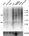
Northern blot analysis of WI-12393 with RNA isolated from different human tissues revealed a low expression in several adult tissues. The lower arrow indicates the 1.7-kb transcript expressed in all tissues examined; the upper arrow indicates a 4.4-kb spanning mRNA. In skeletal muscle, an additional isoform of ∼7.5 kb is visible. On the right side, the positions of bands of a size marker are given in kilobases.
Figure  4
Figure 4
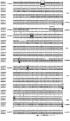
Alignment of three genomic sequences for the most-conserved WI-12393–derived duplicons found in 17q11.2 (BACs R-271K11 and R-640N20) and 19p13.2 (BAC 2189E23). Only those sections (125241–125480, 142289–142585, and 144061–144480) that cover breakpoint fusions and the binding sites for the primers DJ2290 and DJ2314 (140825–140860 and 144733–144793) are depicted. The locations of the forward primer, DJ2290, and the backward primer, DJ2314, which were designed to amplify fusion fragments from total DNA of patients are marked by horizontal arrows above and below the respective sequences. The nucleotides are numbered according to the completely sequenced clone R-271K11 (AC005562) at the beginning and end of each section. Every fifth and tenth nucleotide position is marked by one or two dots above the first row of the alignments. The asterisks below the aligned sequences show positions at which one or more sequences vary. The last unique nucleotide from the proximal (middle row) and distal (distal row) duplicon that border the fusion segment in the patients ULM, COS, WIN, and PFA (right) are shown in white on a black background; all three nucleotides at that position are boxed.
Figure  5
Figure 5
PCR analysis of the fusion between the proximal and distal duplicon on 17q11.2. The primers DJ2290 and DJ2314 were designed specifically to prime the proximal and distal duplicon sequence (see fig. 4). Thus, only certain patients (COS, PFA, and WIN), who had a proximal breakpoint between position 140852 and 144761 (numbering according to AC005562) and a distal breakpoint within the same homologous segment on R-640N20, show a ∼4-kb amplified product that was identified by agarose gel electrophoresis. The breakpoints of patients WEH and KAE have not been identified at the sequence level. N1–N5 are DNA samples from unaffected control individuals.
Figure  6
Figure 6
FISH analysis of metaphase chromosomes and interphase cells of the patient ULM, who had a 17q11.2 microdeletion, and of GGO, PTR, and PPY. The probes were BACs spanning the distal (R-640N20) or the proximal duplicon (R-271K11) (green signals) in combination with the differentially labeled single-copy BAC 542B22 (red signal) located between the duplicons. In all panels, the main hybridization signals of the duplicon BACs are indicated by white arrows, whereas the sites of weak cross-hybridization of these BACs are indicated by white arrowheads. The hybridization sites of BAC 542B22 are emphasized by red arrows. A, BAC R-640N20 (green signal) and BAC 542B22 (red signal) hybridized to metaphase chromosomes and an interphase nucleus of a patient (ULM) with NF1. On the normal chromosome 17, a strong signal of R-640N20 is observed, whereas on the chromosome 17 with deletions, only a reduced signal of this BAC is seen at 17q11.2 (white arrows). BAC 542B22 hybridizes only to the normal chromosome 17 (red arrow). Weak cross-hybridization signals of BAC R-640N20 are visible at 17q21-q24, 19p12-p13.1, and 3q13.3-3q21 (white arrowheads). A, inset, an interphase nucleus of patient ULM with 542B22 (red arrow) flanked by R-640N20 (green signals, white arrows). A reduced signal of R-640N20 is visible in another region of the nucleus derived from the deleted chromosome 17. B, Reciprocal translocation between the GGO chromosomes orthologous to human chromosomes 17 and 5 (GGO chromosomes 4 and 19). Strong signals of BAC R-271K11 (white arrows) and a weaker signal of the single-copy BAC 542B22 (red arrows) are seen at GGO 4p11-p12 in the region orthologous to human 17q11-q12. A weak signal of BAC R-271K11 has been observed at 4p16-17 (data not shown) and at 20p12-p13, which is orthologous to human 19p12-p13.1 (white arrowheads). B, inset, BAC R-271K11 flanking BAC 542B22 on both sites in an interphase cell. C, BAC R-271K11 (white arrows, green signals) visible at PTR chromosome 19p11-p12, owing to a pericentric inversion and cohybridizing there with BAC 542B22 (red arrows). Weak cross-hybridization signals of BAC R-271K11 are observed at PTR 19q14-q22 and at PTR 20p12-13, orthologous to human 17q21-24 and human 19p12-p13.1, respectively (white arrowheads). C, inset, an interphase cell of PTR, in which the single-copy BAC 542B22 (red signal) is flanked by BAC R-271K11 (green signals). D, BAC R-640N20 (white arrows) cohybridized with single-copy BAC 542B22 (red arrow) to metaphase chromosomes of the PPY. A strong hybridization signal of BAC R-640N20 and a weaker signal of BAC 542B22 are visible at PPY 19q11-q12, orthologous to human 17q11-q12. Additionally, weak signals of BAC R-640N20 are seen at PPY 19q14-q22 and at PPY 20p12-p13. D, inset, an interphase cell of PPY, in which the BAC 542B22 (red signal) is flanked by BAC R-640N20 (green signals).

References

Electronic-Database Information

    1. GenBank, http://www.ncbi.nlm.nih.gov/Genbank (for IMAGE clone y174c12)
    1. Human Genome, http://www.ncbi.nlm.nih.gov/genome/guide/human (for draft sequences of the human genome)
    1. Institute for Genome Research, http://www.tigr.org (for draft sequences of the human genome)
    1. Online Mendelian Inheritance in Man (OMIM), http://www.ncbi.nlm.nih.gov/Omim (for NF1 [MIM 162200]) - PubMed

References

    1. Ars E, Serra E, Garcia J, Kruyer H, Gaona A, Lazaro C, Estivill X (2000) Mutations affecting mRNA splicing are the most common molecular defects in patients with neurofibromatosis type 1. Hum Mol Genet 9:237–247 - PubMed
    1. Boerkoel CF, Inoue K, Reiter LT, Warner LE, Lupski JR (1999) Molecular mechanisms for CMT1A duplication and HNPP deletion. Ann N Y Acad Sci 883:22–35 - PubMed
    1. Cnossen MH, van der Est MN, Breuning MH, van Asperen CJ, Breslau-Siderius EJ, van der Ploeg AT, de Goede-Bolder A, van den Ouweland AM, Halley DJ, Niermeijer MF (1997) Deletions spanning the neurofibromatosis type 1 gene: implications for genotype-phenotype correlations in neurofibromatosis type 1? Hum Mutat 9:458–464 - PubMed
    1. Colman SD, Williams CA, Wallace MR (1995) Benign neurofibromas in type 1 neurofibromatosis (NF1) show somatic deletions of the NF1 gene. Nat Genet 11:90–92 - PubMed
    1. Dorschner MO, Sybert VP, Weaver M, Pletcher BA, Stephens K (2000) NF1 microdeletion breakpoints are clustered at flanking repetitive sequences. Hum Mol Genet 9:35–46 - PubMed

Publication types

Associated data