. 2001 Jul 23;154(2):275-81.
doi: 10.1083/jcb.200105046.

Regulation of limb patterning by extracellular microfibrils

Affiliations

Regulation of limb patterning by extracellular microfibrils

E Arteaga-Solis et al. J Cell Biol. .

Abstract

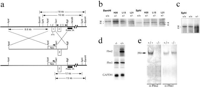
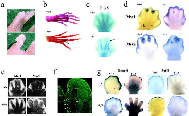
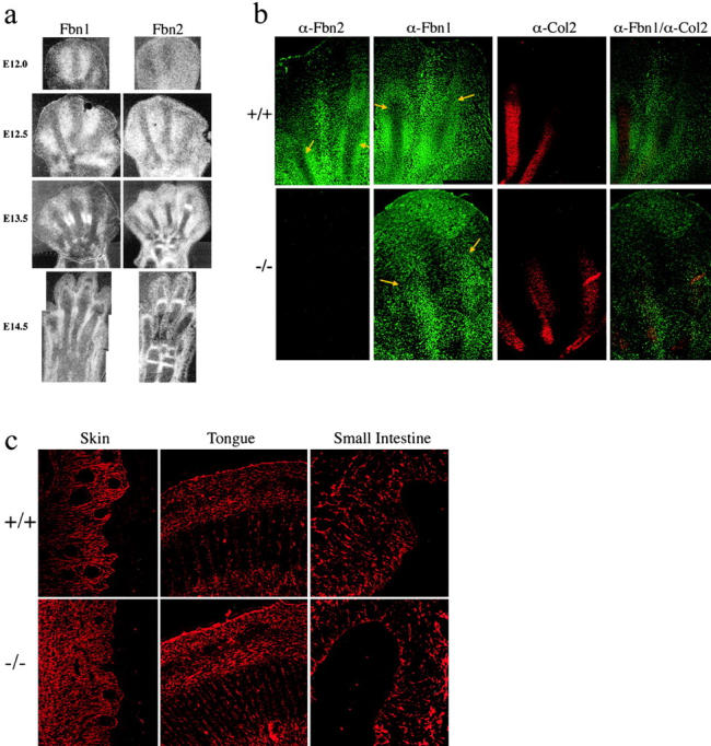
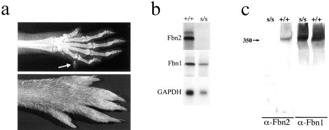
To elucidate the contribution of the extracellular microfibril-elastic fiber network to vertebrate organogenesis, we generated fibrillin 2 (Fbn2)-null mice by gene targeting and identified a limb-patterning defect in the form of bilateral syndactyly. Digit fusion involves both soft and hard tissues, and is associated with reduced apoptosis at affected sites. Two lines of evidence suggest that syndactily is primarily due to defective mesenchyme differentiation, rather than reduced apoptosis of interdigital tissue. First, fusion occurs before appearance of interdigital cell death; second, interdigital tissues having incomplete separation fail to respond to apoptotic clues from implanted BMP-4 beads. Syndactyly is associated with a disorganized matrix, but with normal BMP gene expression. On the other hand, mice double heterozygous for null Fbn2 and Bmp7 alleles display the combined digit phenotype of both nullizygotes. Together, these results imply functional interaction between Fbn2-rich microfibrils and BMP-7 signaling. As such, they uncover an unexpected relationship between the insoluble matrix and soluble factors during limb patterning. We also demonstrate that the Fbn2- null mutation is allelic to the recessive shaker-with-syndactyly (sy) locus on chromosome 18.


Figures

Figure 1.
Figure 1.
Schematic illustration of Fbn2 gene targeting. (a) From top to bottom: restriction map of the targeted genomic region which indicates the relative positions of exons 1 and 2 (□) and probe 3 A (▪), as well as the sizes of relevant DNA fragments; targeting vector with the arrow signifying the transcriptional orientation of the neo gene (□); null Fbn2 allele with the predicted sizes of mutant BamHI and SphI fragments. (b) Southern hybridization of BamHI and SphI-digested DNA from wild-type (+/+) and correctly targeted (+/−) ES clones. (c) Southern hybridization of SphI-digested tail DNA from the chimeric progeny demonstrating germ line transmission of the Fbn2 mutation in one animal (+/−). (d) Northern hybridizations to Fbn1, Fbn2, and GAPDH of RNA from wild-type (+/+) and mutant (−/−) newborn lungs. (e) Western analysis of conditioned media from wild-type (+/+) and mutant (−/−) primary fibroblast cultures with antibodies against Fbn1 and 2 (α-Fbn1 and α-Fbn2); size of the fibrillins (350 kD, arrow) was estimated against the migration of protein standards (not shown).
Figure 2.
Figure 2.
Analysis of Fbn2 mutant limbs. (a) Forelimbs of wild-type (+/+) and mutant (−/−) newborn mice showing contractures of the wrist and metacarpal joints. (b) Skeletal preparation of adult hindlimbs of wild-type (+/+) and mutant (−/−) mice with arrow pointing to hard tissue syndactily in the latter. (c) Staining of cartilaginous elements of E13.5 hindlimbs of wild-type (+/+) and mutant (−/−) embryos with arrow pointing to digit fusion in the latter. (d) Whole-mount hybridizations to Msx probes of E13.5 wild-type (+/+) and mutant (−/−) hindlimbs with implanted BMP-4–coated beads. (e) In situ hybridizations to Msx probes of wild-type (+/+) and mutant (−/−) E13.5 hindlimbs. (f) In situ TUNEL assay of E13.5 Fbn2 −/− hindlimb with arrows pointing to apoptotic signals; note the fewer number of dying cells in the interdigital region destined to fuse, compared with the interdigital ray that will regress and lead to digit separation. (g) Whole-mount in situ hybridizations to Bpm-4 and Fgf-8 of E11.5-E13.5 hindlimbs of wild-type (+/+) and mutant (−/−) embryos.
Figure 3.
Figure 3.
Fbn expression and microfibrillar morphology in mutant Fbn2 mice. (a) In situ hybridization to Fbn1 and Fbn2 probes of E12.0-14.5 hindlimbs of wild-type (+/+) and mutant (−/−) embryos. (b) Immunofluorescence of wild-type (+/+) and mutant (−/−) E13.5 hindlimbs with antibodies against Fbn1 (α-Fbn1), Fbn2 (α-Fbn2), and type II collagen (α-Col 2). Arrows in Fbn-immunostained samples point to the developing digital (chondrogenic) rays that are intensively stained by the type II collagen antibody. Note the distinct organization of the microfibrillar network in the interdigital and digital rays of the wild-type autopod that is lost in the mutant samples. The disorganized architecture of the mutant matrix can be best appreciated in the superimposition of the α-Fbn1 and α-Col2 images. (c) Immunofluorescence of E13.5 skin, tongue, and small intestine from wild-type (+/+) and mutant (−/−) embryos.
Figure 4.
Figure 4.
Analysis of Fbn2 +/−/Bmp-7 +/− and syfp/syfp mice. (a) Limb patterning defects in double heterozygous Fbn2/Bmp-7–null mice. The x-ray on top shows an extra digit, whereas the picture at the bottom documents soft tissue syndactily in adult hindlimbs. (b) Northern hybridization of RNA from wild-type (+/+) and syfp/syfp(s/s) newborn mice. (c) Western analysis of conditioned media from wild type (+/+) and syfp/syfp(s/s) primary fibroblast cultures; size of fibrillins (350 kD, arrow) was estimated against the migration of protein standards (not shown).

References

    1. Chen, Y., and X. Zhao. 1998. Shaping limbs by apoptosis. J. Exp. Zool. 282:691–702. - PubMed
    1. Christian, J.L. 2000. BMP, Wnt and Hedgehog signals: how far can they go? Curr. Opin. Cell Biol. 12:244–249. - PubMed
    1. Dahna, R.D., and J.F. Fallon. 2000. Interdigital regulation of digit identity and homeotic transformation by modulated BMP signaling. Science. 289:438–441. - PubMed
    1. Dudley, A.T., Lyons K.M., and Robertson E.J. 1995. A requirement for bone morphogenetic protein-7 during development of the mammalian kidney and eye. Genes Dev. 9:2795–2807. - PubMed
    1. Flaumenhaft, R., and D.B. Rifkin. 1991. Extracellular matrix regulation of growth factor and protease activity. Curr. Opin. Cell Biol. 3:817–823. - PubMed

Publication types

MeSH terms