. 2001 Jul 23;154(2):345-54.
doi: 10.1083/jcb.200102032.

Novel p62dok family members, dok-4 and dok-5, are substrates of the c-Ret receptor tyrosine kinase and mediate neuronal differentiation

Affiliations

Novel p62dok family members, dok-4 and dok-5, are substrates of the c-Ret receptor tyrosine kinase and mediate neuronal differentiation

J Grimm et al. J Cell Biol. .

Abstract

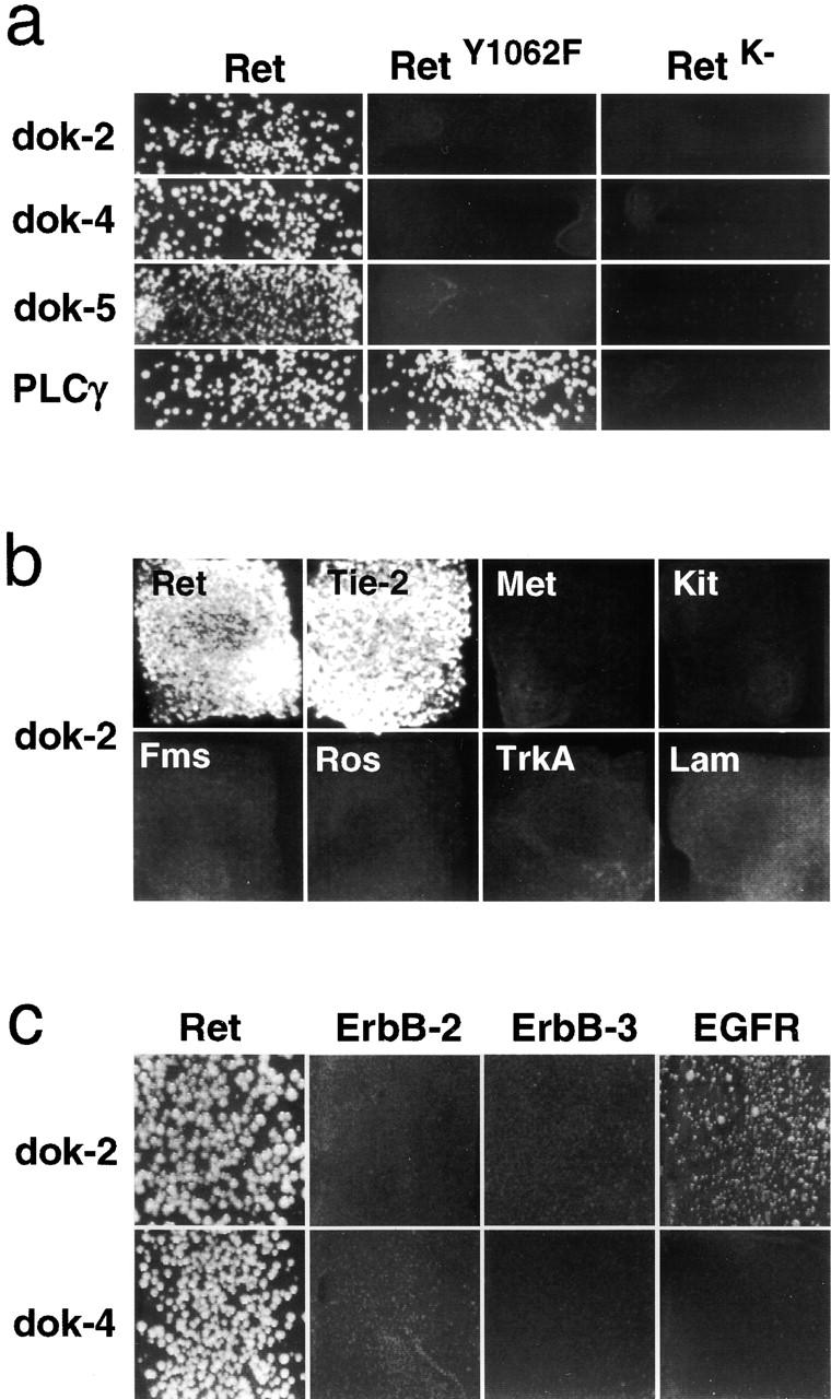
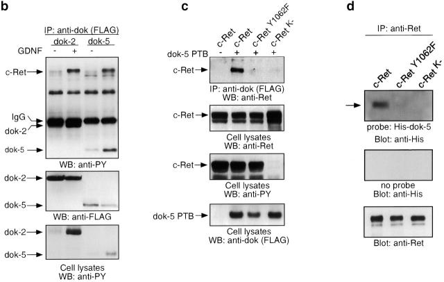
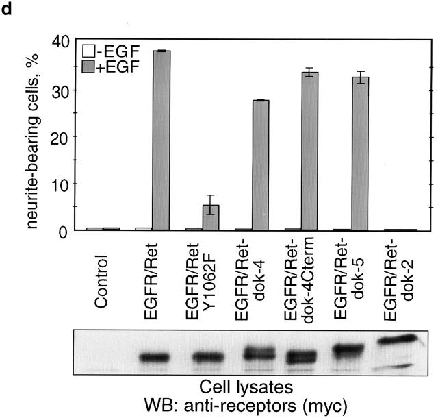
Docking proteins are substrates of tyrosine kinases and function in the recruitment and assembly of specific signal transduction molecules. Here we found that p62dok family members act as substrates for the c-Ret receptor tyrosine kinase. In addition to dok-1, dok-2, and dok-3, we identified two new family members, dok-4 and dok-5, that can directly associate with Y1062 of c-Ret. Dok-4 and dok-5 constitute a subgroup of dok family members that is coexpressed with c-Ret in various neuronal tissues. Activated c-Ret promotes neurite outgrowth of PC12 cells; for this activity, Y1062 in c-Ret is essential. c-Ret/dok fusion proteins, in which Y1062 of c-Ret is deleted and replaced by the sequences of dok-4 or dok-5, induce ligand-dependent axonal outgrowth of PC12 cells, whereas a c-Ret fusion containing dok-2 sequences does not elicit this response. Dok-4 and dok-5 do not associate with rasGAP or Nck, in contrast to p62dok and dok-2. Moreover, dok-4 and dok-5 enhance c-Ret-dependent activation of mitogen-activated protein kinase. Thus, we have identified a subclass of p62dok proteins that are putative links with downstream effectors of c-Ret in neuronal differentiation.


Figures

Figure 1.
Figure 1.
Interaction of dok family members with c-Ret and other receptor tyrosine kinases in the yeast two-hybrid system. Growth of yeast on selective medium. (a) Dok-2, -4, and -5 interact with wild-type c-Ret, but not with c-Ret receptors harboring a Y1062F mutation or an inactive kinase (Ret K, K758M; Liu et al., 1996). (b) Dok-2 interacts with c-Ret and Tie-2, but not with other receptor tyrosine kinases tested. Lam, lamin. (c) Dok-2, but not dok-4, interacts weakly with the EGFR but not other members of the EGFR family.
Figure 2.
Figure 2.
The dok protein family and newly identified dok members. (a) Domain structure of dok family members. PH and PTB domains are marked grey and black, and the positions of tyrosine residues (Y) and PXXP motifs (P) are indicated. (b and c) Deduced amino acid sequences of mouse dok-4 and dok-5, respectively. PH domains are boxed and PTB domains are underlined. (d) Phylogenetic tree of dok family members. Sequences of the PTB domains were aligned.
Figure 2.
Figure 2.
The dok protein family and newly identified dok members. (a) Domain structure of dok family members. PH and PTB domains are marked grey and black, and the positions of tyrosine residues (Y) and PXXP motifs (P) are indicated. (b and c) Deduced amino acid sequences of mouse dok-4 and dok-5, respectively. PH domains are boxed and PTB domains are underlined. (d) Phylogenetic tree of dok family members. Sequences of the PTB domains were aligned.
Figure 3.
Figure 3.
Expression of dok family members and c-Ret in mouse embryos. Whole-mount in situ hybridization of E12.5 and E13.5 mouse embryos followed by semithin sectioning. (a, d, and g–i) Labeling with a dok-4–specific probe. Dok-4–positive cells are observed in the ventral part of the spinal cord (sc), the dorsal root ganglia (drg), the trigeminal (tg) and geniculate ganglia (ge), and in endothelia (arrowheads). g–i are sections through lung, tail, and kidney, respectively. (b and e) Labeling with a dok-5–specific probe. (c, f, and j) Labeling with a c-Ret–specific probe. Panel j shows expression in the buds of the ureter epithelium in the kidney. (k) Whole-mount in situ hybridization with a dok-2–specific probe. Dok-2 is specifically expressed in islands of cells within the embryonal liver (li). Bars, 0.2 mm.
Figure 4.
Figure 4.
Northern blotting of dok and c-Ret transcripts in adult mouse tissues. cDNA probes specific for dok-4 (a), dok-5 (b), dok-2 (c), c-Ret (d), or actin were used. The positions of the transcripts are indicated by arrows.
Figure 5.
Figure 5.
Interaction and phosphorylation of dok family members by c-Ret and Tie-2 in mammalian cells. (a) Coimmunoprecipitation of c-Ret with dok family members in 293 cells. Flag-tagged doks and c-Ret were transfected, and immune complexes were precipitated and immunoblotted with anti-Ret or anti-PY antibodies. Expression and phosphorylation of dok family members and c-Ret is shown in the lower gels. (b) Dok-2 and dok-5 are tyrosine phosphorylated by endogenous c-Ret upon GDNF stimulation in Neuro 2A cells. Cells stably expressing GFRα1 were used. (c) Coimmunoprecipitation of the PTB domain of dok-5 (amino acids 109–261) and c-Ret constructs. Expression levels and phophorylation are shown in the lower gels. (d) Far Western blot demonstrating direct interaction of dok-5 with c-Ret but not mutant c-Ret. (e) A dok-5–specific antibody recognizes dok-5 in transfected 293 cells and in preparations of spinal cord and dorsal root ganglia, which also express c-Ret (top). The anti–dok-5 antibody coimmunoprecipitates phosphorylated c-Ret in extracts of spinal cords and dorsal root ganglia (bottom). (f) Coimmunoprecipitation of Tie-2 and dok-4 in 293 cells. Flag-tagged dok-4 and endothelial receptor tyrosine kinases were transfected, and immune complexes were precipitated and immunoblotted with anti-PY antibodies. Expression of phosphorylated receptors and dok-4 is shown in the lower gels.
Figure 5.
Figure 5.
Interaction and phosphorylation of dok family members by c-Ret and Tie-2 in mammalian cells. (a) Coimmunoprecipitation of c-Ret with dok family members in 293 cells. Flag-tagged doks and c-Ret were transfected, and immune complexes were precipitated and immunoblotted with anti-Ret or anti-PY antibodies. Expression and phosphorylation of dok family members and c-Ret is shown in the lower gels. (b) Dok-2 and dok-5 are tyrosine phosphorylated by endogenous c-Ret upon GDNF stimulation in Neuro 2A cells. Cells stably expressing GFRα1 were used. (c) Coimmunoprecipitation of the PTB domain of dok-5 (amino acids 109–261) and c-Ret constructs. Expression levels and phophorylation are shown in the lower gels. (d) Far Western blot demonstrating direct interaction of dok-5 with c-Ret but not mutant c-Ret. (e) A dok-5–specific antibody recognizes dok-5 in transfected 293 cells and in preparations of spinal cord and dorsal root ganglia, which also express c-Ret (top). The anti–dok-5 antibody coimmunoprecipitates phosphorylated c-Ret in extracts of spinal cords and dorsal root ganglia (bottom). (f) Coimmunoprecipitation of Tie-2 and dok-4 in 293 cells. Flag-tagged dok-4 and endothelial receptor tyrosine kinases were transfected, and immune complexes were precipitated and immunoblotted with anti-PY antibodies. Expression of phosphorylated receptors and dok-4 is shown in the lower gels.
Figure 6.
Figure 6.
Dok-4 and dok-5 mediate c-Ret–dependent neurite outgrowth. PC12 cells were transfected with retroviruses expressing constructs which encode the extracellular EGFR domain and intracellular c-Ret/dok fusions, and neurite outgrowth was monitored after EGF treatment. (a) Cells expressing an EGFR/c-Ret hybrid or a hybrid containing a Y1062F mutation. (b) Cells expressing EGFR/c-Ret/dok-4 fusions lacking Y1062 of c-Ret, or a construct only containing the COOH terminus of dok-4. (c) Cells expressing EGFR/c-Ret/dok-5 or EGFR/c-Ret/dok-2 fusions. (d) Quantification of neurite outgrowth and a Western blot of hybrid receptor expression of experiments a–c. Activated Ret induces neurite outgrowth, but not when Y1062 is mutated. Also, Ret fusions with dok-4 and dok-5, but not dok-2, promote axonal outgrowth. For this activity, only the COOH terminus of dok-4 downstream of the PTB domain is required.
Figure 6.
Figure 6.
Dok-4 and dok-5 mediate c-Ret–dependent neurite outgrowth. PC12 cells were transfected with retroviruses expressing constructs which encode the extracellular EGFR domain and intracellular c-Ret/dok fusions, and neurite outgrowth was monitored after EGF treatment. (a) Cells expressing an EGFR/c-Ret hybrid or a hybrid containing a Y1062F mutation. (b) Cells expressing EGFR/c-Ret/dok-4 fusions lacking Y1062 of c-Ret, or a construct only containing the COOH terminus of dok-4. (c) Cells expressing EGFR/c-Ret/dok-5 or EGFR/c-Ret/dok-2 fusions. (d) Quantification of neurite outgrowth and a Western blot of hybrid receptor expression of experiments a–c. Activated Ret induces neurite outgrowth, but not when Y1062 is mutated. Also, Ret fusions with dok-4 and dok-5, but not dok-2, promote axonal outgrowth. For this activity, only the COOH terminus of dok-4 downstream of the PTB domain is required.
Figure 7.
Figure 7.
Dok-4 and dok-5 do not associate with rasGAP or Nck, and mediate MAP kinase and Elk-1 transactivation. (a) Coimmunoprecipitation of dok family members and rasGAP or Nck in 293 cells in the presence and absence of c-Ret. Flag-tagged doks were transfected, and immune complexes with endogenous rasGAP and Nck were precipitated and immunoblotted. Phosphorylation of dok family members by c-Ret is shown in the lower gel. (b) Western blot demonstrating activation of Erk1/2 by fusion constructs of EGFR/c-Ret with dok-4 and dok-5 but not dok-2. PC12 cells from experiment shown in Fig. 6 were used, and Erk-1/2 phosphorylation was determined by a specific antibody. (c) Elk-1 transactivation by fusion constructs of EGFR/c-Ret with dok-4 and dok-5 but not dok-2. Neuro 2A cells were transiently transfected with the indicated constructs together with Elk-1/Gal4 and Gal4-luciferase reporter plasmids. Cells were treated with EGF for 5 h and Luciferase activity was then determined. (d) Inhibition of c-Ret–induced Elk-1 activation by dok-2. 293 cells were transfected with c-Ret, increasing amounts of dok-2 and reporter plasmids. Cells were stimulated for 5 h with 50 ng/ml GDNF and 0.5 μg/ml soluble GFRα1. (e) Inhibition of MEN2A Ret induced activation of Elk-1 by dok-2 but not dok-5. 293 cells were transfected with Ret C634R, increasing amounts of dok-2 or dok-5, and reporter plasmids. Luciferase activity was determined 48 h posttransfection.
Figure 7.
Figure 7.
Dok-4 and dok-5 do not associate with rasGAP or Nck, and mediate MAP kinase and Elk-1 transactivation. (a) Coimmunoprecipitation of dok family members and rasGAP or Nck in 293 cells in the presence and absence of c-Ret. Flag-tagged doks were transfected, and immune complexes with endogenous rasGAP and Nck were precipitated and immunoblotted. Phosphorylation of dok family members by c-Ret is shown in the lower gel. (b) Western blot demonstrating activation of Erk1/2 by fusion constructs of EGFR/c-Ret with dok-4 and dok-5 but not dok-2. PC12 cells from experiment shown in Fig. 6 were used, and Erk-1/2 phosphorylation was determined by a specific antibody. (c) Elk-1 transactivation by fusion constructs of EGFR/c-Ret with dok-4 and dok-5 but not dok-2. Neuro 2A cells were transiently transfected with the indicated constructs together with Elk-1/Gal4 and Gal4-luciferase reporter plasmids. Cells were treated with EGF for 5 h and Luciferase activity was then determined. (d) Inhibition of c-Ret–induced Elk-1 activation by dok-2. 293 cells were transfected with c-Ret, increasing amounts of dok-2 and reporter plasmids. Cells were stimulated for 5 h with 50 ng/ml GDNF and 0.5 μg/ml soluble GFRα1. (e) Inhibition of MEN2A Ret induced activation of Elk-1 by dok-2 but not dok-5. 293 cells were transfected with Ret C634R, increasing amounts of dok-2 or dok-5, and reporter plasmids. Luciferase activity was determined 48 h posttransfection.

Similar articles

Cited by

References

    1. Alberti, L., M.G. Borrello, S. Ghizzoni, F. Torriti, M.G. Rizzetti, and M.A. Pierotti. 1998. Grb2 binding to the different isoforms of Ret tyrosine kinase. Oncogene. 17:1079–1087. - PubMed
    1. Arighi, E., L. Alberti, F. Torriti, S. Ghizzoni, M.G. Rizzetti, G. Pelicci, B. Pasini, I. Bongarzone, C. Piutti, M.A. Pierotti, and M.G. Borrello. 1997. Identification of Shc docking site on Ret tyrosine kinase. Oncogene. 14:773–782. - PubMed
    1. Asai, N., H. Murakami, T. Iwashita, and M. Takahashi. 1996. A mutation at tyrosine 1062 in MEN2A-Ret and MEN2B-Ret impairs their transforming activity and association with shc adaptor proteins. J. Biol. Chem. 271:17644–17649. - PubMed
    1. Avantaggiato, V., N.A. Dathan, M. Grieco, N. Fabien, D. Lazzaro, A. Fusco, A. Simeone, and M. Santoro. 1994. Developmental expression of the RET protooncogene. Cell Growth Differ. 5:305–311. - PubMed
    1. Behrens, J., J.P. von Kries, M. Kuhl, L. Bruhn, D. Wedlich, R. Grosschedl, and W. Birchmeier. 1996. Functional interaction of β-catenin with the transcription factor LEF-1. Nature. 382:638–642. - PubMed

MeSH terms

Substances