. 2001 Jul 23;154(2):415-26.
doi: 10.1083/jcb.200103035.

mua-3, a gene required for mechanical tissue integrity in Caenorhabditis elegans, encodes a novel transmembrane protein of epithelial attachment complexes

Affiliations

mua-3, a gene required for mechanical tissue integrity in Caenorhabditis elegans, encodes a novel transmembrane protein of epithelial attachment complexes

M Bercher et al. J Cell Biol. .

Abstract

Normal locomotion of the nematode Caenorhabditis elegans requires transmission of contractile force through a series of mechanical linkages from the myofibrillar lattice of the body wall muscles, across an intervening extracellular matrix and epithelium (the hypodermis) to the cuticle. Mutations in mua-3 cause a separation of the hypodermis from the cuticle, suggesting this gene is required for maintaining hypodermal-cuticle attachment as the animal grows in size postembryonically. mua-3 encodes a predicted 3,767 amino acid protein with a large extracellular domain, a single transmembrane helix, and a smaller cytoplasmic domain. The extracellular domain contains four distinct protein modules: 5 low density lipoprotein type A, 52 epidermal growth factor, 1 von Willebrand factor A, and 2 sea urchin-enterokinase-agrin modules. MUA-3 localizes to the hypodermal hemidesmosomes and to other sites of mechanically robust transepithelial attachments, including the rectum, vulva, mechanosensory neurons, and excretory duct/pore. In addition, it is shown that MUA-3 colocalizes with cytoplasmic intermediate filaments (IFs) at these sites. Thus, MUA-3 appears to be a protein that links the IF cytoskeleton of nematode epithelia to the cuticle at sites of mechanical stress.


Figures
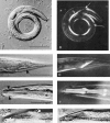
Figure 1.
Figure 1.
DIC and polarized light micrographs of mua-3 animals. (A) DIC micrograph of a mua-3(rh169) animal showing typical curled posture. (B) Same animal as in A visualized by polarized light, a muscle band that has detached from the ventral body wall is visible as a bright birefringent band (arrows) that has collapsed dorsally. (C) DIC micrograph of mua-3(rh195) homozygote showing localized separation of tissues from the cuticle at tail (arrowhead). (D) Same as C under polarized light showing separation of ventral body wall muscles from tip of tail. (E) DIC micrograph of mua-3(rh195) homozygote showing localized separation of tissues from cuticle in head region (arrowheads). (F) Same as C under polarized light showing rearward retraction of the body wall muscles from the area of tissue separation. (G and H) Enlargements of area of tissue separation, two different focal planes of same animal as in E. Note retracted muscles (arrowhead in G) about the region of separation. The large blister labeled a appears to due to separation of the apical hypodermal membrane from the cuticle. Bars: (A–F) 100 μm; (G and H) 10 μm.
Figure 2.
Figure 2.
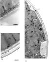
mua-3 is required for attachment between the apical hypodermal surface and cuticle. (A and B) TEM micrographs of adult wild-type and mua-3(rh195) body wall in intact muscle quadrants. Body wall muscle is indicated by m, hypodermis h, and the basal layer of the cuticle by bc. In the mutant an obviously substantial gap (asterisk) between apical hypodermis and cuticle can be observed. (C) TEM micrograph of rh195 mutant body wall in region of muscle detachment. Large gaps indicated by (asterisk) are observed between apical hypodermis and basal cuticle. Hypodermis (h) remains tightly apposed to muscle (m) in the detachment region. A portion of the hypodermis (region between arrows) has become decompressed in the detachment region, whereas immediately under the gaps it is still compressed. Bars, 0.5 μm.
Figure 8.
Figure 8.
Genetic (top) and physical (middle and bottom) maps of the mua-3 region. The vertical lines on the bottom map represent the location of restriction sites in the DNA sequence. The extents of F55E6, T20G5, and pOT22 relative to these sites are shown. The location of the open reading frames that comprise mua-3 are shown as a gray bar. F55E6 and pOT22 rescue mua-3 mutant animals, T20G5 does not.
Figure 3.
Figure 3.
Structures RNA and protein from mua-3 gene. (A) RNA transcript structure as determined from RT-PCR–generated cDNA (EMBL/GenBank/DDBJ accession no. AAD29428). The relative positions and sizes of exons are shown, and the splicing joins are indicated by Vs. The 5′ end transsplices to SL1. (B) The domain/module structure of the MUA-3 protein as deduced from the predicted amino acid sequence. The two letter module labels conform to those used in Hutter et al., (2000), see footnotes.
Figure 4.
Figure 4.
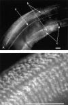
Localization of MUA-3 to hemidesmosome zone of hypodermis in animals stained with anti–MUA-3 antibodies. (A) Immunofluorescence in hypodermis-bordering muscle quadrants using MUA-3 antibody recognized with a FITC secondary antibody. a, ALM; b, hemidesmosome region of hypodermis; c, gaps where nerve processes cross between hypodermis and muscle. (B) Enlargement of hemidesmosome region of hypodermis. Bars, 10 μm.
Figure 6.
Figure 6.
MUA-3 colocalizes with cytoplasmic IFs at hypodermal hemidesmosomes. It is also associated with MUA-3 at other sites of MUA-3 localization in animals double stained with anti-p70 (IF) and MUA-3 antibodies. (A) IFs associated with hypodermal hemidesmosomes visualized with anti-p70 in an adult. (B) Same animal stained for MUA-3. (C) Merge of A and B. (D) p70 localization in threefold embryo. Embryo is folded on itself: t indicates location of tail; the dashed line shows location of fold between posterior and central body regions, the anterior region is folded under the posterior end and not in field of view. Ventral- and dorsal-staining stripes in hypodermis are visible and indicated by arrowheads and an arrow, respectively. (E) Same embryo showing MUA-3 localization. Note stripe pattern similar to p70, indicated by arrowheads and an arrow. (F) Merge of E and F. (G) Duct and pore cells showing p70 IF localization. Ventral is down, dashed line indicates location of body wall (not visible), arrows indicate pore cell (note lack of staining in hollow lumen), and the arrowhead indicates the duct cell. (H) Same, showing MUA-3 localization. Note that MUA-3 is located closer to the lumenal cuticle and extends closer to body wall than the p70, especially apparent in the pore cell (arrows). Arrowhead indicates duct cell. (I) Merge of G and H. Bars, 10 μm.
Figure 5.
Figure 5.
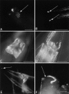
Localization of MUA-3 to nonmuscle body wall muscle sites. (A) Amphids, indicated by arrow. (B) Phasmid sensilla in tail (arrow). (C and D) Rectum, shown in two focal planes, posterior to upper right. The staining forms a collar completely surrounding the rectal valve, where the sphincter muscle sits. Hypodermis bordering body wall muscle can also be seen in D. (E) Side view rectum, again note collar of staining surrounding rectal valve. Arrow indicates phasmid. (F) The excretory duct cell and pore cells. Arrow indicates consistent gap in MUA-3 pattern seen at pore cell/duct cell boundary. Bars, 10 μm.
Figure 7.
Figure 7.
In mua-3 mutants hemidesmosomes show normal assembly and patterning. (A and B) L4 stage mua-3(rh169) doubly labeled for IFs (p70) (A) and MUA-3 (B). Note presence of organized IFs (between arrows), despite disorganized MUA-3 localization. (C) L2 stage mua-3(rh169) stained with hemidesmosomal marker MH5. Arrows indicate region of separation of MH5 staining from the cuticle. Dotted rectangle indicates area of enlargement in D. (D) Different mua-3(rh169) animal also stained with MH5. MH5 staining separates from cuticle at arrowhead and runs through interior of animal (arrowheads). (E) Same animal as in E under DIC illumination shows muscle, visible as detached band (arrowheads), separating from body wall at arrow. Note that MH5 in E localizes with the muscle, suggesting that the basal hypodermis remains attached to the musculature. (F) Same animal as D, enlarged view of area indicated in C showing that MH5 localization is normal where muscles remain attached to the body wall. (G) Age-matched wild-type animal, showing normal MH5 pattern Bars: (A–E) 10 μm; (F and G) 1 μm.

References

    1. Barstead, R.J., and R.H. Waterston. 1991. Vinculin is essential for muscle function in the nematode. J. Cell Biol. 114:715–724. - PMC - PubMed
    1. Baum, P.D., and G. Garriga. 1997. Neuronal migrations and axon fasciculation are disrupted in ina-1 integrin mutants. Neuron. 19:51–62. - PubMed
    1. Bork, P., and L. Patty. 1995. The SEA module: a new extracellular domain associated with O-glycosylation. Prot. Sci. 4:1421–1425. - PMC - PubMed
    1. Bucher, E.A., and I.S. Greenwald. 1991. A genetic mosaic screen of essential zygotic genes in Caenorhabditis elegans. Genetics. 128:281–292. - PMC - PubMed
    1. Campbell, K.P. 1995. Three muscular dystrophies: loss of cytoskeletal-extracellular matrix linkage. Cell. 80:675–679. - PubMed

Publication types

MeSH terms

Associated data