. 2001 Jul 23;154(2):447-58.
doi: 10.1083/jcb.200103069.

Identification and characterization of a novel extracellular matrix protein nephronectin that is associated with integrin alpha8beta1 in the embryonic kidney

Affiliations

Identification and characterization of a novel extracellular matrix protein nephronectin that is associated with integrin alpha8beta1 in the embryonic kidney

R Brandenberger et al. J Cell Biol. .

Abstract

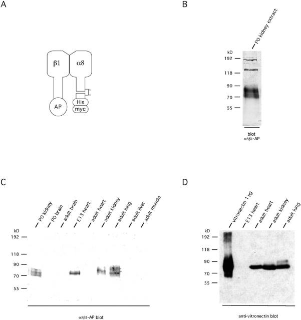
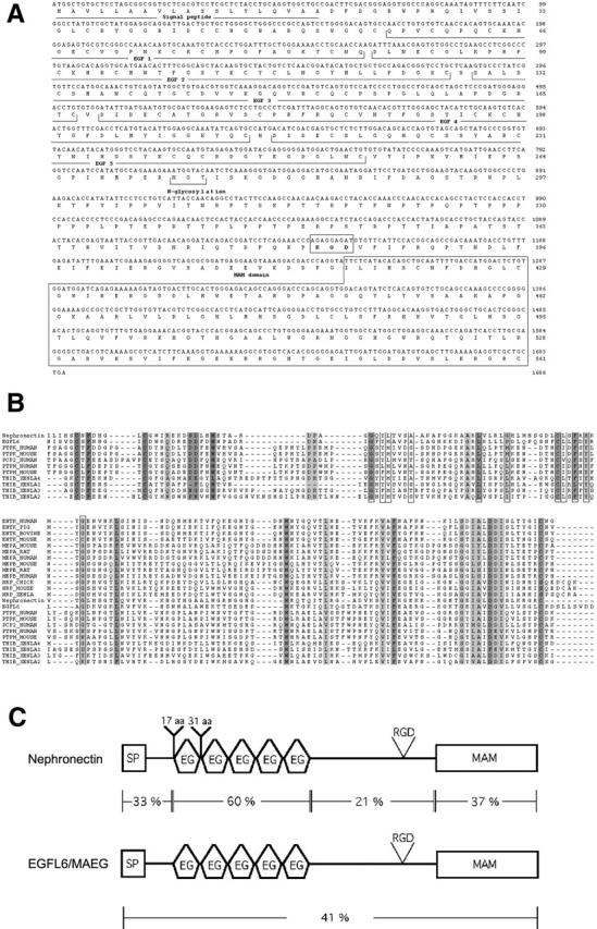
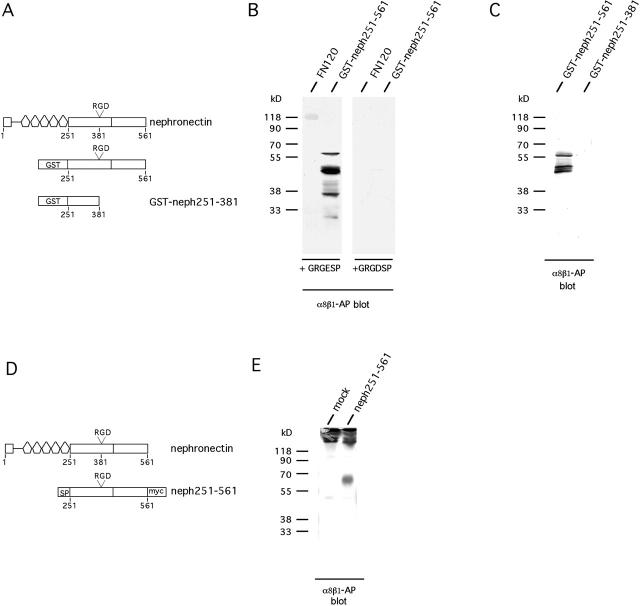
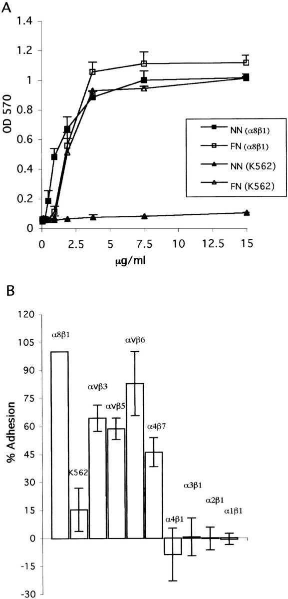
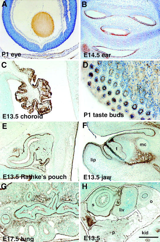
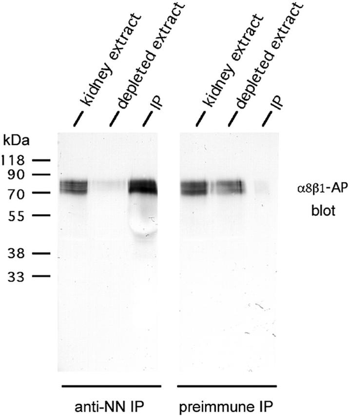
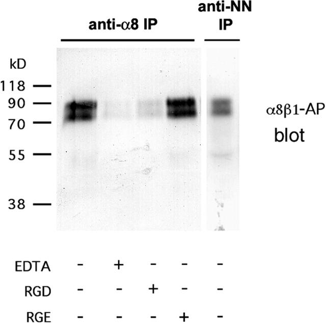
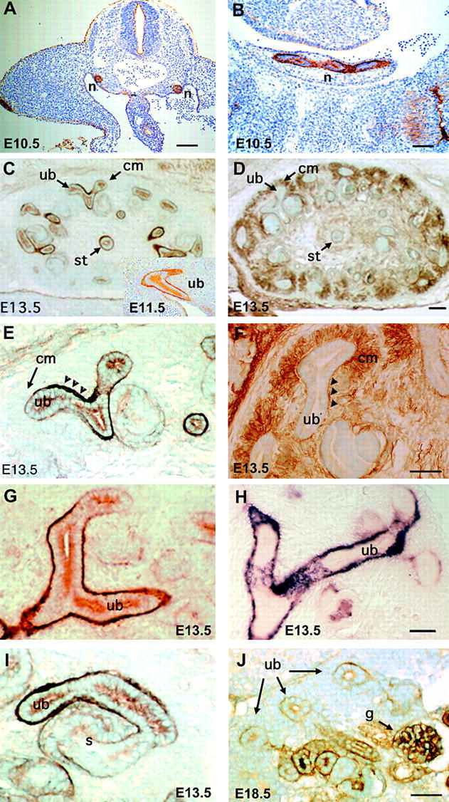
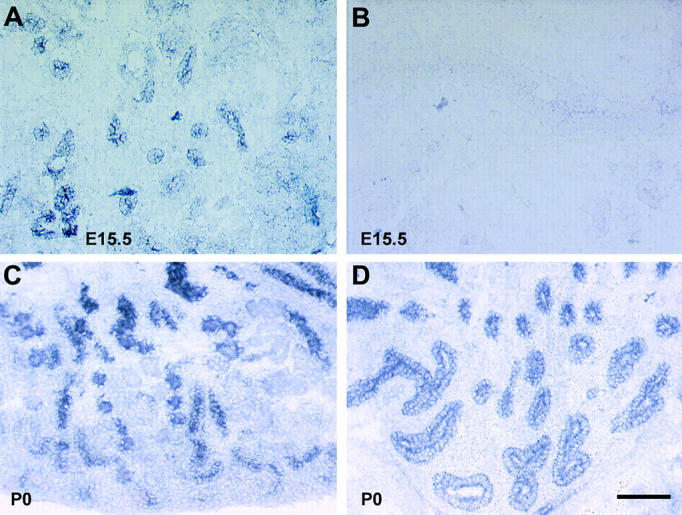
The epithelial-mesenchymal interactions required for kidney organogenesis are disrupted in mice lacking the integrin alpha8beta1. None of this integrin's known ligands, however, appears to account for this phenotype. To identify a more relevant ligand, a soluble integrin alpha8beta1 heterodimer fused to alkaline phosphatase (AP) has been used to probe blots and cDNA libraries. In newborn mouse kidney extracts, alpha8beta1-AP detects a novel ligand of 70-90 kD. This protein, named nephronectin, is an extracellular matrix protein with five EGF-like repeats, a mucin region containing a RGD sequence, and a COOH-terminal MAM domain. Integrin alpha8beta1 and several additional RGD-binding integrins bind nephronectin. Nephronectin mRNA is expressed in the ureteric bud epithelium, whereas alpha8beta1 is expressed in the metanephric mesenchyme. Nephronectin is localized in the extracellular matrix in the same distribution as the ligand detected by alpha8beta1-AP and forms a complex with alpha8beta1 in vivo. Thus, these results strongly suggest that nephronectin is a relevant ligand mediating alpha8beta1 function in the kidney. Nephronectin is expressed at numerous sites outside the kidney, so it may also have wider roles in development. The approaches used here should be generally useful for characterizing the interactions of novel extracellular matrix proteins identified through genomic sequencing projects.


Figures

Figure 1.
Figure 1.
Strategy to identify a novel ligand for integrin α8β1. (A) Schematic representation of soluble integrin α8β1-AP heterodimer. α8β1-AP protein was expressed as described (Denda et al., 1998a). The soluble α8β1-AP has the same binding characteristics as the transmembrane protein (Denda et al., 1998a,b). AP, secreted AP; His, polyhistidine tag; myc, c-myc epitope tag. (B) The soluble α8β1-AP recognizes several different proteins in kidney extracts. Extracts were prepared from postnatal day 0 (P0) mouse kidneys, separated on 7% SDS-PAGE, transferred to nitrocellulose, and probed with α8β1-AP in the presence of 2 mM Mn2+. The most prominent potential ligands for α8β1-AP have molecular masses of 70–90 kD. (C) Screening of mouse tissues for the presence of a 70–90-kD ligand(s) for α8β1-AP. Embryonic day 13 (E13) heart, adult kidney, and lung contain a 70–90-kD ligand(s) similar to newborn (P0) kidney. (D) Embryonic day 13 heart tissue does not contain detectable amounts of VN, a known ligand for α8β1, as compared with other tissues positive for the 70–90-kD ligand. Therefore, E13 heart was chosen as the tissue to screen for a novel ligand for α8β1-AP.
Figure 2.
Figure 2.
Nephronectin, a novel extracellular matrix protein. (A) Mouse nephronectin cDNA nucleotide sequence and deduced amino acid sequence. The putative signal peptide, the EGF repeats, the RGD motif, N-glycosylation sites, and the MAM homology region are indicated. (B) Alignment of the MAM domain. MAM domains were aligned using ClustalW 1.8. The boxed amino acids are the consensus residues of the prosite signature. Shaded amino acids are residues conserved in ≥70% of the proteins. (C) Overall homology of nephronectin to EGFL6 (Yeung et al., 1999). The two proteins share the same overall organization and sequence of domains. The least conserved domain is the one containing the RGD motif.
Figure 3.
Figure 3.
The RGD-containing region in nephronectin is required for binding by α8β1. (A) Schematic representation of E. coli fusion proteins. GST-neph251-561 contains the COOH-terminal part of nephronectin from immediately after the EGF repeats to the end of the protein, whereas GST-neph251-381 contains the fragment from the end of the EGF repeats to just in front of the RGD motif. α8β1-AP binds in an RGD-inhibitable manner to nephronectin. 2 μg of FN, GST-neph251-561, or GST-neph251-381 were separated on a 12% SDS-PAGE, transferred to nitrocellulose, and probed with α8β1-AP in the presence of 2 mM Mg2+. (B) GRGDSP or GRGESP peptides were added at 50 μg/ml. Note that, under the conditions used, binding of α8β1-AP to FN is weaker than to GST-neph251-561. This blot was developed longer than the blot in C to show the FN binding. Therefore, more degradation products of GST-neph251-561 are seen than in C. (C) The RGD region of nephronectin is required for binding of α8β1-AP to nephronectin. A GST fusion protein lacking the RGD-containing region and more COOH-terminal sequences does not bind the α8β1-AP. (D) Schematic representation of fusion protein expressed in COS7 cells. Neph251-561 contains an IgG signal peptide, a fragment of nephronectin extending from the COOH terminus of the EGF repeats to the end of the protein with a myc/His tag at the COOH terminus. SP, Ig κ signal peptide; myc, myc/his tag. (E) Nephronectin fragment expressed in eukaryotic cells binds to α8β1-AP. Neph251-561 protein was transiently expressed in COS-7 cells. Then, the supernatant was harvested and immunoprecipitated with anti-myc antibody. Immunoprecipitates were separated by 12% nonreducing SDS-PAGE, transferred to nitrocellulose, and probed with α8β1-AP. mock, empty plasmid negative control. The strong band at the top of the gel represents nonreduced 9E10 Ig.
Figure 4.
Figure 4.
Adhesion of K562 cells, and K562 expressing specific additional integrins, to nephronectin. (A) K562 and K562 expressing α8β1 were allowed to adhere to increasing concentrations, 0.06–15.0 μg/ml, of either FN or amino acids 251–561 of nephronectin (NN) purified from CHO cell–conditioned medium. Experiments were carried out in the presence of 1 mM Mn2+. (B) Adhesion of K562 cells expressing indicated additional integrin heterodimers to 2 μg/ml of a GST fusion protein containing amino acids 251–561 of nephronectin in the presence of 1 mM Mn2+ and the anti-α5 mAb BIIG2 to inhibit α5β1-mediated adhesion. Adhesion of the parental K562 cells that express α5β1 to these two substrates was measured in the absence of the anti-α5 mAb. For all lines, OD values for wells coated with GST alone were subtracted. Adhesion of α8β1-expressing K562 cells was defined as 100% and adhesion of other cells is expressed as a percentage of that value.
Figure 9.
Figure 9.
Expression of nephronectin in various tissues and organs. (A) Eye of a P1 animal. Note uniform expression of nephronectin in the lens. (B) Ear of an E14.5 embryo. Note expression of nephronectin in basal lamina surrounding epithelium and on apical surface of epithelia. (C) Choroid plexus of an E13.5 embryo. Nephronectin is localized in basal lamina and is strongly expressed by epithelial cells in this organ. (D) Tongue of a P1 animal. Nephronectin is expressed in developing taste buds. Lighter expression is also present in muscle. (E) Rathke's pouch at E13.5. Nephronectin is present in the basal lamina surrounding this organ that develops into the pituitary gland. (F) Jaw of E13.5 animal. In cross section, extensive expression of nephronectin can be observed in the basal laminae underlying the skin and oral epithelium. Nephronectin also surrounds a developing incisor (t). Lip and Meckel's cartilage (mc) are indicated. (G) E13.5 lung. Nephronectin is expressed in the basal lamina surrounding the branching epithelia in this organ. In contrast to embryonic kidney, nephronectin is also present in the surrounding mesenchyme. (H) Expression of nephronectin in various organs of E13.5 embryo. Nephronectin expression is prominent in the developing stomach (s) and oesophagus (o). In contrast, very little expression is present in a lobe of the liver (liv). Light expression is observed in the pancreas (p). Expression in kidney (k) epithelia is obvious at bottom right. Bar: (F) 267 μm; (A and H) 200 μm; (C, E, and G) 100 μm; (B) 90 μm; (D) 50 μm.
Figure 5.
Figure 5.
Nephronectin is the protein(s) of 70–90 kD recognized by α8β1-AP in kidney extracts. Newborn mouse kidney extracts were immunoprecipitated with antinephronectin antiserum or preimmune serum. The extract, depleted extract, and the immunoprecipitate were analyzed in blots using α8β1-AP. Antinephronectin depleted virtually all α8β1-AP binding activity from the kidney extract, whereas the preimmune serum did not reduce the activity at 70–90 kD responsible for α8β1-AP binding.
Figure 6.
Figure 6.
Coimmunoprecipitation of α8β1 with nephronectin. Newborn mouse kidney extracts were immunoprecipitated with antiintegrin α8 serum or antinephronectin serum in the presence or absence of 10 mM EDTA, with 200 μg/ml GRGDSP or GRGESP peptides, as indicated. Precipitates were separated by 12% SDS-PAGE, were transferred to nitrocellulose, and were probed with α8β1-AP. The band(s) corresponding to nephronectin (compare with the antinephronectin immunoprecipitation) are present in anti-α8 immunoprecipitates. Addition of EDTA leads to the disappearance of the nephronectin from the α8 immunoprecipitates. Inclusion of GRGDSP but not GRGESP strongly reduces the amount of nephronectin in the anti-α8 immunoprecipitates.
Figure 7.
Figure 7.
Expression of nephronectin, α8β1, and the α8β1-AP ligand. (A and B) Expression of nephronectin in E10.5 embryo. In coronal section (A), prominent expression is observed in the mesonephric duct within the urogenital ridge and in ectoderm surrounding the embryo. A sagittal section (B) illustrates expression in the developing mesonephric duct. (C) Expression of nephronectin in E13.5 kidney, as detected using antinephronectin. Note prominent expression of nephronectin at the interfaces between ureteric bud epithelial cells and surrounding mesenchyme. Expression of nephronectin is also strong in the stalk of the ureter. An insert in lower right corner of C illustrates nephronectin expression in the ureteric bud at E11.5. (D) Expression of the integrin α8 in E13.5 kidney visualized with anti-α8. α8 is expressed in the mesenchyme surrounding developing branches of the ureteric bud. α8 also is concentrated at the interface between the metanephric mesenchyme and ureteric epithelial cells. α8 is not expressed at detectable levels in the ureteric epithelial cells. (E) Higher resolution pattern of expression of nephronectin in E13.5 kidney. Note the strong expression in the basal lamina between the ureteric bud and metanephric mesenchyme. A condensing comma-shaped body of mesenchyme expresses little or no nephronectin. (F) Higher resolution illustration of expression of α8 in E13.5 kidney. Note strong expression of α8 in the metanephric mesenchyme and the absence of expression in the ureteric bud epithelial cells. (G and H) Comparison of expression patterns of nephronectin (G) and the ligand-bound by α8β1-AP (H) in E13.5 kidney. Note that both have similar patterns of expression in branching ureteric buds. (I) Expression of nephronectin continues to be high in the ureteric bud–derived portion of a tubule created by fusion of condensing mesenchyme with a ureteric bud in E13.5 kidney. Expression appears to be initiating in the S-shaped portion of this tubule derived from the condensing mesenchyme. (J) In the E18.5 kidney, nephronectin is expressed in maturing tubules and in differentiating glomeruli. n, mesonephric duct; cm, condensing mesenchyme; ub, ureteric bud; st, ureteric stalk; c, comma-shaped body; s, S-shaped body; g, glomerulus. Bars: (C and D) 250 μm; (A and E–J) 100 μm; (B) 50 μm.
Figure 8.
Figure 8.
In situ analysis of nephronectin mRNA in E15.5 and P0 kidney. Nephronectin mRNA is present in branching epithelia of the developing kidney at E15.5 (A) and P0 (C and D). A view of the P0 kidney cortex is shown in C. Note the many condensing mesenchymal structures not expressing nephronectin mRNA. A view of the P0 pelvic region of the kidney is shown in D. Incubation of sections with a sense probe as a control at E15.5 (B) or P1 (not shown) resulted in virtually no reaction product. Bar, 100 μm.

Comment in

References

    1. Aufderheide, E., R. Chiquet-Ehrismann, and P. Ekblom. 1987. Epithelial–mesenchymal interactions in the developing kidney lead to expression of tenascin in the mesenchyme. J. Cell Biol. 105:599–608. - PMC - PubMed
    1. Blystone, S.D., I.L. Graham, F.P. Lindberg, and E.J. Brown. 1994. Integrin αVβ3 differentially regulates adhesive and phagocytic functions of the fibronectin receptor α5β1. J Cell Biol. 127:1129–1137. - PMC - PubMed
    1. Bossy, B., E. Bossy-Wetzel, and L.F. Reichardt. 1991. Characterization of the integrin α8 subunit: a new integrin β1 associated subunit, which is prominently expressed on axons and on cells in contact with basal laminae in chick embryos. EMBO J. 10:2375–2385. - PMC - PubMed
    1. Buchner G, U. Orfanelli, N. Quaderi, M.T. Bassi, G. Andolfi, A. Ballabio, and B. Franco. 2000. Identification of a new EGF-repeat-containing gene from human Xp22: a candidate for developmental disorders. Genomics. 65:16–23. - PubMed
    1. Denda, S., U. Müller, K.L. Crossin, H.P. Erickson, and L.F. Reichardt. 1998. a. Utilization of a soluble integrin-alkaline phosphatase chimera to characterize integrin α8β1 receptor interactions with tenascin: murine α8β1 binds to the RGD site in tenascin-c fragments, but not to native tenascin-c. Biochemistry. 37:5464–5474. - PMC - PubMed

Publication types

MeSH terms