. 2001 Jul 23;154(2):459-68.
doi: 10.1083/jcb.200103103.

Epidermal growth factor (EGF)-like repeats of human tenascin-C as ligands for EGF receptor

Affiliations

Epidermal growth factor (EGF)-like repeats of human tenascin-C as ligands for EGF receptor

C S Swindle et al. J Cell Biol. .

Abstract

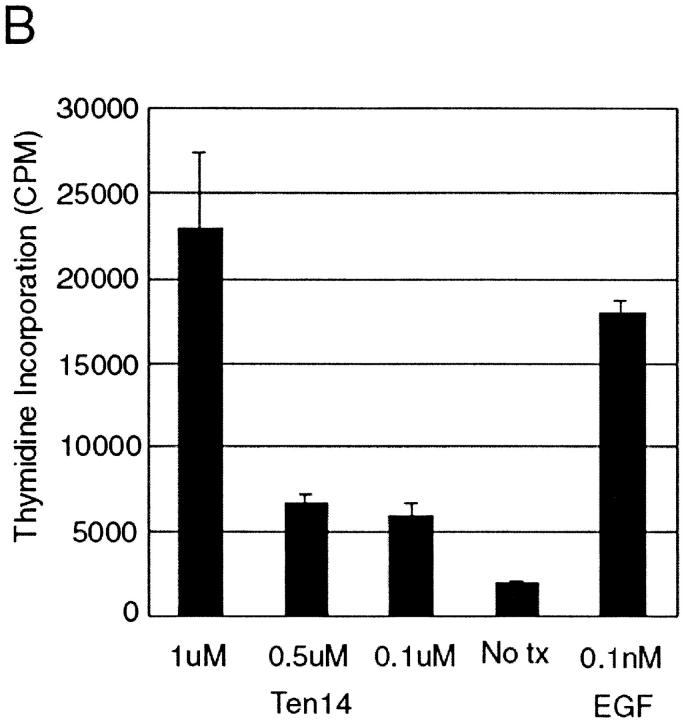
Signaling through growth factor receptors controls such diverse cell functions as proliferation, migration, and differentiation. A critical question has been how the activation of these receptors is regulated. Most, if not all, of the known ligands for these receptors are soluble factors. However, as matrix components are highly tissue-specific and change during development and pathology, it has been suggested that select growth factor receptors might be stimulated by binding to matrix components. Herein, we describe a new class of ligand for the epidermal growth factor (EGF) receptor (EGFR) found within the EGF-like repeats of tenascin-C, an antiadhesive matrix component present during organogenesis, development, and wound repair. Select EGF-like repeats of tenascin-C elicited mitogenesis and EGFR autophosphorylation in an EGFR-dependent manner. Micromolar concentrations of EGF-like repeats induced EGFR autophosphorylation and activated extracellular signal-regulated, mitogen-activated protein kinase to levels comparable to those induced by subsaturating levels of known EGFR ligands. EGFR-dependent adhesion was noted when the ligands were tethered to inert beads, simulating the physiologically relevant presentation of tenascin-C as hexabrachion, and suggesting an increase in avidity similar to that seen for integrin ligands upon surface binding. Specific binding to EGFR was further established by immunofluorescence detection of EGF-like repeats bound to cells and cross-linking of EGFR with the repeats. Both of these interactions were abolished upon competition by EGF and enhanced by dimerization of the EGF-like repeat. Such low affinity behavior would be expected for a matrix-"tethered" ligand; i.e., a ligand which acts from the matrix, presented continuously to cell surface EGF receptors, because it can neither diffuse away nor be internalized and degraded. These data identify a new class of "insoluble" growth factor ligands and a novel mode of activation for growth factor receptors.


Figures

Figure 1.
Figure 1.
Tenascin-C EGF–like repeats stimulate cell mitogenesis. (A) WT (black) or M (open) NR6 cells were exposed to the EGF-like repeat proteins, and 3H-thymidine incorporation was assessed. EGF (1 nM) and serum (1%) were used as positive controls (for WT and M NR6 cells, respectively). The EGF-like repeat proteins were used at the following concentrations: left bars are 2 uM for 1/2; 1 uM for 9/10; 4 uM for 11/12/13; and 3 uM for 14, and at 10 and 1% of that level for each concentration (middle and right bars). (B) Mitogenesis assay performed in serum-free media on WT NR6 cells with decreasing concentrations of Ten14 as described. No tx designates cells not exposed to ligand. Values are the mean ±SD (performed in triplicate) for one experiment representative of three experiments.
Figure 1.
Figure 1.
Tenascin-C EGF–like repeats stimulate cell mitogenesis. (A) WT (black) or M (open) NR6 cells were exposed to the EGF-like repeat proteins, and 3H-thymidine incorporation was assessed. EGF (1 nM) and serum (1%) were used as positive controls (for WT and M NR6 cells, respectively). The EGF-like repeat proteins were used at the following concentrations: left bars are 2 uM for 1/2; 1 uM for 9/10; 4 uM for 11/12/13; and 3 uM for 14, and at 10 and 1% of that level for each concentration (middle and right bars). (B) Mitogenesis assay performed in serum-free media on WT NR6 cells with decreasing concentrations of Ten14 as described. No tx designates cells not exposed to ligand. Values are the mean ±SD (performed in triplicate) for one experiment representative of three experiments.
Figure 2.
Figure 2.
Tenascin EGF–like repeats activate the EGFR kinase cascade. WT (top) and M (bottom) NR6 cells were treated with EGF-like repeat proteins and activation of EGFR signaling was assessed. (A) EGFR autophosphorylation was determined by antiphosphotyrosine immunoblotting of immunoprecipitated EGFR after treatment with EGF or EGF-like repeats 1/2 (5 uM), 11/12/13 (1 uM), or 14 (6 uM). Immunoblotting with an antibody to EGFR demonstrated equal loading (data not shown). (B) ERK MAP kinase activation was assessed by immunoblotting for dually phosphorylated p44/p42 ERK, indicative of activated ERK. The cells were treated with various concentrations of the EGF-like repeats (5 uM for 1/2; 2 uM for 9/10; 1 uM for 11/12/13; and 6 uM for 14) and at 50 and 10% of those levels. EGF and serum were used as positive controls (for WT and M NR6 cells, respectively). Numbers below a lane represent relative values of intensity for pEGFR or p42 in each lane for that experiment as determined by densitometry. In both panels, an experiment representative of at least three determinations are shown.
Figure 3.
Figure 3.
Differential detection of activation of the EGFR kinase cascade. (A and B) WT NR6 cells were treated for 5 min with decreasing concentrations of a high affinity (EGF, kd ∼2 nM; left) or lower affinity (Y13G-EGF, kd ∼100 nM; right; Reddy et al., 1996a) EGFR ligand. Activation status of EGFR was determined by immunoblotting with the PY20 antiphosphotyrosine antibody of 175 kD (A), and ERK MAP kinase activation was assessed by immunoblotting for dually phosphorylated p44/p42 ERK, indicative of activated ERK (B). no tx designates cells not exposed to ligand. (C) WT NR6 cells were treated as in B with various concentrations of EGF or 2 uM tenascin 14 fragment in quiescence media or serum-free media. The two different concentrations of Ten14 are from different preparations. ERK MAP kinase activation was assessed as in B. In A–C, concentration in nM of EGFR ligand used for treatment is indicated. (D and E) Augmentation of EGFR phosphorylation by decreased attenuation mechanisms in WT NR6 cells. (D) WT NR6 cells were treated for 30 min with EGF (0.1 or 0.01 nM) or tenascin 14 fragment (2 uM) in the presence of 0.1 mM sodium vanadate; EGFR activation status was determined as in A. (E) EGFR ligands were tethered to PEO latexes and tested for the ability to induce EGFR phosphorylation. WT NR6 cells were exposed to EGF (5 min at 10 or 1 nM), bead complexes containing tenascin 14 fragments (30, 120, and 240 min), bead complexes containing EGF (30 min of 20- and 5-ul beads), or control beads (30 min); EGFR activation status was determined as in A. Numbers below a lane represent relative values of intensity for pEGFR or p42 in each lane for that experiment as determined by densitometry. In all panels a representative experiment is shown of at least three determinations.
Figure 3.
Figure 3.
Differential detection of activation of the EGFR kinase cascade. (A and B) WT NR6 cells were treated for 5 min with decreasing concentrations of a high affinity (EGF, kd ∼2 nM; left) or lower affinity (Y13G-EGF, kd ∼100 nM; right; Reddy et al., 1996a) EGFR ligand. Activation status of EGFR was determined by immunoblotting with the PY20 antiphosphotyrosine antibody of 175 kD (A), and ERK MAP kinase activation was assessed by immunoblotting for dually phosphorylated p44/p42 ERK, indicative of activated ERK (B). no tx designates cells not exposed to ligand. (C) WT NR6 cells were treated as in B with various concentrations of EGF or 2 uM tenascin 14 fragment in quiescence media or serum-free media. The two different concentrations of Ten14 are from different preparations. ERK MAP kinase activation was assessed as in B. In A–C, concentration in nM of EGFR ligand used for treatment is indicated. (D and E) Augmentation of EGFR phosphorylation by decreased attenuation mechanisms in WT NR6 cells. (D) WT NR6 cells were treated for 30 min with EGF (0.1 or 0.01 nM) or tenascin 14 fragment (2 uM) in the presence of 0.1 mM sodium vanadate; EGFR activation status was determined as in A. (E) EGFR ligands were tethered to PEO latexes and tested for the ability to induce EGFR phosphorylation. WT NR6 cells were exposed to EGF (5 min at 10 or 1 nM), bead complexes containing tenascin 14 fragments (30, 120, and 240 min), bead complexes containing EGF (30 min of 20- and 5-ul beads), or control beads (30 min); EGFR activation status was determined as in A. Numbers below a lane represent relative values of intensity for pEGFR or p42 in each lane for that experiment as determined by densitometry. In all panels a representative experiment is shown of at least three determinations.
Figure 4.
Figure 4.
Inhibition of EGFR activation prevents signaling from EGF-like repeats. (A) WT NR6 cells were treated with the tenascin EGF–like repeats at concentrations that activate ERK MAP kinase (5 uM for 1/2; 1 uM for 11/12/13; and 6 uM for 14). The cells were treated with the EGF-like repeat proteins in the absence (−) or presence (+) of the EGFR-specific pharmacologic inhibitor PD153035. no tx represents no ligand. (B) WT NR6 cells were treated with Ten14 (1 uM) or EGF (0.01 nM) in the presence (+) or absence (−) of an antibody specific for the extracellular domain of EGFR (Clone 528; Calbiochem) under serum-free conditions. (C) B82 cells expressing WT EGFR were challenged with tenascin 14 repeats (2 uM) under serum-free conditions. Numbers below a lane represent relative values of intensity for p42 in each lane for that experiment as determined by densitometry. Shown are one of three experiments.
Figure 5.
Figure 5.
Beads presenting tethered tenascin 14 fragments bind to EGFR. WT NR6 cells were treated for 4 h at 37°C with beads tethered with either tenascin 14 or EGF or control beads. Cells were washed three times with iced PBS and visualized by phase-contrast microscopy (top). In parallel, cells were treated with 5 ug/ml anti-EGFR antibody (clone 528; Sunada et al., 1986; Turner et al., 1996) for 15 min before addition of beads and continuing throughout the 4 h (bottom). Beads appear as bright, refractile, and translucent spheres mainly above the plane of the cells. Shown is one of three experiments.
Figure 6.
Figure 6.
Tenascin 14 binds to EGFR-expressing WT NR6 cells. (A) WT NR6 cells were quiesced for 24 h and exposed to various concentrations of monomeric or dimerized ligand to visualize binding to EGFR. Ten14 and mEGF-His6 expressed from the same vector (pRSETA; Invitrogen) as Ten14 were incubated overnight for dimerization with monoclonal anti-HisG (Invitrogen) antibody that recognized the NH2-terminal poly His epitope of mEGF-His6 and Ten14. This was at concentrations of 50 nM for mEGF-His6 and 25 nM for monoclonal anti-HisG antibody and 2 uM Ten14 with 0.63 uM of the antibody (Ab) to increase affinity to receptor. Ligands were incubated for 10 min at room temperature before fixation. 1:500 of goat anti–mouse antibody conjugated to Oregon green was used as secondary antibody before visualizing by fluorescence microscopy and captured at a constant exposure by a SPOT II CCD camera. a, mEGF-His6 (50 nM); b, mEGF-His6 (50 nM) dimerized with primary antibody (25 nM); c, cells were preincubated with EGF (100 nM) for 5 min to compete for EGFR with mEGF-His6 (50 nM) dimerized with antibody (25 nM); d, Ten14 (2 uM); e, Ten14 (2 uM) dimerized with primary antibody(0.63 uM); f, cells preincubated with EGF (100 nM) for 5 min with Ten14 (2 uM) preincubated with 0.63 uM antibody subsequently added. (B) Each cell in four randomly selected fields were outlined and measured for luminosity as compared with background. The data are the mean ±SE of an average of >25 cells per experimental condition. Statistical analyses were performed via Student's t test. Double asterisk represents P < 0.01. Shown is one representative of two sets of experiments.
Figure 6.
Figure 6.
Tenascin 14 binds to EGFR-expressing WT NR6 cells. (A) WT NR6 cells were quiesced for 24 h and exposed to various concentrations of monomeric or dimerized ligand to visualize binding to EGFR. Ten14 and mEGF-His6 expressed from the same vector (pRSETA; Invitrogen) as Ten14 were incubated overnight for dimerization with monoclonal anti-HisG (Invitrogen) antibody that recognized the NH2-terminal poly His epitope of mEGF-His6 and Ten14. This was at concentrations of 50 nM for mEGF-His6 and 25 nM for monoclonal anti-HisG antibody and 2 uM Ten14 with 0.63 uM of the antibody (Ab) to increase affinity to receptor. Ligands were incubated for 10 min at room temperature before fixation. 1:500 of goat anti–mouse antibody conjugated to Oregon green was used as secondary antibody before visualizing by fluorescence microscopy and captured at a constant exposure by a SPOT II CCD camera. a, mEGF-His6 (50 nM); b, mEGF-His6 (50 nM) dimerized with primary antibody (25 nM); c, cells were preincubated with EGF (100 nM) for 5 min to compete for EGFR with mEGF-His6 (50 nM) dimerized with antibody (25 nM); d, Ten14 (2 uM); e, Ten14 (2 uM) dimerized with primary antibody(0.63 uM); f, cells preincubated with EGF (100 nM) for 5 min with Ten14 (2 uM) preincubated with 0.63 uM antibody subsequently added. (B) Each cell in four randomly selected fields were outlined and measured for luminosity as compared with background. The data are the mean ±SE of an average of >25 cells per experimental condition. Statistical analyses were performed via Student's t test. Double asterisk represents P < 0.01. Shown is one representative of two sets of experiments.
Figure 7.
Figure 7.
Tenascin 14 can be cross-linked to EGFR. Ten14 (2 uM) and mEGF (mEGF-His6; 10 nM) were bound and chemically cross-linked to quiesced WT NR6 fibroblasts and immunoprecipitated from ensuing lysates with anti-HisG. Presence of EGFR (A) or insulin receptor β chain was assessed by immunoblotting with respective antibodies. In lanes 1 and 2, cells were preincubated with 100 nM EGF for 5 min and throughout cross-linking as a competitive ligand. In lane 5, cells were exposed to and cross-linked with antibody alone and lysate was immunoprecipitated. From left to right: 1, EGF/mEGF-His6 (pretreatment with EGF and addition of mEGF-His6 ligand); 2, EGF/Ten14 (pretreatment with EGF and addition of Ten14 ligand); 3, mEGF-His6 (10 nM); 4, Ten14 (2 uM); 5, IgG (anti-HisG antibody); 6, No tx (no treatment with ligand); and 7, cell lysate (WT NR6 lysate). Shown is a representative of two experiments.

References

    1. Akiyama, S.K., and K.M. Yamada. 1985. Synthetic peptides competitively inhibit both direct binding to fibroblasts and functional biological assays for the purified cell-binding domain of fibronectin. J. Biol. Chem. 260:4492–4500. - PubMed
    1. Anklesaria, P., J. Teixido, M. Laiho, J.H. Pierce, J.S. Greenberger, and J. Massague. 1990. Cell adhesion mediated by binding of membrane-anchored transforming growth factor alpha to epidermal growth factor receptors promotes cell proliferation. Proc. Natl. Acad. Sci. USA. 87:3289–3293. - PMC - PubMed
    1. Ashcroft, G.S., M.A. Horan, and M.W. Ferguson. 1995. The effects of ageing on cutaneous wound healing in mammals. J. Anat. 187:1–26. - PMC - PubMed
    1. Banerjee, P., D.J. Irvine, A.M. Mayes, and L.G. Griffith. 2000. Polymer latexes for controlling cell adhesion and receptor-mediated interactions. J. Biomed. Mater. Res. 50:331–339. - PubMed
    1. Bhalla, U.S., and R. Iyengar. 1999. Emergent properties of networks of biological signaling pathways. Science. 283:381–387. - PubMed

Publication types

MeSH terms