. 2001 Aug 1;29(15):3258-69.
doi: 10.1093/nar/29.15.3258.

Molecular evolution of the homeodomain family of transcription factors

Affiliations

Molecular evolution of the homeodomain family of transcription factors

S Banerjee-Basu et al. Nucleic Acids Res. .

Abstract

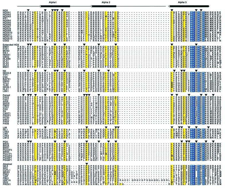
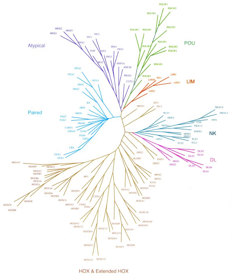
The homeodomain family of transcription factors plays a fundamental role in a diverse set of functions that include body plan specification, pattern formation and cell fate determination during metazoan development. Members of this family are characterized by a helix-turn-helix DNA-binding motif known as the homeodomain. Homeodomain proteins regulate various cellular processes by specifically binding to the transcriptional control region of a target gene. These proteins have been conserved across a diverse range of species, from yeast to human. A number of inherited human disorders are caused by mutations in homeodomain-containing proteins. In this study, we present an evolutionary classification of 129 human homeodomain proteins. Phylogenetic analysis of these proteins, whose sequences were aligned based on the three-dimensional structure of the homeodomain, was performed using a distance matrix approach. The homeodomain proteins segregate into six distinct classes, and this classification is consistent with the known functional and structural characteristics of these proteins. An ancestral sequence signature that accurately describes the unique sequence characteristics of each of these classes has been derived. The phylogenetic analysis, coupled with the chromosomal localization of these genes, provides powerful clues as to how each of these classes arose from the ancestral homeodomain.


Figures

Figure 1
Figure 1
Multiple sequence alignment of the homeodomain classes. The abbreviations for each protein correspond to those in Table 1. The amino acid residues that are absolutely conserved across all homeodomain classes are shown in white with a blue background and the residues that are conservatively substituted are highlighted in yellow. For each of the homeodomain classes, the conserved residues are indicated by a downward arrow. The numbering scheme at the top of the figure refers to the amino acid positions within the 60 residue canonical homeodomain. The positions of the three α-helices found in the X-ray structure of engrailed (8) are shown above the figure. ALSCRIPT v.2.04 was used to format the alignment (85).
Figure 2
Figure 2
Phylogenetic relationships between the members of the homeodomain protein family. Unrooted neighbor joining trees were generated using the PHYLIP Phylogeny Inference Package v.3.5c (23). TREETOOL, a freestanding tree editor and formatter, was used to generate the tree diagram. The homeodomain classes are color coded and the abbreviations used here correspond to those in the alignment presented in Figure 1.
Figure 3
Figure 3
Sequence signature for each of the homeodomain classes. Signatures are based on the multiple sequence alignment shown in Figure 1 and indicate positions within the homeodomain that are absolutely conserved within each class. The sequence signatures presented here unambiguously identify members within each homeodomain class. The numbering scheme at the top of the figure refers to the amino acid positions within the 60 residue canonical homeodomain.
Figure 4
Figure 4
Chromosomal distribution of the human homeobox gene family. The homeobox genes are color coded corresponding to the phylogenetic classes shown in Figure 2. The chromosomal map positions of the homeobox genes were obtained from LocusLink (http://www.ncbi.nlm.nih.gov/LocusLink/index.html). The centromere is shown in dark blue. The four HOX clusters, HOXA, HOXB, HOXC and HOXD, are distributed on chromosomes 7, 17, 12 and 2, respectively.

References

    1. Scott M.P. and Weiner,A.J. (1984) Structural relationships among genes that control development: sequence homology between the Antennapedia, Ultrabithorax and fushi tarazu loci of Drosophila. Proc. Natl Acad. Sci. USA, 81, 4115–4119. - PMC - PubMed
    1. McGinnis W., Levine,M.S., Hafen,E., Kuroiwa,A. and Gehring,W.J. (1984) A conserved DNA sequence in homoeotic genes of the Drosophila Antennapedia and bithorax complexes. Nature, 308, 428–433. - PubMed
    1. Gehring W.J., Affolter,M. and Burglin,T. (1994) Homeodomain proteins. Annu. Rev. Biochem., 63, 487–526. - PubMed
    1. Ceska T., Lamers,M., Monaci,P., Nicosia,A., Cortese,R. and Suck,D. (1993) The X-ray structure of an atypical homeodomain present in the rat liver transcription factor LFB1/HNF1 and implications for DNA binding. EMBO J., 12, 1805–1810. - PMC - PubMed
    1. Dekker N., Cox,M., Boelens,R., Verrijzer,C., van der Vliet,P. and Kaptein,R. (1993) Solution structure of the POU-specific DNA-binding domain of Oct-1. Nature, 362, 852–855. - PubMed

MeSH terms