. 2001 Jul 31;98(16):9277-82.
doi: 10.1073/pnas.161272698. Epub 2001 Jul 24.

IL-7 differentially regulates cell cycle progression and HIV-1-based vector infection in neonatal and adult CD4+ T cells

Affiliations

IL-7 differentially regulates cell cycle progression and HIV-1-based vector infection in neonatal and adult CD4+ T cells

V Dardalhon et al. Proc Natl Acad Sci U S A. .

Abstract

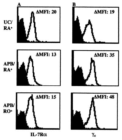
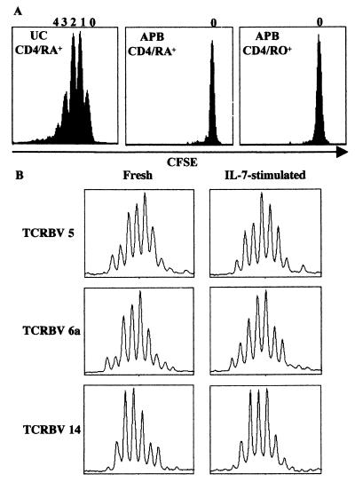
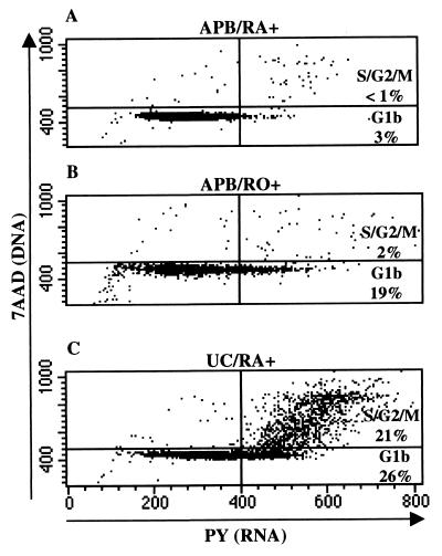
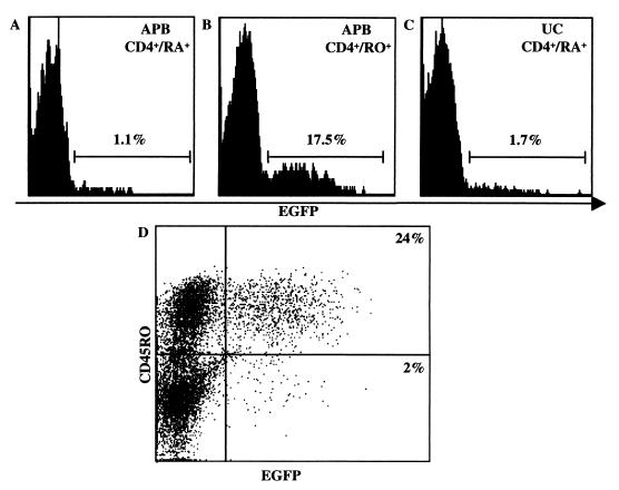
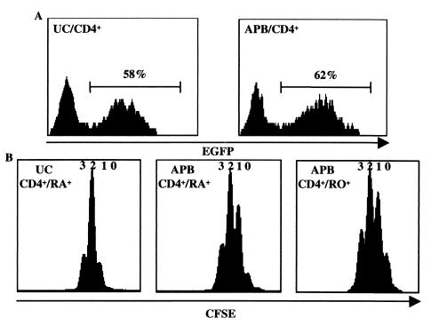
Differences in the immunological reactivity of umbilical cord (UC) and adult peripheral blood (APB) T cells are poorly understood. Here, we show that IL-7, a cytokine involved in lymphoid homeostasis, has distinct regulatory effects on APB and UC lymphocytes. Neither naive nor memory APB CD4(+) cells proliferated in response to IL-7, whereas naive UC CD4(+) lymphocytes underwent multiple divisions. Nevertheless, both naive and memory IL-7-treated APB T cells progressed into the G(1b) phase of the cell cycle, albeit at higher levels in the latter subset. The IL-7-treated memory CD4(+) lymphocyte population was significantly more susceptible to infection with an HIV-1-derived vector than dividing CD4(+) UC lymphocytes. However, activation through the T cell receptor rendered UC lymphocytes fully susceptible to HIV-1-based vector infection. These data unveil differences between UC and APB CD4(+) T cells with regard to IL-7-mediated cell cycle progression and HIV-1-based vector infectivity. This evidence indicates that IL-7 differentially regulates lymphoid homeostasis in adults and neonates.


Figures

Figure 1
Figure 1
Expression of the IL-7R differs on UC and APB CD4+ T cells. Naive (RA+) and memory (RO+) CD4+ resting T lymphocytes were isolated from UC and APB by negative selection. Expression of the IL-7Rα (A) and γc chains (B) on the purified CD4+ T cell subsets was analyzed on a FACScalibur with phycoerythrin-conjugated monoclonal antibodies (open histograms). The Ig isotype control (filled histograms) and the change in mean fluorescence intensity (ΔMFI) are indicated in each histogram. Data are representative of results obtained in four independent experiments.
Figure 2
Figure 2
IL-7 drives the proliferation of naive UC but not APB CD4+ T cells. (A) Naive CD4+ UC T lymphocytes (CD4/RA+) and naive as well as memory CD4+ APB T lymphocytes (CD4/RA+ and CD4/RO+, respectively) were purified, labeled with CFSE, and cultured in vitro in the presence of IL-7 (10 ng/ml). After 6 days, cells were analyzed for CFSE intensity by flow cytometry. The numbers shown above the peaks indicate the number of cell divisions. Data are representative of results obtained with CD4+ T cells isolated from eight UC and APB donors. (B) TCR complementary determining region 3 (CDR3) size distribution (Immunoscope profiles) of freshly isolated and IL-7-stimulated CD4+ UC T cells (9 days). Products were generated by reverse transcription PCR with 24 different TCRBV and 1 constant β consensus primer (Cb), followed by a run-off reaction with a fluorescent Cb primer. Examples of three representative TCRBV subfamilies from freshly isolated and IL-7-stimulated CD4+ UC lymphocytes are shown.
Figure 3
Figure 3
Comparison of cell cycle entry in naive CD4+ APB, memory CD4+ APB, and naive CD4+ UC lymphocytes after IL-7 stimulation. Naive CD4+ APB (A), memory CD4+ APB (B), and naive CD4+ UC (C) cell populations were analyzed for cell cycle progression after stimulation with IL-7. DNA and RNA levels were measured by 7AAD and pyronin Y (PY) staining, respectively. In each dot blot, cells to the left of the inserted vertical line have not yet entered the G1b phase of the cell cycle. The percentages of cells in the G1b and S/G2/M stages are indicated. Data are representative of IL-7-induced cell cycle progression after 6–10 days of in vitro culture and were obtained in four independent experiments.
Figure 4
Figure 4
Memory CD4+ APB lymphocytes are significantly more susceptible to infection with an HIV-1-based vector. Naive APB (A), memory APB (B), and naive UC CD4+ T cells (C) were stimulated in culture with IL-7 for 4 days. Cells were then infected with VSV-G-pseudotyped HIV-1-derived virions expressing the EGFP transgene (corresponding to 50 ng p24/ml) for a 12-h period. Cells were analyzed by flow cytometry 2 days after infection, and the percentage of EGFP+ cells is indicated in each histogram. Data are representative of results obtained in six separate experiments. (D) To compare the infection of naive and memory T cells directly, nonsorted APB T cells were stimulated with IL-7 and infected as described above. After 2 days, infection in the memory and naive subsets was assessed by simultaneously monitoring EGFP fluorescence and CD45RO expression with an ECD-conjugated antibody. The percentage of EGFP+ infected memory (CD45RO+) and naive (CD45RO) T lymphocytes is indicated in the upper right and the lower right quadrant, respectively.
Figure 5
Figure 5
HIV-1-based vector infection and proliferation in UC and APB CD4+ T cells after TCR activation. (A) CD4+ UC and APB lymphocytes were stimulated with immobilized anti-CD3 and anti-CD28 antibodies for 2 days and then infected with VSV-G-pseudotyped HIV-1 virions during a 12-h period. EGFP expression was analyzed 48 h later. (B) CFSE-labeled naive UC, naive APB, and memory APB CD4+ T cells were stimulated for 3 days as indicated, and the number of generations of dividing cells was assessed by flow cytometry. Data are representative of results obtained in four independent experiments.

References

    1. Hou S, Hyland L, Ryan K W, Portner A, Doherty P C. Nature (London) 1994;369:652–654. - PubMed
    1. Lau L L, Jamieson B D, Somasundaram T, Ahmed R. Nature (London) 1994;369:648–652. - PubMed
    1. Brocker T. J Exp Med. 1997;186:1223–1232. - PMC - PubMed
    1. Bruno L, von Boehmer H, Kirberg J. Eur J Immunol. 1996;26:3179–3184. - PubMed
    1. Takeda S, Rodewald H R, Arakawa H, Bluethmann H, Shimizu T. Immunity. 1996;5:217–228. - PubMed

Publication types