. 2001 Jul 31;98(16):9038-43.
doi: 10.1073/pnas.161278998. Epub 2001 Jul 24.

The subunit structure and catalytic mechanism of the Bacillus subtilis DNA repair enzyme spore photoproduct lyase

Affiliations

The subunit structure and catalytic mechanism of the Bacillus subtilis DNA repair enzyme spore photoproduct lyase

R Rebeil et al. Proc Natl Acad Sci U S A. .

Abstract

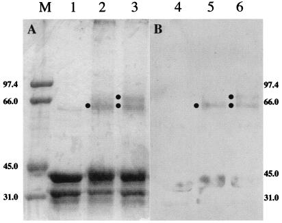
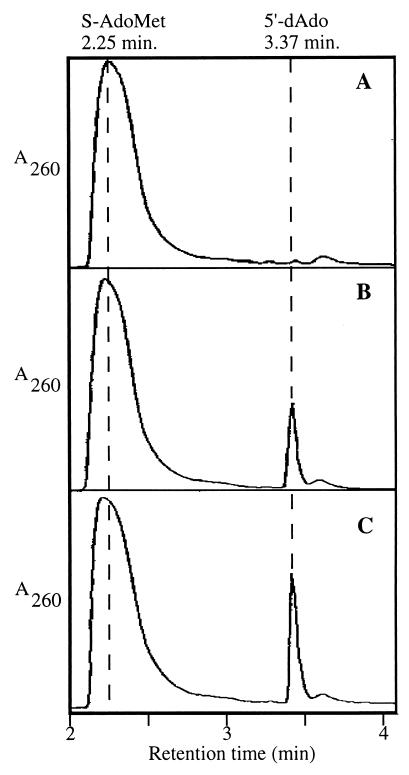
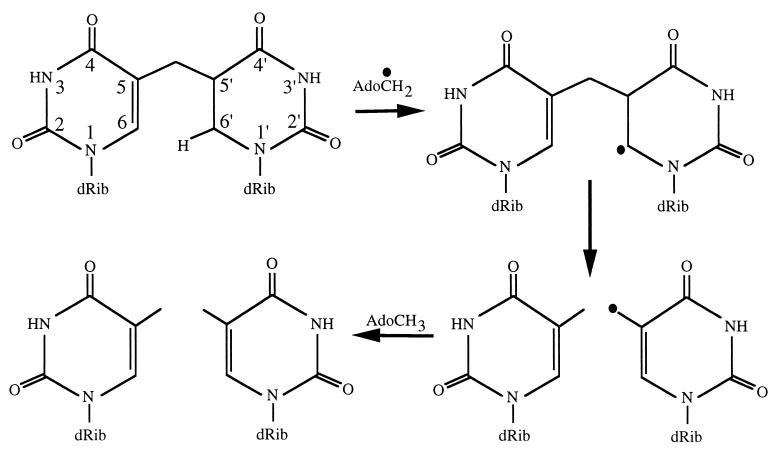
The major DNA photoproduct of dormant, UV-irradiated Bacillus subtilis spores is the thymine dimer 5-thyminyl-5,6-dihydrothymine [spore photoproduct (SP)]. During spore germination, SP is reversed to two intact thymines in situ by the DNA repair enzyme SP lyase, an S-adenosylmethionine (S-AdoMet)-dependent iron-sulfur ([Fe-S]) protein encoded by the splB gene. In the present work, cross-linking, SDS/PAGE, and size exclusion chromatography revealed that SplB protein dimerized when incubated with iron and sulfide under anaerobic reducing conditions. SplB isolated under aerobic conditions generated an EPR spectrum consistent with that of a partially degraded [3Fe-4S] center, and reduction of SplB with dithionite shifted the spectrum to that of a [4Fe-4S] center. Addition of S-AdoMet to SplB converted some of the [4Fe-4S] centers to an EPR-silent form consistent with electron donation to S-AdoMet. HPLC and electrospray ionization MS analyses showed that SP lyase cleaved S-AdoMet to generate 5'-deoxyadenosine. The results indicate that (i) SP lyase is a homodimer of SplB; (ii) dimer formation is coordinated by a [4Fe-4S] center; and (iii) the reduced [4Fe-4S] center is capable of donating electrons to S-AdoMet to generate a 5'-adenosyl radical that is then used for the in situ reversal of SP. Thus, SP lyase belongs to the "radical SAM" superfamily of enzymes that use [Fe-S] centers and S-AdoMet to generate adenosyl radicals to effect catalysis. SP lyase is unique in being the first and only DNA repair enzyme known to function via this novel enzymatic mechanism.


Figures

Figure 1
Figure 1
Analysis of cross-linked SplB by SDS/PAGE. (10His)SplB was treated as described in Experimental Procedures, denatured, and electrophoresed through 10% SDS/PAGE. (A) Coomassie blue-stained gel. (B) Western blot. Lanes 1 and 4, untreated; lanes 2 and 5, cross-linked with DMS; lanes 3 and 6, cross-linked with DMS after incubation under conditions favoring [Fe-S] cluster formation. Filled circles indicate positions of cross-linked species at a molecular mass of ≈64 kDa and ≈72 kDa. The lane marked M contains molecular weight markers. Sizes are in kilodaltons.
Figure 2
Figure 2
Analysis of (10His)SplB by size exclusion chromatography. (10His)SplB was chromatographed through Biogel P-100, immediately after its purification by Ni-NTA chromatography (A) and after incubation under conditions to reconstitute [Fe-S] clusters (B). The y axes (A280) in A and B are scaled identically. (C) Results of colorimetric iron assays (16) performed on fractions from the experiment depicted in B.
Figure 3
Figure 3
EPR analysis of (10His)SplB. EPR spectra of (10His)SplB isolated under aerobic conditions (protein concentration 6 mg/ml; A); (10His)SplB treated with 10 mM dithionite (protein concentration 12 mg/ml; B); and (10His)SplB treated with 10 mM dithionite and 1 mM S-AdoMet (protein concentration 12 mg/ml; C). In all three traces, relative signal strength (y axes) are depicted on identical scales.
Figure 4
Figure 4
HPLC analysis of S-AdoMet conversion to 5′-dAdo by (10His)SplB. Standard SP lyase reactions were performed as described in Experimental Procedures and analyzed by HPLC. Reactions contained no (10His)SplB (A); 400 μg (10His)SplB (B); and 400 μg (10His)SplB and 1.63 μg B. subtilis SP-containing chromosomal DNA (C). The vertical dashed lines denote the RT at which purified S-AdoMet and 5′-dAdo elute from the column. In all three traces, the y axes (A260) are depicted on identical scales.
Figure 5
Figure 5
ESI-MS analysis of the peak (RT = 3.37 min) from Fig. 4. Displayed are the primary ESI-MS spectra of the unknown compound (A) and its fragmentation product (B).
Figure 6
Figure 6
Proposed SP cleavage mechanism of SP lyase. See text and ref. for details.

References

    1. Setlow P. Comments Mol Cell Biophys. 1988;5:253–264.
    1. Setlow P. Annu Rev Microbiol. 1995;49:29–54. - PubMed
    1. Nicholson W L, Munakata N, Horneck G, Melosh H J, Setlow P. Microbiol Mol Biol Rev. 2000;64:548–572. - PMC - PubMed
    1. Donnellan J E, Setlow R B. Science. 1965;149:308–310. - PubMed
    1. Varghese A J. Biochem Biophys Res Commun. 1970;38:484–490. - PubMed

Publication types

LinkOut - more resources