. 2001 Jul 31;98(16):9133-8.
doi: 10.1073/pnas.171286998. Epub 2001 Jul 24.

Presence of mitochondria-type ribosomes outside mitochondria in germ plasm of Drosophila embryos

Affiliations

Presence of mitochondria-type ribosomes outside mitochondria in germ plasm of Drosophila embryos

R Amikura et al. Proc Natl Acad Sci U S A. .

Abstract

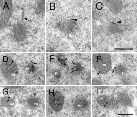
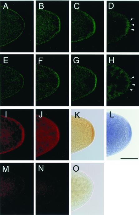
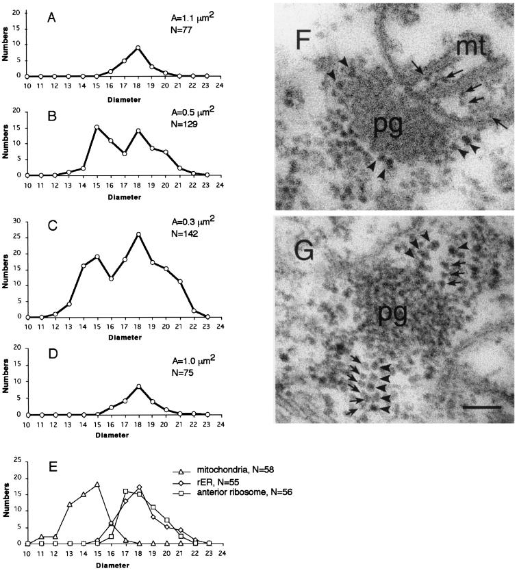
Mitochondrially encoded large and small ribosomal RNAs (mtlrRNA and mtsrRNA) are transported out of mitochondria to polar granules, the distinctive organelles of germ plasm in Drosophila. Reduction of the extramitochondrial mtlrRNA amount leads to the failure of embryos to form the germ-line progenitors, or pole cells, suggesting that mtlrRNA, along with mtsrRNA, functions on the polar granules to specify the germ line. In this study, we provide several lines of evidence showing that there are mitochondria-type ribosomes on the polar granules during a short period before pole cell formation. Our ultrastructural analysis reveals that these ribosomes include both mitochondrial rRNAs and at least two mitochondrial ribosomal proteins (S12 and L7/L12). Furthermore, these ribosomes are integrated into well developed polysomes on the surface of polar granules. We propose that translation dependent on mitochondria-type ribosomes is an important mechanism underlying germ-line formation.


Figures

Figure 1
Figure 1
Presence of mitochondrial rRNAs and ribosomal proteins in the polar granule polysomes at stage 2. (A) An electron micrograph showing well developed polysomes (arrow) on the surface of polar granules. (B and C) Electron micrographs of sections hybridized with probes for mtlrRNA (B) and mtsrRNA (C). Signals were arranged linearly from the surface of polar granules (arrowheads). (Scale bar in C = 0.25 μm.) (D–G) Distribution of S12-EGFP (D), L7/L12-EGFP (E), L7/L12-HA (F), and endogenous S12 (G). Signals were detected at the periphery of polar granules (arrows), as well as within mitochondria (arrowheads). (I) Distribution of endogenous S12 in polar plasm of early stage-1 embryo. S12 was detected within mitochondria (arrow) but not on polar granules. This distribution pattern was essentially identical to the patterns of the tagged ribosomal proteins (data not shown). Without induction of the tagged proteins, we could not detect signals for L7/L12-EGFP (H) and S12 EGFP and L7/L12-HA (data not shown). mt, mitochondria; pg, polar granules. (Scale bar in I = 0.2 μm.)
Figure 2
Figure 2
Distribution of mitochondrial ribosomal proteins in oocytes and early embryos. (A–H) Developmental changes in the distribution of S12-EGFP (A–D) and L7/L12-EGFP (E–H) detected by EGFP fluorescence. (A and E) Mature oocytes, (B and F) stage-1 embryos, (C and G) stage-2 embryos, and (D and H) stage-3 embryos. (I–L) Immunohistochemical detection of the Gal4-induced S12-EGFP (I) and L7/L12-EGFP (J) by an anti-GFP antibody and L7/L12-HA by an anti-HA antibody (K). Without induction by Gal4, no staining for S12-EGFP (M), L7/L12-EGFP (N), or L7/L12-HA (O) was observed. (L) A wild-type embryo stained with anti-S12 antibody. (I–O) Stage-2 embryos. The stages of the embryos were determined by the number and the location of nuclei stained by propidium iodide (22) referred to by Campos-Ortega and Hartenstein (13). (Scale bar = 50 μm.)
Figure 3
Figure 3
Two types of ribosomes in the polar-granule polysomes. (A–D) The average number of ribosomes in a unit area (0.3 μm2) is plotted against its diameter. Diameters of the ribosomes around polar granules were measured in stage-14 oocytes (A), stage-1 embryos (B), stage-2 embryos (C), and in pole cells of stage-4 embryos (D). In A–D, total area examined (A) and total number of the ribosomes counted (N) are shown. (E) The number of ribosomes within mitochondria, in the anterior region and on the surface of rough endoplasmic reticulum, are plotted against their diameters. (F and G) Electron micrographs of sections through polar granules. (F) In stage-1 embryos, polar granules are intimately associated with mitochondria, and no ribosomes were found at the boundary between these organelles. Arrows point to ribosomes within a mitochondrion. (G) In stage-2 embryos, the smaller ribosomes (arrows) are integrated into the polar granule-polysomes. Arrowheads (F and G) point to the larger ribosomes. pg, polar granules; mt, mitochondria. (Scale bar = 0.1 μm.)

References

    1. Beam H W, Kessel R G. Int Rev Cytol. 1974;39:413–479. - PubMed
    1. Eddy E M. Int Rev Cytol. 1975;43:229–280. - PubMed
    1. Kobayashi S, Okada M. Development (Cambridge, UK) 1989;107:733–774. - PubMed
    1. Iida T, Kobayashi T. Proc Natl Acad Sci USA. 1998;95:11274–11278. - PMC - PubMed
    1. Kobayashi S, Amikura R, Okada M. Science. 1993;260:1521–1524. - PubMed

Publication types

LinkOut - more resources