. 2001 Jun;124(3):346-52.
doi: 10.1046/j.1365-2249.2001.01510.x.

Enhanced expression and production of monocyte chemoattractant protein-1 in myocarditis

Affiliations

Enhanced expression and production of monocyte chemoattractant protein-1 in myocarditis

K Fuse et al. Clin Exp Immunol. 2001 Jun.

Abstract

Monocyte chemoattractant protein-1 (MCP-1) is a member of the C-C chemokine family that has been shown to play a major role in the migration of monocytes and T cells to an inflammatory focus. To clarify the role of MCP-1 in the pathogenesis of myocarditis, we have examined the expression of MCP-1 in rat hearts with experimental autoimmune myocarditis (EAM), and have also measured serum levels of MCP-1 in patients with histology-proven acute myocarditis. Lewis rats were immunized with cardiac myosin and were killed 9, 12, 15, 18, 21, 24, 27, 30, 33, 36, 42 and 56 days after immunization. Large mononuclear cells in the myocardial interstitium were stained with an anti-MCP-1 antibody. mRNA of MCP-1 increased in the hearts of EAM rats from days 15--27 as shown by quantitative reverse transcription-polymerase chain reaction. Serum MCP-1 levels of the rats with EAM were significantly elevated from days 15--24. In the clinical study, serum levels of MCP-1 in 24 patients with acute myocarditis at the time of admission (165.2 +/- 55.8 pg/ml) were significantly (P = 0.0301) elevated compared with those of 20 healthy volunteers (61.8 +/- 10.7 pg/ml). Serum MCP-1 levels of 8 fatal cases (371.8 +/- 145.2 pg/ml) were significantly (P = 0.0058) higher than those of 16 cases who survived (65.5 +/- 12.8 pg/ml). In conclusions, MCP-1 may play an important role in the pathogenesis of human acute myocarditis as well as in the progression of rat EAM.


Figures
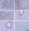
Fig. 1
Fig. 1
Immunohistochemical staining for MCP-1 in the hearts of EAM rats on (A) day 21 and (B) day 24. Large mononuclear cells in the myocardial interstitium (arrows) were stained with an anti-MCP-1 antibody. (C) On day 42, focal infiltration of mononuclear cells was still observed, however, only a few MCP-1 positive cells were found (arrow). However, in cardiac myocytes, MCP-1 was not stained. (D) On day 18, in intact cardiac myocytes, vascular endothelial cells and vascular smooth muscle cells, MCP-1 was not stained. Controls were not stained (E) with an anti-MCP-1 antibody in normal rat heart and (F) without an anti-MCP-1 antibody in the heart of EAM on day 21. Bar indicates 50 μm.
Fig. 2
Fig. 2
mRNA levels of MCP-1 were increased over 10 fold in the hearts of EAM rats from days 15–27 compared with those of the normal control rats (NC), and peaked over 60 fold on day 24. The vertical line represents the copy numbers of mRNA in the same dilution samples. The horizontal line represents the days from immunization. Each bar represents mean ± SEM in three to four separate experiments. *P < 0.05 versus NC.
Fig. 3
Fig. 3
Serum levels of MCP-1 in EAM rats significantly increased from days 15–24 compared with those of the normal control rats (NC) and peaked on day 18. After day 30, serum levels of MCP-1 fell to within or below normal range. *P < 0.05 versus NC.
Fig. 4
Fig. 4
Serum MCP-1 levels of the healthy control group (n = 20) and the myocarditis group (n = 24). The mean serum MCP-1 level in the myocarditis group (165·2 ± 55·8 pg/ml) was significantly higher than that of the healthy control group (48·2 ± 3·9 pg/ml). The solid bar and closed circles indicates mean ± SEM. *P < 0.0301.
Fig. 5
Fig. 5
Serum MCP-1 levels of the fatal group (n = 8) and the survival group (n = 16) in patients with acute myocarditis. The serum levels of MCP-1 in the fatal group (371·8 ± 145·2 pg/ml) were significantly higher than those of the group who survived (61·8 ± 10·7 pg/ml). *P < 0.0058. Solid bar and closed circle indicate mean ± SEM.

References

    1. Rollins BJ. Monocyte chemoattractant protein 1: a potential regulator of monocyte recruitment in inflammatory disease. Mol Med Today. 1996;2:198–204. - PubMed
    1. Adams DH, Lloyd AR. Chemokines: leukocyte recruitment and activation cytokines. Lancet. 1997;349:490–5. - PubMed
    1. Rollins BJ, Yoshimura T, Leonard EJ, Pober JS. Cytokine-activated human endothelial cells synthesize and secrete a monocyte chemoattractant, MCP-1/JE. Am J Pathol. 1990;136:1229–33. - PMC - PubMed
    1. Rollins BJ, Pober JS. Interleukin-4 induces the synthesis and secretion of MCP-1/JE by human endothelial cells. Am J Pathol. 1991;138:1315–9. - PMC - PubMed
    1. Shyy Y-J, Li Y-S, Kolattukudy PE. Activation of MCP-1 gene expression is mediated through multiple signaling pathways. Biochem Biophys Res Commun. 1993;192:693–9. - PubMed

Publication types