High frequency of circulating HBcAg-specific CD8 T cells in hepatitis B infection: a flow cytometric analysis
- PMID: 11472405
- PMCID: PMC1906072
- DOI: 10.1046/j.1365-2249.2001.01561.x
High frequency of circulating HBcAg-specific CD8 T cells in hepatitis B infection: a flow cytometric analysis
Abstract
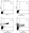
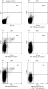
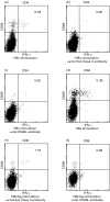
Viral antigen-specific T cells are important for virus elimination. We studied the hepatitis B virus (HBV)-specific T cell response using flow cytometry. Three phases of HBV infection were studied: Group A, HBeAg (+) chronic hepatitis; Group B, HBeAb (+) HBV carrier after seroconversion; and Group C, HBsAb (+) phase. Peripheral T cells were incubated with recombinant HB core antigen (HBcAg), and intracytoplasmic cytokines were analysed by flow cytometry. HBcAg-specific CD4 and CD8 T cells were identified in all three groups and the number of IFN-gamma-positive T cells was greater than TNF-alpha-positive T cells. The frequency of IFN-gamma-positive CD4 and CD8 T cells was highest in Group C, compared with Groups A and B. No significant difference in the HBcAg-specific T cell response was observed between Group A and Group B. The HBcAg-specific CD8 T cell response was diminished by CD4 depletion, addition of antibody against human leucocyte antigen (HLA) class I, class II or CD40L. Cytokine-positive CD8 T cells without HBcAg stimulation were present at a high frequency (7 of 13 cases) in Group B, but were rare in other groups. HBcAg-specific T cells can be detected at high frequency by a sensitive flow cytometric analysis, and these cells are important for controlling HBV replication.
Figures


 Group A;
  Group B; ▪ Group C.


Similar articles
-
Lentiviral vector encoding ubiquitinated hepatitis B core antigen induces potent cellular immune responses and therapeutic immunity in HBV transgenic mice.Immunobiology. 2016 Jul;221(7):813-21. doi: 10.1016/j.imbio.2016.01.015. Epub 2016 Feb 2. Immunobiology. 2016. PMID: 26874581
-
Hepatitis B core antigen-specific IFN-gamma production of peripheral blood mononuclear cells in patients with chronic hepatitis B virus infection.J Immunol. 1989 Jun 1;142(11):4006-11. J Immunol. 1989. PMID: 2469730
-
Adenovirus Vector Harboring the HBcAg and Tripeptidyl Peptidase II Genes Induces Potent Cellular Immune Responses In Vivo.Cell Physiol Biochem. 2017;41(2):423-438. doi: 10.1159/000456579. Epub 2017 Jan 27. Cell Physiol Biochem. 2017. PMID: 28214886
-
Activation of a heterogeneous hepatitis B (HB) core and e antigen-specific CD4+ T-cell population during seroconversion to anti-HBe and anti-HBs in hepatitis B virus infection.J Virol. 1995 Jun;69(6):3358-68. doi: 10.1128/JVI.69.6.3358-3368.1995. J Virol. 1995. PMID: 7538172 Free PMC article.
-
HBcAg-specific IL-21-producing CD4+ T cells are associated with relative viral control in patients with chronic hepatitis B.Scand J Immunol. 2013 Nov;78(5):439-46. doi: 10.1111/sji.12099. Scand J Immunol. 2013. PMID: 23957859
Cited by
-
HBcAg-specific CD8 T cells play an important role in virus suppression, and acute flare-up is associated with the expansion of activated memory T cells.J Clin Immunol. 2003 May;23(3):223-32. doi: 10.1023/a:1023366013858. J Clin Immunol. 2003. PMID: 12797544
-
Reduced hepatitis B virus (HBV)-specific CD4+ T-cell responses in human immunodeficiency virus type 1-HBV-coinfected individuals receiving HBV-active antiretroviral therapy.J Virol. 2005 Mar;79(5):3038-51. doi: 10.1128/JVI.79.5.3038-3051.2005. J Virol. 2005. PMID: 15709024 Free PMC article.
-
Monocytic MDSCs homing to thymus contribute to age-related CD8+ T cell tolerance of HBV.J Exp Med. 2022 Apr 4;219(4):e20211838. doi: 10.1084/jem.20211838. Epub 2022 Mar 7. J Exp Med. 2022. PMID: 35254403 Free PMC article.
-
rt269I Type of Hepatitis B Virus (HBV) Polymerase versus rt269L Is More Prone to Mutations within HBV Genome in Chronic Patients Infected with Genotype C2: Evidence from Analysis of Full HBV Genotype C2 Genome.Microorganisms. 2021 Mar 15;9(3):601. doi: 10.3390/microorganisms9030601. Microorganisms. 2021. PMID: 33803998 Free PMC article.
-
Hepatitis B and circadian rhythm of the liver.World J Gastroenterol. 2022 Jul 21;28(27):3282-3296. doi: 10.3748/wjg.v28.i27.3282. World J Gastroenterol. 2022. PMID: 36158265 Free PMC article. Review.
References
-
- Tiollais P, Pourcel C, Dejean A. The hepatitis B virus. Nature. 1985;317:489–95. - PubMed
-
- Beasley RP, Lin C-C, Hwang L-Y, Chien C-S. Hepatocellular carcinoma and hepatitis B virus. A prospective study of 22, 7. 07 men in Taiwan Lancet. 1981;21:1129–32. - PubMed
-
- Nayersina R, Folwer P, Guilhot S, et al. HLA A2 restricted cytotoxic T lymphocyte responses to multiple hepatitis B surface antigen epitopes during hepatitis B virus infection. J Immunol. 1993;150:4659–71. - PubMed
Publication types
MeSH terms
Substances
LinkOut - more resources
Full Text Sources
Other Literature Sources
Medical
Research Materials

