

Functional heterogeneity of anti-endothelial cell antibodies
- PMID: 11472414
- PMCID: PMC1906088
- DOI: 10.1046/j.1365-2249.2001.01528.x
Functional heterogeneity of anti-endothelial cell antibodies
Abstract
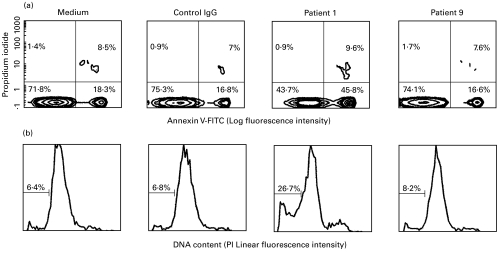
While it has been claimed that some anti-endothelial cell antibodies (AECA) activate EC, there is also evidence that others trigger apoptosis. To address the issue of whether activation is a prerequisite for AECA-mediated apoptosis of EC, 23 AECA-positive sera were evaluated for their ability to induce activation and/or apoptosis. Activation was defined as an over-expression of E-selectin and intercellular adhesion molecule 1. Optical microscopy, annexin V binding, hypoploid cell enumeration, and determination of poly (ADP-ribose) polymerase cleavage-related products were used to assess apoptosis. Four functional profiles were defined: 10 sera promoted activation and apoptosis (act+/apo+), one was act+/apo-, six act-/apo+, and the remaining six act-/apo-. The reduced membrane expression of thrombomodulin was associated with apoptosis, rather than activation. Caspase-3 was implicated in the two models of apoptosis, the ratios of several survival proteins to Bax decreased, regardless of the ability of apo+ AECA to activate the cells, while radical oxygen species did not appear to be involved. Furthermore, it occurred that macrophages engulfed EC treated with apoptosis-promoting AECA, but not those incubated with AECA that did not induce apoptosis. Hence, AECA represent an extremely heterogeneous family of autoantibodies, not only because of the variety of their target antigens, but also the subsequent diversity of their effects.
Figures


Similar articles
-
Anti-endothelial cell antibodies from lupus patients bind to apoptotic endothelial cells promoting macrophage phagocytosis but do not induce apoptosis.Rheumatology (Oxford). 2005 Jul;44(7):879-84. doi: 10.1093/rheumatology/keh633. Epub 2005 Apr 12. Rheumatology (Oxford). 2005. PMID: 15827042
-
IgG anti-endothelial cell autoantibodies from patients with systemic lupus erythematosus or systemic vasculitis stimulate the release of two endothelial cell-derived mediators, which enhance adhesion molecule expression and leukocyte adhesion in an autocrine manner.Arthritis Rheum. 1999 Apr;42(4):631-40. doi: 10.1002/1529-0131(199904)42:4<631::AID-ANR5>3.0.CO;2-X. Arthritis Rheum. 1999. PMID: 10211876
-
The binding of some human antiendothelial cell antibodies induces endothelial cell apoptosis.J Clin Invest. 1998 May 15;101(10):2029-35. doi: 10.1172/JCI2261. J Clin Invest. 1998. PMID: 9593758 Free PMC article.
-
Are autoantibodies triggering endothelial cell apoptosis really pathogenic?Autoimmun Rev. 2009 Jun;8(7):605-10. doi: 10.1016/j.autrev.2009.02.005. Epub 2009 Feb 12. Autoimmun Rev. 2009. PMID: 19393202 Review.
-
Pathogenic role of anti-endothelial cell antibodies in systemic vasculitis.Wien Klin Wochenschr. 2000 Aug 25;112(15-16):660-4. Wien Klin Wochenschr. 2000. PMID: 11020952 Review.
Cited by
-
Genetic and molecular biology of systemic lupus erythematosus among Iranian patients: an overview.Auto Immun Highlights. 2021 Jan 30;12(1):2. doi: 10.1186/s13317-020-00144-y. Auto Immun Highlights. 2021. PMID: 33516274 Free PMC article. Review.
-
A novel autoantibody against fibronectin leucine-rich transmembrane protein 2 expressed on the endothelial cell surface identified by retroviral vector system in systemic lupus erythematosus.Arthritis Res Ther. 2012 Jul 2;14(4):R157. doi: 10.1186/ar3897. Arthritis Res Ther. 2012. PMID: 22747982 Free PMC article.
-
Effects of anti-endothelial cell antibodies in leprosy and malaria.Infect Immun. 2004 Jan;72(1):301-9. doi: 10.1128/IAI.72.1.301-309.2004. Infect Immun. 2004. PMID: 14688109 Free PMC article.
-
[Immunological Aspects after Lung Transplantation].Zentralbl Chir. 2025 Jun;150(3):295-305. doi: 10.1055/a-2590-9933. Epub 2025 May 13. Zentralbl Chir. 2025. PMID: 40359989 Free PMC article. Review. German.
-
Autoantibodies to vascular smooth muscle are pathogenic for vasculitis.Am J Pathol. 2005 Jun;166(6):1851-60. doi: 10.1016/S0002-9440(10)62494-7. Am J Pathol. 2005. PMID: 15920169 Free PMC article.
References
-
- Meroni PL, Youinou P. Endothelial cell antibodies. In: Peter JB, Shoenfeld Y, editors. Autoantibodies. Amsterdam: Elsevier; 1996. p. 245.
-
- Damianovich M, Gilburd B, George J, et al. Pathogenic role of anti-endothelial cell antibodies in vasculitis. An idiotypic experimental model. J Immunol. 1996;156:4946–51. - PubMed
Publication types
MeSH terms
Substances
LinkOut - more resources
Full Text Sources
Research Materials
