Selective recruitment of CCR6-expressing cells by increased production of MIP-3 alpha in rheumatoid arthritis
- PMID: 11472439
- PMCID: PMC1906097
- DOI: 10.1046/j.1365-2249.2001.01542.x
Selective recruitment of CCR6-expressing cells by increased production of MIP-3 alpha in rheumatoid arthritis
Abstract
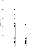
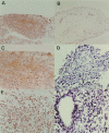
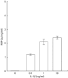
Infiltration of various types of leucocytes has been shown to play a crucial role in the pathogenesis of rheumatoid arthritis (RA). Macrophage inflammatory protein-3 alpha (MIP-3 alpha) is a recently identified chemokine which is a selective chemoattractant for leucocytes such as memory T cells, naïve B cells and immature dendritic cells. In this study, we investigated the expression of MIP-3 alpha and its specific receptor CCR6 in the inflamed joints of patients with RA. Increased amounts of MIP-3 alpha were found by ELISA in synovial fluids (SF) of patients with RA. MIP-3 alpha was apparently detected in all synovial tissue specimens of RA patients (n = 6), but it could not be detected in that of osteoarthritis (OA) patients (n = 4). Expression of MIP-3 alpha was detected especially in the sublining layer, and infiltrating mononuclear cells in RA synovial tissue. Gene expression of MIP-3 alpha was also found in six out of 11 RA-synovial fluid cells by RT-PCR. Cultured synovial fibroblasts derived from either RA or OA patients were capable of producing MIP-3 alpha in response to IL-1 beta and TNFalpha in vitro. Furthermore, expression of CCR6 was found in infiltrating mononuclear cells in the cellular clusters and around the vessels of RA synovial tissue. These findings indicate that increased production of MIP-3 alpha may contribute to the selective recruitment of CCR6-expressing cells in RA.
Figures


References
-
- Feldmann M, Brennan FM, Maini RN. Rheumatoid arthritis. Cell. 1996;85:307–10. - PubMed
-
- Rollins BJ. Chemokines. Blood. 1997;90:909–28. - PubMed
-
- Baggiolini M. Chemokines and leukocyte traffic. Nature. 1998;392:565–8. - PubMed
-
- Sallusto F, Lanzavecchia A, Macky CR. Chemokines and chemokine receptors in T-cell priming and Th1/Th2-mediated responses. Immunol Today. 1998;19:568–73. - PubMed
-
- Ward SG, Bacon K, Westwick J. Chemokines and T lymphocytes: More than an attraction. Immunity. 1998;9:1–11. - PubMed
Publication types
MeSH terms
Substances
LinkOut - more resources
Full Text Sources
Other Literature Sources
Medical

