. 2001 Aug;67(8):3665-70.
doi: 10.1128/AEM.67.8.3665-3670.2001.

Bacillus spore inactivation methods affect detection assays

Affiliations

Bacillus spore inactivation methods affect detection assays

J L Dang et al. Appl Environ Microbiol. 2001 Aug.

Abstract

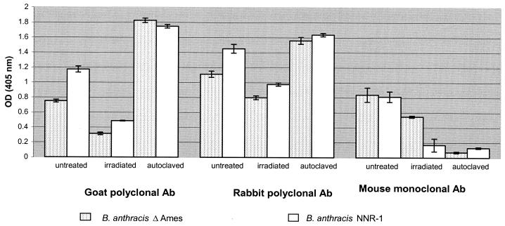
Detection of biological weapons is a primary concern in force protection, treaty verification, and safeguarding civilian populations against domestic terrorism. One great concern is the detection of Bacillus anthracis, the causative agent of anthrax. Assays for detection in the laboratory often employ inactivated preparations of spores or nonpathogenic simulants. This study uses several common biodetection platforms to detect B. anthracis spores that have been inactivated by two methods and compares those data to detection of spores that have not been inactivated. The data demonstrate that inactivation methods can affect the sensitivity of nucleic acid- and antibody-based assays for the detection of B. anthracis spores. These effects should be taken into consideration when comparing laboratory results to data collected and assayed during field deployment.


Figures

FIG. 1
FIG. 1
Direct ELISA analysis of B. anthracis ΔAmes and NNR1 spore preparations following inactivation. Serial dilutions of the ΔAmes spores were performed with PBS and tested by direct ELISA with goat, rabbit, or mouse antibodies as shown. Irradiation of spore preparations resulted in a decreased ELISA signal for all antibodies tested. Autoclave treatment resulted in a decrease in ELISA signal when ΔAmes and NNR1 spores were probed with the mouse MAb BF1. In contrast to the MAb, both of the PAb showed an increase in signal following thermal inactivation.
FIG. 2
FIG. 2
Direct ELISA analysis of the supernatants from 107-CFU/ml dilutions of B. anthracis ΔAmes spores. Spores were pelleted by centrifugation, and the supernatant was removed. Serial dilutions of the ΔAmes spore supernatants were performed with PBS and assayed by direct ELISA using goat, rabbit, or mouse antibodies as shown.
FIG. 3
FIG. 3
Untreated, autoclaved, or inactivated ΔAmes or NNR1 spores were stained with mouse MAb AB2 and BF1 or goat antibody to spores. Fluorescence staining was measured by flow cytometry. The representative forward- and side-scatter profiles for treated and untreated spore samples are shown in the left panel. In the right panel of histograms, positive staining is revealed by the bold lines and negative control staining is shown by the light lines. It can be seen that both autoclaving and irradiation destroy the BF1 as well as the AB2 epitope. Under the same conditions of spore inactivation, the goat PAb staining remains intact.
FIG. 4
FIG. 4
TaqMan PCR analysis targeting the capA gene of Bacillus spore preparations. Each tube was subjected to PCR amplification in triplicate with the Applied Biosystems model 7700 using equivalent numbers of spores from the indicated cell lines. The data are plotted as the relative change in total fluorescence (ΔRn) versus the cycle number. A, untreated B. anthracis ΔAmes spores; B, irradiated B. anthracis ΔAmes spores; C, autoclaved B. anthracis ΔAmes spores; D, untreated B. thuringiensis subsp. kurstaki spores; E, untreated B. subtilis 1031 spores; F, untreated B. cereus 6E1 spores.
FIG. 5
FIG. 5
TaqMan PCR analysis targeting the lef gene of Bacillus spore preparations. The data are plotted as the relative change in total fluorescence (ΔRn) versus the cycle number. A, untreated B. anthracis ΔAmes spores; B, irradiated B. anthracis ΔAmes spores; C, autoclaved B. anthracis ΔAmes spores; D, untreated NNR1 B. anthracis spores; E, untreated B. subtilis spores; F, untreated B. cereus 6E1 spores; G, untreated B. thuringiensis subsp. kurstaki spores. H, double-distilled water.

References

    1. Beyer W, Glöckner P, Otto J, Böhm R. A nested PCR method for the detection of Bacillus anthracis in environmental samples collected from former tannery sites. Microbiol Res. 1995;150:179–186. - PubMed
    1. Cano R J, Borucki M K. Revival and identification of bacterial spores in 25- to 40-million-year-old Dominican amber. Science. 1995;268:1060–1063. - PubMed
    1. Green B D, Battisti L, Koehler T M, Thorne C B, Ivins B E. Demonstration of a capsule plasmid in Bacillus anthracis. Infect Immun. 1985;49:291–297. - PMC - PubMed
    1. Hacker J, Blum-Oehler G, Mühldorfer I, Tschäpe H. Pathogenicity islands of virulent bacteria: structure, function and impact on microbial evolution. Mol Microbiol. 1997;23:1089–1097. - PubMed
    1. Heid C A, Stevens J, Livak K J, Williams P M. Real time quantitative PCR. Genome Res. 1996;6:986–994. - PubMed

Publication types

MeSH terms