. 2001 Sep 28;276(39):36446-53.
doi: 10.1074/jbc.M104386200. Epub 2001 Jul 30.

Functional analysis of the four DNA binding domains of replication protein A. The role of RPA2 in ssDNA binding

Affiliations

Functional analysis of the four DNA binding domains of replication protein A. The role of RPA2 in ssDNA binding

S A Bastin-Shanower et al. J Biol Chem. .

Abstract

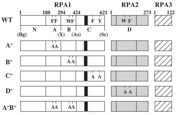
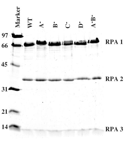
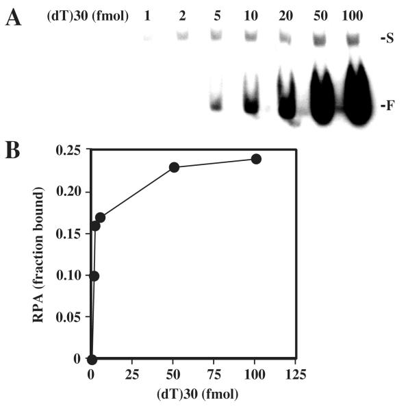
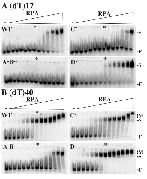
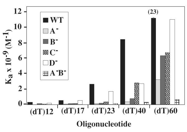
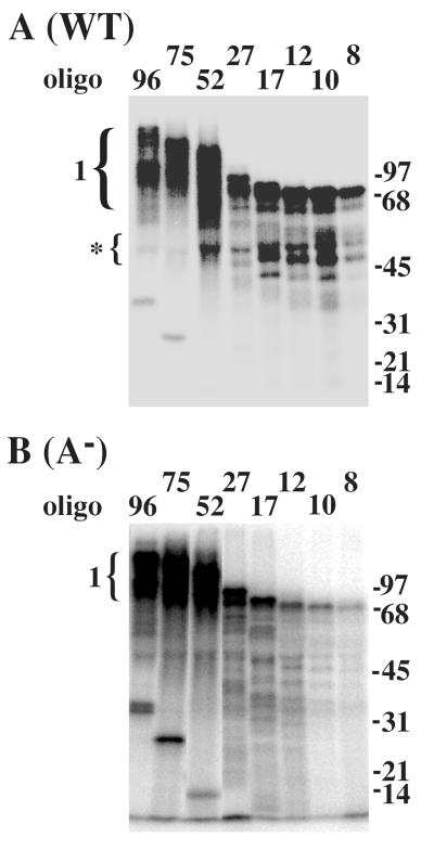
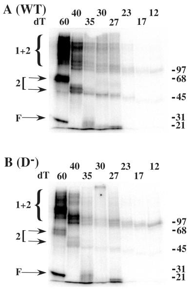
Replication Protein A (RPA), the heterotrimeric single-stranded DNA (ssDNA)-binding protein of eukaryotes, contains four ssDNA binding domains (DBDs) within its two largest subunits, RPA1 and RPA2. We analyzed the contribution of the four DBDs to ssDNA binding affinity by assaying recombinant yeast RPA in which a single DBD (A, B, C, or D) was inactive. Inactivation was accomplished by mutating the two conserved aromatic stacking residues present in each DBD. Mutation of domain A had the most severe effect and eliminated binding to a short substrate such as (dT)12. RPA containing mutations in DBDs B and C bound to substrates (dT)12, 17, and 23 but with reduced affinity compared with wild type RPA. Mutation of DBD-D had little or no effect on the binding of RPA to these substrates. However, mutations in domain D did affect the binding to oligonucleotides larger than 23 nucleotides (nt). Protein-DNA cross-linking indicated that DBD-A (in RPA1) is essential for RPA1 to interact efficiently with substrates of 12 nt or less and that DBD-D (RPA2) interacts efficiently with oligonucleotides of 27 nt or larger. The data support a sequential model of binding in which DBD-A is responsible for the initial interaction with ssDNA, that domains A, B, and C (RPA1) contact 12-23 nt of ssDNA, and that DBD-D (RPA2) is needed for RPA to interact with substrates that are 23-27 nt in length.


Figures

Fig. 1
Fig. 1. Heterotrimeric RPA proteins used in this study
The domain structure of wt RPA (Top) or the indicated mutant RPA protein is illustrated schematically. The RPA1 subunit consists of an N-terminal domain (N) and DBDs -A, -B, and -C while RPA 2 consists of DBD-D. RPA3 is wt in all RPA complexes. The amino acid residues comprising the domains of RPA1 are: N, 1 - 179; A, 180 - 294; B, 295 to 415; and C, 416 - 621. DBD-D is defined as amino acids 40-174 of RPA2. The positions of conserved aromatic residues within each DBD are indicated using single letter code. The following mutations are indicated: A, F238A, F269A; B, W360A, F385A; C, F537A, Y586A; and D, W101A, F143A. The unique restriction sites created in the wt RFA1 plasmid and their positions relative to the DBDs are presented: Bg, BglII; X, XhoI; As, Asp718; Sc, SacII; S, SalI; Bs, BsiWI. The black box located within DBD-C denotes the zinc-finger motif extending from position 486 to 508.
Fig. 2
Fig. 2. Purified RPA complexes
ScRPA proteins were expressed in E.coli and purified as described in the Experimental Procedures. Two μg of wt RPA or the indicated mutant was resolved on by SDS-17% PAGE and visualized with Coommassie blue. The positions of RPA1 (69 kDa), RPA2 (36 kDa) and RPA3 (13 kDa) are indicated. The molecular mass standards are indicated in kDa.
Fig. 3
Fig. 3. Determining the fraction of active RPA
(A) 8.7 fmol of purified wt RPA was incubated with the indicated amount of 32P-labeled (dT)30 and the reactions were resolved on a 6% nondenaturing polyacrylamide gel. The positions of singly liganded (S) and unbound (F) substrate are indicated. (B) The radioactivity corresponding to free DNA and protein-DNA complex in (A) was quantitated using liquid scintillation counting. The fraction of RPA in the bound form was then calculated and plotted as a function of input DNA.
Fig. 4
Fig. 4. Gel mobility shift assays of wt and mutant RPA
Increasing amounts of wt or the indicated mutant RPA (0, 1, 2, 6, 25, 50, 100, 250, 500, 1000, 2000, 6000, 20000 or 60000 pg) were incubated with 2 fmols of radiolabeled (dT)17 (A) or (dT)40 (B). The reactions were then resolved on a 6% nondenaturing polyacrylamide gel. The first lane of each gel (−), is a negative control reaction containing no protein. The positions of RPA-DNA complexes (S, singly-liganded; M, multiply-liganded) and unbound oligonucleotide (F) are indicated. The asterisk indicates a binding reaction containing equimolar amounts of RPA and substrate.
Fig. 5
Fig. 5. Comparison of association constants (Ka)
The apparent binding constants (Ka) of wt or the indicated mutant RPA are presented graphically as a function of substrate size. All data is taken from Table 2.
Fig. 6
Fig. 6. DBD-A is required for binding short oligonucleotides
One pmol of wild type (A) or mutant (B) RPA was incubated with one pmol of the indicated radiolabeled oligonucleotide. Following UV-crosslinking, the reactions were denatured and incubated with antiserum against RPA1. The reactions were then incubated with protein A beads to precipitate the RPA1-DNA complexes which were resolved by SDS-PAGE and analyzed with a phosphorimager. The numbered bracket indicates the position of RPA1 crosslinked to the indicated oligonucleotide. The bracket with an asterisk indicates binding by RPA1 break-down products. Oligonucleotides of 17 - 96 nt are of random sequence while those of 8 - 12 nt are oligo(dT). Unbound oligonucleotide can be observed in the 96, 75 and 52 lanes at molecular weights of 32, 25 and 14 kDa, respectively. Molecular mass standards are indicated in kDa.
Fig. 7
Fig. 7. Optimal binding by RPA2 requires 40 to 60 nt of ssDNA
0.4 pmol of wild type (A) or mutant (B) RPA was incubated with an equimolar amount of radiolabeled oligo(dT) of the indicated length. The reactions were UV-crosslinked, denatured and incubated with antiserum against RPA2. The reactions were then incubated with protein A beads to precipitate the RPA2-DNA complexes which were then resolved by SDS-PAGE and visualized with a phosphorimager. Arrows (2) indicate the position of RPA2 singly crosslinked to DNA. The brackets (2 + 1) indicate the position of RPA2 bound to RPA1 and DNA. Unbound oligonucleotide (F) and molecular mass standards (kDa) are indicated.

References

    1. Wold MS. Annu. Rev. Biochem. 1997;66:61–91. - PubMed
    1. Wold MS, Kelly T. Proc. Natl. Acad. Sci. USA. 1988;85:2523–2527. - PMC - PubMed
    1. Fairman MP, Stillman B. EMBO J. 1988;7:1211–1218. - PMC - PubMed
    1. Wobbe CR, Weissbach L, Borowiec JA, Dean FB, Murakami Y, Bullock P, Hurwitz J. Proc. Natl. Acad. Sci. USA. 1987;84:1834–1838. - PMC - PubMed
    1. Heyer WD, Rao MR, Erdile LF, Kelly TJ, Kolodner RD. EMBO J. 1990;9:2321–2329. - PMC - PubMed

Publication types