. 2001 Jul 31;98(16):9056-61.
doi: 10.1073/pnas.161280798.

The Calpha ---H...O hydrogen bond: a determinant of stability and specificity in transmembrane helix interactions

Affiliations

The Calpha ---H...O hydrogen bond: a determinant of stability and specificity in transmembrane helix interactions

A Senes et al. Proc Natl Acad Sci U S A. .

Abstract

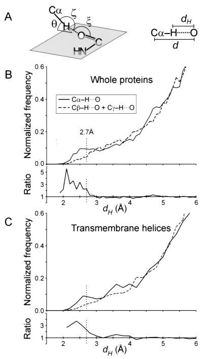
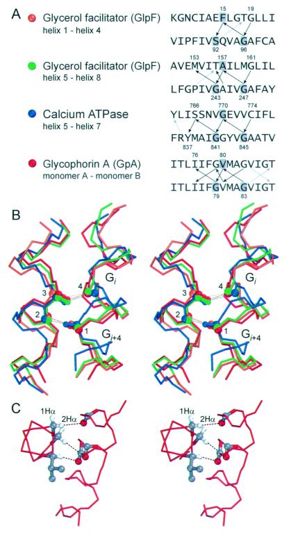
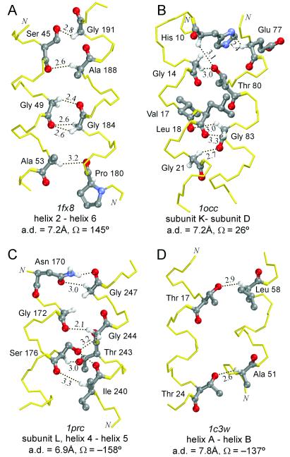
The Calpha---H...O hydrogen bond has been given little attention as a determinant of transmembrane helix association. Stimulated by recent calculations suggesting that such bonds can be much stronger than has been supposed, we have analyzed 11 known membrane protein structures and found that apparent carbon alpha hydrogen bonds cluster frequently at glycine-, serine-, and threonine-rich packing interfaces between transmembrane helices. Parallel right-handed helix-helix interactions appear to favor Calpha---H...O bond formation. In particular, Calpha---H...O interactions are frequent between helices having the structural motif of the glycophorin A dimer and the GxxxG pair. We suggest that Calpha---H...O hydrogen bonds are important determinants of stability and, depending on packing, specificity in membrane protein folding.


Figures

Figure 1
Figure 1
(A) Definition of the geometrical parameters of the Cα—H⋅⋅⋅O hydrogen bond. Nomenclature according to Derewenda et al. (14). The ideal values (14, 18) are as follows: H—O distance, dH ≤ 2.7 Å; Cα—O distance, d ≤ 3.8 Å; Cα—H—O angle, ζ = 180°; H—O—C angle, ξ = 120°; elevation angle, θ = 0° (angle between the Cα—H vector and the amide plane). (B) Distribution of hydrogen to acceptor distances (dH) for all Cα—H donor groups (solid line), compared with that of Cβ—H + Cγ—H groups (dashed line), in the 11 membrane proteins structures. The ratio between the two curves is also shown (Cα/Cβγ). The control set is similarly sized and is composed by the Cβ—H groups of all Ile, Leu, Val, Met, Phe, and Tyr residues and the Cγ—H of Leu and Cγ1—H of Ile. Contacts below 2.7 Å have an overall frequency of occurrence that is 3 times higher for Cα donors than for the Cβγ control set. (C) Analogous distribution as in B, but limited to the subset of residues in helical transmembrane segments.
Figure 2
Figure 2
(A) Distribution of interhelical packing angles (Ω). Interhelical packing angles were calculated as the angle between the local helical axes at the point of minimal axial distance. L-H, left-handed; R-H, right-handed; blue, all helix–helix interactions; red, helix–helix interactions with at least two Cα—H⋅⋅⋅O contacts to backbone or side-chain oxygen atoms; yellow, helix–helix interactions with at least two backbone-to-backbone Cα—H⋅⋅⋅O⩵C contacts. (B) Distribution of interhelical axial distances among the same three categories as for A.
Figure 3
Figure 3
Parallel right-handed helix–helix interactions with extended networks of Cα—H⋅⋅⋅O contacts (GpA-like motifs). (A) Schematic representation of the structure of the glycerol facilitator (GlpF, Protein Database ID 1fx8), the calcium ATPase (1eul), and GpA (model 19 in 1afo). The page is parallel to the plane of the membrane. The color coding corresponds to the interactions shown in B. The interaction between helices 2 and 6 in GlpF (yellow) is antiparallel and is shown in Fig. 4A. Some extramembranous regions of the calcium ATPase were removed for clarity. (B) Apparent networks of Cα—H⋅⋅⋅O hydrogen bonds at the interface of four right-handed helix–helix interactions. Some side-chain atoms have been removed for clarity. In the GpA homodimer (red) each interaction occurs symmetrically on both sides but only one set of interactions is shown for clarity. Apparent hydrogen bonds are denoted with dots (⋅⋅⋅) and the distance (dH) in Å is indicated. The interhelical axial distance (a.d.) and packing angle (Ω) of the helix–helix interactions are also indicated.
Figure 4
Figure 4
(A) Connectivity of the networks of apparent Cα—H⋅⋅⋅O hydrogen bonds in the four parallel right-handed GpA-like motifs. The arrows show the interactions in the donor-to-acceptor direction. Black arrows: backbone-to-backbone Cα—H⋅⋅⋅O bonds; gray arrows: backbone-to-side chain bonds. Backbone-to-backbone Cα—H⋅⋅⋅O⩵C contacts occur between an amino acid residue on one helix and two residues spaced at i, i + 4 on the opposite helix in all structures. The three residues involved in the alignment in B are shaded in the sequences. (B) Superimposition of the four structures aligned using the carbonyl oxygen (4) at i and the Cα (1) at i + 4 of the GxxxG (SxxxG) motifs on the helix on the right; and the Cα (3) and carbonyl oxygen (2) of the residue that interacts with the two Gly residues with apparent Cα—H⋅⋅⋅O bonds. The overall rms deviation of the superimposition calculated on the backbone atoms of 13 residues on each helix is 1.6 Å. (C) The 2Hα of glycine residues is oriented roughly in the same direction of the Hα of the residue at i + 1 and i − 3. When a GxxxG motif is present a potential interaction interface arises. The example shows the interaction of Gly-79, Val-80, and Gly-83 of GpA that donate to carbonyl oxygen atoms spaced at i, i + 3 and i + 4 on the opposite monomer.
Figure 5
Figure 5
Additional helix–helix interactions with multiple Cα—H⋅⋅⋅O hydrogen bonds at various interhelical packing angles. (A) Antiparallel right-handed interaction from the GlpF. (B) Parallel left-handed interaction from bovine heart cytochrome c oxidase. (C) Antiparallel left-handed interaction from the photosynthetic reaction center. (D) Antiparallel left-handed interaction with apparent backbone-to-side-chain Cα—H⋅⋅⋅O bonds from bacteriorhodopsin.

References

    1. Hendsch Z S, Tidor B. Protein Sci. 1994;3:211–226. - PMC - PubMed
    1. Hendsch Z S, Tidor B. Protein Sci. 1999;8:1381–1392. - PMC - PubMed
    1. Sheinerman F B, Norel R, Honig B. Curr Opin Struct Biol. 2000;10:153–159. - PubMed
    1. Popot J L, Engelman D M. Biochemistry. 1990;29:4031–4037. - PubMed
    1. Deisenhofer J, Epp O, Sinning I, Michel H. J Mol Biol. 1995;246:429–457. - PubMed

Publication types

LinkOut - more resources