. 2001 Jul 31;98(16):9086-91.
doi: 10.1073/pnas.161290898.

ATP transduces signals from ASGM1, a glycolipid that functions as a bacterial receptor

Affiliations

ATP transduces signals from ASGM1, a glycolipid that functions as a bacterial receptor

N McNamara et al. Proc Natl Acad Sci U S A. .

Abstract

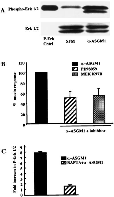
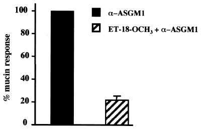
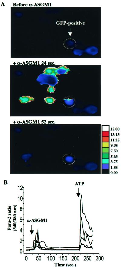
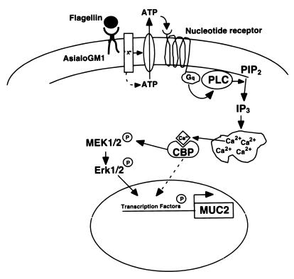
The flagella of the Gram-negative bacterium Pseudomonas aeruginosa serve not only for motility but also to bind bacteria to the host cell glycolipid asialoGM1 (ASGM1) through the protein flagellin. This interaction triggers defensive responses in host cells. How this response occurs is unclear because ASGM1 lacks transmembrane and cytoplasmic domains and there is little information about the downstream effectors that connect ASGM1 ligation to the initiation of host defense responses. Here, we show that ASGM1 ligation promotes ATP release from the host cell, followed by autocrine activation of a nucleotide receptor. This response links ASGM1 to cytoplasmic signaling molecules and results in activation of phospholipase C, Ca(2+) mobilization, phosphorylation of a mitogen-activated protein kinase (Erk 1/2), and activation of mucin transcription. These results indicate that bacterial interaction with host cells can trigger autocrine nucleotide signaling and suggest that agents affecting nucleotide receptors may modulate host responses to bacteria.


Figures

Figure 1
Figure 1
Mucin (MUC 2) transcription is stimulated by ligation of the flagellin receptor, ASGM1. In A and B, HM3 cells stably transfected with a MUC 2 promoter-driven luciferase reporter were studied. Promoter activity is reported as the fold increase observed in stimulated cells vis-a-vis cells exposed to serum-free medium. (A) Stimulation with flagellin purified from strain PAO1. Proteolysis followed by boiling abolished activity. (B) Stimulation with an antibody directed against the flagellin receptor, asialoGM1 (α-ASGM1). Stimulation was for 4 h before cell lysis. Exposure of cells to an approximately equal concentration of an unrelated polyclonal antibody [CCAAT/enhancer binding protein α (α-c/ebp)] had no effect. (C) RNase protection assay was performed by using a MUC 2 gene-specific probe and a control cyclophilin probe to show MUC 2 mRNA up-regulation by purified flagellin (Flag) and ASGM1 antibody (α-ASGM1). SFM, serum-free medium.
Figure 2
Figure 2
ASGM1-mucin induction is calcium dependent. (A) Ratiometric calcium imaging of islands of HM3 cells loaded with Fura-2/AM shows increases in the 340/380-nm excitation fluorescence ratio (510 nm emission) after exposure to a (1:40) dilution of αASGM1. This increase in calcium mobilization peaks 20–24 s after antibody application. (B) HM3 cells stably transfected with a MUC 2 promoter-driven luciferase reporter gene were pretreated with BAPTA/AM (30 μM, 45 min) or thapsigargin (400 nM, 30 min) and then stimulated with a (1:100) dilution of ASGM1 antibody in the continued presence of the drug. After 4 h, cells were lysed and assayed for luciferase activity. Data are presented as the percentage of the mucin response, where 100% is assigned to cells treated with ASGM1 antibody in the presence of vehicle alone. Data are the mean ± SD of triplicate determinations from three to six separate experiments.
Figure 3
Figure 3
Ligation of flagellin receptor activates the MAP kinase cascade, which is required for mucin induction. (A) HM3 cells grown in six-well plates were lysed with SDS sample buffer 5 min after the addition of agonist antibody, α-ASGM1. Equal amounts of lysate were run on a 12% SDS gel, transferred to nitrocellulose, immunoblotted with a phospho-specific Erk 1/2 antibody, and visualized by chemiluminescence. Blots were stripped and reprobed with Erk 1/2 antibody to show equal loading. P-Erk Cntrl, positive control from vendor. (B) Effect of MEK 1/2 inhibitor PD98059 (37 μM, 30 min) and dominant negative mutant MEK K97R on MUC 2 promoter activity. (c) Inhibition of Erk phosphorylation by the calcium chelator, BAPTA/AM (30 μM, 45 min). Fold increase expressed as the ratio of phosphorylated Erk to baseline Erk 1/2 as determined by densitometry.
Figure 4
Figure 4
ASGM1 mucin induction depends on PLC. Effect of the phosphatidylinositol-specific phospholipase C (PI-PLC) inhibitor ET-18-OCH3 (30 μM, 45 min) on HM3 cells stably transfected with a MUC 2 promoter-driven luciferase reporter and stimulated with agonist antibody.
Figure 5
Figure 5
ASGM1/mucin induction depends on a P2Y nucleotide receptor. (A) Involvement of a nucleotide receptor in the flagellin/mucin response demonstrated using the GPCR inhibitor suramin (250 μM, 45 min, in the presence of Triton X-100, 0.01%), the G-protein antagonist GDPβS (1 mM, 60 min, in the presence of Triton X-100, 0.01%), P2Y inhibitors [Reactive Blue-2 (100 μM, 45 min) and Acid Blue 129 (100 μM, 45 min)], and P2X inhibitors [PPADS (100 μM, 45 min) and NF023 (300 μM, 45 min)]. (B) Measurement of ATP in the extracellular medium of HM3 cells grown to 70% confluence and stimulated with 1:40 dilution of α-ASGM1 either alone or in the presence of 2.5 units/ml apyrase. (C) Luciferase reporter assay. HM3 MUC 2 cells pretreated with ATP permeability inhibitors, gadolinium (200 μM, 45 min), or glybenclamide (100 μM, 45 min) were then stimulated with α-ASGM1 antibody (1:100) for 4 h. Data are the mean ± SD values of triplicate determinations from three to six separate experiments.
Figure 6
Figure 6
Fura-2 measurements of [Ca2+]i in cocultured cells. (A) Pseudocolor images of HEK293 cells cotransfected with GFP and P2Y2 (HEK-P2Y2-GFP, ATP “sensors”) in coculture with NCI H292 cells (ATP “releasers”) at three different time points. Representative HEK-P2Y2-GFP cell identified retrospectively by its GFP label is circled and shows an increase in intracellular Ca2+ 24 s after the addition of α-ASGM1 (1:40), which recovers by 52 s (typical of 28 cells, n = 5 experiments). (B) Fura-2 ratios showing increased [Ca2+]i 20 s after the addition of α-ASGM1 in ATP “sensing,” GFP-labeled HEK293 cells, followed by stimulation with 100 μM ATP as a control. Cells were imaged in the same high-power (×20) field while in coculture with ATP “releasers,” NCI H292 cells.
Figure 7
Figure 7
Cartoon depicting events in flagellin-triggered host cell signaling. Flagellin binds to asialoGM1, a membrane glycolipid, which causes the extracellular release of ATP, which then binds to a nucleotide receptor. Downstream events include G-protein activation, the cleavage of PIP2 by PLC, the formation of IP3, Ca2+ mobilization, the phosphorylation of MEK 1/2 and Erk 1/2 [via an unknown calcium binding protein (CBP)], and mucin (MUC 2) transcription. Flagellin-induced signaling bifurcates after Ca2+ mobilization, with ≈50% of the response giving rise to downstream events that are Erk dependent whereas the remaining 50% is Erk independent.

References

    1. Shapiro L. Cell. 1995;80:525–527. - PubMed
    1. DiMango E, Zar H J, Bryan R, Prince A. J Clin Invest. 1995;96(5):2204–2210. - PMC - PubMed
    1. Gewirtz A T, Simon P O, Jr, Schmitt C K, Taylor L J, Hagedorn C H, O'Brien A D, Neish A S, Madara J L. J Clin Invest. 2001;107:99–109. - PMC - PubMed
    1. Feldman M, Bryan R, Rajan S, Scheffler L, Brunnert S, Tang H, Prince A. Infect Immun. 1998;66:43–51. - PMC - PubMed
    1. Li J D, Feng W, Gallup M, Kim J H, Gum J, Kim Y, Basbaum C. Proc Natl Acad Sci USA. 1998;95:5718–5723. - PMC - PubMed

Publication types

MeSH terms

LinkOut - more resources