. 2001 Jul 31;98(16):9271-6.
doi: 10.1073/pnas.161290298.

An HIV-1 transgenic rat that develops HIV-related pathology and immunologic dysfunction

Affiliations

An HIV-1 transgenic rat that develops HIV-related pathology and immunologic dysfunction

W Reid et al. Proc Natl Acad Sci U S A. .

Abstract

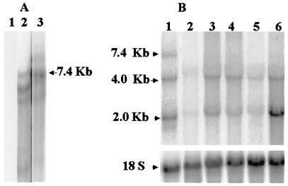
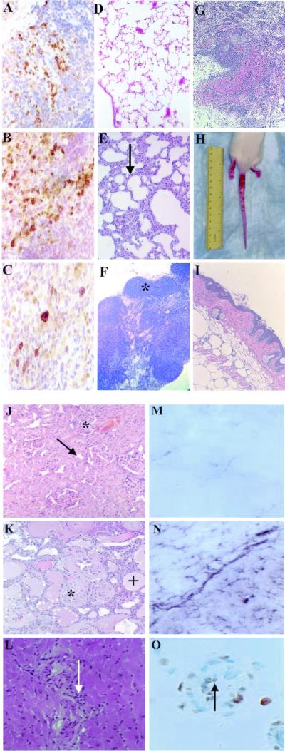
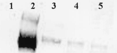
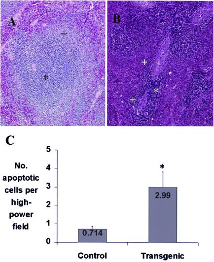
We report, to our knowledge, the first HIV type 1 (HIV-1) transgenic (Tg) rat. Expression of the transgene, consisting of an HIV-1 provirus with a functional deletion of gag and pol, is regulated by the viral long terminal repeat. Spliced and unspliced viral transcripts were expressed in lymph nodes, thymus, liver, kidney, and spleen, suggesting that Tat and Rev are functional. Viral proteins were identified in spleen tissue sections by immunohistochemistry and gp120 was present in splenic macrophages, T and B cells, and in serum. Clinical signs included wasting, mild to severe skin lesions, opaque cataracts, neurological signs, and respiratory difficulty. Histopathology included a selective loss of splenocytes within the periarterial lymphoid sheath, increased apoptosis of endothelial cells and splenocytes, follicular hyperplasia of the spleen, lymphocyte depletion of mesenteric lymph nodes, interstitial pneumonia, psoriatic skin lesions, and neurological, cardiac, and renal pathologies. Immunologically, delayed-type hypersensitivity response to keyhole limpet hemocyanin was diminished. By contrast, Ab titers and proliferative response to recall antigen (keyhole limpet hemocyanin) were normal. The HIV-1 Tg rat thus has many similarities to humans infected with HIV-1 in expression of viral genes, immune-response alterations, and pathologies resulting from infection. The HIV-1 Tg rat may provide a valuable model for some of the pathogenic manifestations of chronic HIV-1 diseases and could be useful in testing therapeutic regimens targeted to stages of viral replication subsequent to proviral integration.


Figures

Figure 1
Figure 1
Detection and expression of transgene. (A) Detection of transgene. A Southern blot was performed on EcoRI-digested DNA from tail snips of a normal rat (15 μg; lane 1) or Tg rats with mild (20 μg; lane 2) or very opaque (5 μg; lane 3) cataracts. The blot was hybridized to an α-32P-labeled 7.4-kb EaeΙ-NaeΙ fragment from pEVd1443. (B) Expression of transgene RNA. A Northern blot was performed on total RNA isolate from various tissues and analyzed by Northern blot as described (32). HIV-specific transcripts (7-, 4-, and 2-kb) were detected with an α-32P-labeled 1.3-kbp (BglII/BglII) fragment from pHXB2, containing gp41 and nef coding regions. HIV transcripts were quantified with a Storm 840 PhosphorImager (Molecular Dynamics) and normalized for hybridization to 18S rRNA by using an appropriate probe. RNA was from the axillary (lane 1) and mesenteric (lane 2) lymph nodes, thymus (lane 3), liver (lane 4), kidney (lane 5), and spleen (lane 6) of transgenic rat with highly opaque cataracts. The positions of the 7.0-kb full-length mRNA, 4-kb singly spliced env mRNA, and 2-kb multiply spliced mRNA are indicated.
Figure 2
Figure 2
Pathology and transgene expression. A section of Tg rat spleen was stained for HIV gp120 (A), Nef (B), and Tat (C). Cytoplasmic staining (dark brown) is evident for all three proteins (original magnification = ×60). H&E-stained sections of control (D) and Tg (E) lung are shown (×20). Arrow in E indicates area of interstitial thickening. H&E-stained sections of control (F) and Tg (G) mesenteric lymph node (×10) are shown. Note normal follicle (*, F) and hemorrhage, lymphoid depletion, and vascular proliferation in Tg section (G). Gross tail and foot lesions (H) and H&E-stained Tg skin (×60). Note psoriatic skin lesions with hyperkeratosis and mononuclear cell infiltrate (arrow, I). H&E-stained control kidney shows normal glomerulus (*) and renal tubule (arrow, J); Tg kidney shows focal glomerulosclerosis (*) and tubulointerstitial disease (+, K; ×60). H&E-stained heart section (L) shows myocardial inflammation with mononuclear cell infiltration (arrow) (×60). Astrocytes from normal (M) or Tg (N) brains were stained for glial fibrillary acidic protein (GFAP; ×40). Staining for GFAP was as reported (22), using anti-GFAP Ab (U7038; Dako). Staining of normal brain was limited, but Tg brain was heavily stained, indicating reactive gliosis, a marker for central nervous system damage. (O) Blood vessels in Tg brain were stained with ApopTag (×60); arrow indicates vascular endothelial apoptosis.
Figure 3
Figure 3
Transgene expression in splenocytes. Seven micrograms of protein from extracts of spleen-derived macrophages (lane 3), B cells (lane 4), or T cells (lane 5) was fractionated on a 4–12% NuPage gel (NOVEX, San Diego) and analyzed by Western blot, using a 1:100 mouse anti-HIV-1 gp120 mAb (NEA 9301, NEN). Proteins were visualized by using the enhanced chemiluminescence (ECL) plus Western blotting system (Amersham Pharmacia). Lane 1 contained extract from a normal control spleen, and lane 2 contained 30 ng of recombinant gp120 as a positive control.
Figure 4
Figure 4
Pathology and apoptosis in Tg rat spleen. (A) H&E-stained control spleen. Note T cell region (PALS) (*) and B cell region (marginal zone) (+) (original magnification ×60). (B) H&E-stained Tg rat spleen (×60). Note loss of T cells within the PALS (*). The two + show the marginal zone. (C) Five-microgram tissue sections were assayed in situ for apoptosis by using an ApopTag kit. Spleen sections from 5-month-old male Tg and Fisher 344/NHsd control rats were taken from 3 animals per group, and apoptotic cells from each section were enumerated 3 times at ×400 by using a Nikon Labophot-2 light microscope. The entire tissue section was counted by using a stage micrometer to pick successive nonoverlapping fields. The * shows the statistically significant difference between HIV-1 transgenic rats and age-matched controls.

References

    1. Popovic M, Gartner S. Curr Opin Immunol. 1989;1:516–520. - PubMed
    1. Price R W. Lancet. 1996;348:445–452. - PubMed
    1. Burke A P, Benson W, Ribas J L, Anderson D, Chu W S, Smialek J, Virmani R. Am J Pathol. 1993;142:1701–1713. - PMC - PubMed
    1. Gallo R C, Montagnier L. Sci Am. 1988;259:41–48. - PubMed
    1. Fauci A S. Eur J Immunol. 1993;262:1011–1018.

Publication types