. 2001 Jul 31;98(16):9324-9.
doi: 10.1073/pnas.161271798.

Placental defects and embryonic lethality in mice lacking suppressor of cytokine signaling 3

Affiliations

Placental defects and embryonic lethality in mice lacking suppressor of cytokine signaling 3

A W Roberts et al. Proc Natl Acad Sci U S A. .

Abstract

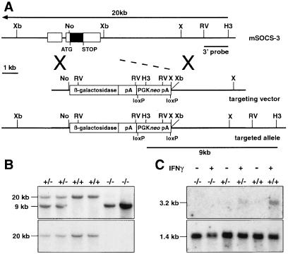
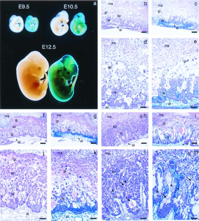
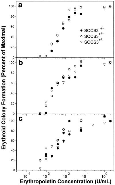
Mice lacking suppressor of cytokine signaling 3 (SOCS3) exhibited embryonic lethality with death occurring between days 11 and 13 of gestation. At this stage, SOCS3(-/-) embryos were slightly smaller than wild type but appeared otherwise normal, and histological analysis failed to detect any anatomical abnormalities responsible for the lethal phenotype. Rather, in all SOCS3(-/-) embryos examined, defects were evident in placental development that would account for their developmental arrest and death. The placental spongiotrophoblast layer was significantly reduced and accompanied by increased numbers of giant trophoblast cells. Delayed branching of the chorioallantois was evident, and, although embryonic blood vessels were present in the labyrinthine layer of SOCS3(-/-) placentas, the network of embryonic vessels and maternal sinuses was poorly developed. Yolk sac erythropoiesis was normal, and, although the SOCS3(-/-) fetal liver was small at day 12.5 of gestation (E12.5), normal frequencies of erythroblasts and hematopoietic progenitor cells, including blast forming unit-erythroid (BFU-E) and, colony forming unit-erythroid (CFU-E) were present at both E11.5 and E12.5. Colony formation for both BFU-E and CFU-E from SOCS3(-/-) mice displayed wild-type quantitative responsiveness to erythropoietin (EPO), in the presence or absence of IL-3 or stem cell factor (SCF). These data suggest that SOCS3 is required for placental development but dispensable for normal hematopoiesis in the mouse embryo.


Figures

Figure 1
Figure 1
Disruption of the SOCS3 locus by homologous recombination. (A) The structure of the murine SOCS3 locus is illustrated with exons as raised boxes and the coding region shaded (Xb, XbaI; No, NotI; X, XhoI; RV, EcoRV; H3, HindIII). In the targeted allele, the coding region was deleted and a cassette fusing the lacZ gene to the SOCS3 ATG and including the selectable PGKneo gene was included. (B) Southern blot of genomic DNA from E11.5 embryos collected from a cross between SOCS3+/− mice, including wild-type (+/+), SOCS3+/−, and SOCS3−/− samples. The DNA was digested with HindIII and hybridized with the 3′ probe, which distinguishes between the targeted (9-kb) and endogenous (20-kb) SOCS3 loci. (C) Northern blot of RNA extracted from wild-type (+/+), SOCS3+/−, and SOCS3−/− primary embryo fibroblasts after stimulation with IFN-γ (+) or saline (−). The blot was probed with a SOCS3 coding region probe followed by a GAPDH probe to confirm RNA integrity.
Figure 2
Figure 2
Placental abnormalities in SOCS3−/− embryos. (a) Viable E9.5, E10.5, and E12.5 SOCS3 null embryos and their wild-type littermates. Embryos were stained for lacZ expression. The SOCS3 null embryos are to the right of their wild-type littermates. (bm) Histological sections of the placentas from these embryos. Placentas were stained for lacZ and then sectioned and further stained with hematoxylin and eosin (H&E). The lacZ-staining pattern was detectable in the H&E-stained sections and was confirmed by staining near-adjacent sections with nuclear fast red (not shown). Temporal profile of placental development in wild-type (b, d, f, h, j, and l) and SOCS3 null (c, e, g, k, i, and m) placentas at E9.5 (be) and E10.5 (f, g, j, and k) and E12.5 (h, i, l, and m) is shown. There is a paucity of spongiotrophoblast in the SOCS3 null placentas. Higher power (d, e, and jm) reveals lacZ staining of the labyrinthine trophoblast and the allantois in the SOCS3 null placentas. Some lacZ staining is also detected in the walls of embryonic vessels penetrating the labyrinth. LacZ staining is not seen in wild-type embryos. At E12.5, embryonic (arrowheads) and maternal (arrows) vessels within the labyrinth are dilated. Al, allantois; gi, trophoblast giant cells; la, labyrinth; lt, labyrinthine trophoblasts; ma, maternal decidua basalis; sp, spongiotrophoblast. Scale bars for b, c, and fI = 100 μm; for d, e, and jm = 10 μm.
Figure 3
Figure 3
Erythropoietin responsiveness of SOCS3−/− erythroid progenitor cells. (a and b) Data from two independent experiments are shown. E11.5 fetal liver cells (5 × 10³) were cultured for 3 days in various concentrations of erythropoietin. Individual results represent means of duplicate cultures, normalized for CFU-E number in supramaximal concentrations of erythropoietin (2 units/ml). The numbers of CFU-E in cultures containing maximal concentrations of erythropoietin ranged between 264 and 684. (c) E11.5 fetal liver cells (104) were cultured for 7 days in triplicate in stem cell factor, IL-3, and various final concentrations of erythropoietin, and the number of BFU-E (including mix-CFC) enumerated as above. The numbers of BFU-E in cultures containing maximal concentrations of erythropoietin ranged between 26 and 50.

References

    1. Hilton D J, Richardson R T, Alexander W S, Viney E M, Willson T A, Sprigg N S, Starr R, Nicholson S E, Metcalf D, Nicola N A. Proc Natl Acad Sci USA. 1998;95:114–119. - PMC - PubMed
    1. Krebs D L, Hilton D J. J Cell Sci. 2000;113:2813–2819. - PubMed
    1. Ihle J N. Nature (London) 1995;377:591–594. - PubMed
    1. Hansen J A, Lindberg K, Hilton D J, Nielsen J H, Billestrup N. Mol Endocrinol. 1999;13:1832–1843. - PubMed
    1. Cohney S J, Sanden D, Cacalano N A, Yoshimura A, Mui A, Migone T S, Johnston J A. Mol Cell Biol. 1999;19:4980–4988. - PMC - PubMed

Publication types