Review
. 2001 Aug 1;20(15):3893-901.
doi: 10.1093/emboj/20.15.3893.

Cell surface monoamine oxidases: enzymes in search of a function

Affiliations
Review

Cell surface monoamine oxidases: enzymes in search of a function

S Jalkanen et al. EMBO J. .

Abstract

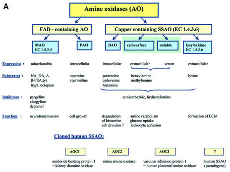
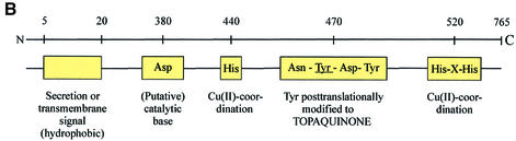
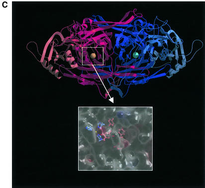
Ectoenzymes with a catalytically active domain outside the cell surface have the potential to regulate multiple biological processes. A distinct class of copper-containing semicarbazide-sensitive monoamine oxidases, expressed on the cell surface and in soluble forms, oxidatively deaminate primary amines. Via transient covalent enzyme-substrate intermediates, this reaction results in production of aldehydes, hydrogen peroxide and ammonium, which are all biologically active substances. The physiological functions of these enzymes have remained unknown, although they have been suggested to be involved in the metabolism of biogenic amines. Recently, new roles have been proposed for these enzymes in regulation of glucose uptake and, even more surprisingly, in leukocyte-endothelial cell interactions. The emerging functions of ectoenzymes in signalling and cell-cell adhesion suggest a novel mode of molecular control of these complex processes.


Figures

None
Fig. 1. (A) The classification of AOs. NA, noradrenaline; DA, dopamine; A, adrenaline; β-PEA, β-phenylethylamine; trypt, tryptamine, ECM, extracellular matrix; AOC, amine oxidase, copper-dependent. (B) The conserved motifs of SSAOs. In the N-terminus either a secretion signal or a transmembrane segment is found. The characteristic positions of the catalytic base, copper-coordinating histidines and the four amino acids-long sequence containing the tyrosine, which is modified to TPQ, as an SSAO sequence signature are shown. The line above the SSAO molecule illustrates the approximate amino acid positions of each important motif (the overall length of SSAO varies and hence the numbers are only approximations). (C) An overall fold of the catalytically active domain (D4) of a human SSAO (VAP-1). The monomers are coloured red and blue. The inset shows a closer view of the active site. The important active site residues are shown under the transparent surface. His520, His522 and His684 (blue) bind to the copper ion (yellow). The other highlighted residues are TPQ, Tyr372, Tyr384 and Asp386 (red). Figure 1C is by the courtesy of Dr Tiina Salminen, Åbo Akademi University, Turku, Finland.
None
Fig. 2. The SSAO reaction and leukocyte extravasation. (A) The catalytic reaction of SSAO. In the reductive half-reaction (1–4) the primary amine group interacts with the TPQ of the enzyme. Then a proton is abstracted by the active-site base (aspartate) and, through a carbanionic intermediate, a product Schiff base is formed. Thereafter, hydrolysis occurs, the product aldehyde is released and the reduced cofactor is left attached to enzyme mainly in an aminoquinol-Cu2+ form. In the oxidative half-reaction (5, 6, 1) the reduced enzyme is recycled to the resting state via an iminoquinol intermediate in a copper- and molecular oxygen-dependent reaction. During this half-reaction, hydrogen peroxide and ammonia are released. (B) The leukocyte extravasation cascade. The different steps of the adhesion cascade and the involvement of VAP-1 are shown. (C) The oligosaccahride modifications of VAP-1 (purple extensions) can bind to an unknown lectin-like molecule (yellow) on lymphocytes. Alternatively, when endothelial VAP-1 uses a lymphocyte surface amine as a substrate, the catalytic reaction results in the formation of a transient covalent bond (step 3 in A) between the two cell types. This enzymatic reaction seems to be involved in the binding during the rolling step. The oligosaccharide and Schiff-base mediated bindings can involve separate molecules on the lymphocyte surface or, if the lectin-type lymphocyte surface molecule also presents the amine to VAP-1, the same molecule may be used in both steps.

References

    1. Barbry P. et al. (1990) Human kidney amiloride-binding protein: cDNA structure and functional expression. Proc. Natl Acad. Sci. USA, 87, 7347–7351. - PMC - PubMed
    1. Bogdan C., Röllinghoff,M. and Diefenbach,A. (2000) Reactive oxygen and reactive nitrogen intermediates in innate and specific immunity. Curr. Opin. Immunol., 12, 64–76. - PubMed
    1. Bono P., Salmi,M., Smith,D.J. and Jalkanen,S. (1998a) Cloning and characterization of mouse vascular adhesion protein-1 reveals a novel molecule with enzymatic activity. J. Immunol., 160, 5563–5571. - PubMed
    1. Bono P., Salmi,M., Smith,D.J., Leppänen,I., Horelli-Kuitunen,N., Palotie,A. and Jalkanen,S. (1998b) Isolation, structural characterization and chromosomal mapping of the mouse vascular adhesion protein-1 gene and promoter. J. Immunol., 161, 2953–2960. - PubMed
    1. Boomsma F., van Dijk,J., Bhaggoe,U.M., Bouhuizen,A.M. and van den Meiracker,A.H. (2000) Variation in semicarbazide-sensitive amine oxidase activity in plasma and tissues of mammals. Comp. Biochem. Physiol. C Toxicol. Pharmacol., 126, 69–78. - PubMed

Publication types

MeSH terms