. 2001 Aug 1;20(15):3967-74.
doi: 10.1093/emboj/20.15.3967.

Altered behavioral rhythms and clock gene expression in mice with a targeted mutation in the Period1 gene

Affiliations

Altered behavioral rhythms and clock gene expression in mice with a targeted mutation in the Period1 gene

N Cermakian et al. EMBO J. .

Abstract

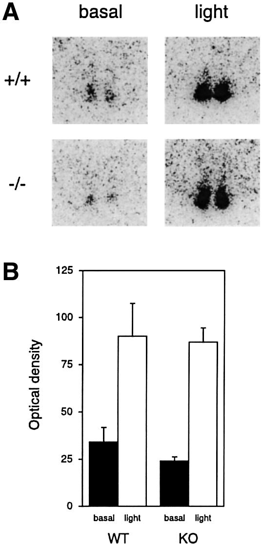
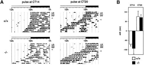
A group of specialized genes has been defined to govern the molecular mechanisms controlling the circadian clock in mammals. Their expression and the interactions among their products dictate circadian rhythmicity. Three genes homologous to Drosophila period exist in the mouse and are thought to be major players in the biological clock. Here we present the generation of mice in which the founding member of the family, Per1, has been inactivated by homologous recombination. These mice present rhythmicity in locomotor activity, but with a period almost 1 h shorter than wild-type littermates. Moreover, the expression of clock genes in peripheral tissues appears to be delayed in Per1 mutant animals. Importantly, light-induced phase shifting appears conserved. The oscillatory expression of clock genes and the induction of immediate-early genes in response to light in the master clock structure, the suprachiasmatic nucleus, are unaffected. Altogether, these data demonstrate that Per1 plays a distinct role within the Per family, as it may be involved predominantly in peripheral clocks and/or in the output pathways of the circadian clock.


Figures

None
Fig. 1. Generation of Per1 knock-out mice. (APer1 gene structure. Numbered boxes are the exons. The position of BamHI (B), HindIII (H) and NcoI (N) restriction sites is shown, as well as the initiator and stop codons. Regions of boxes corresponding to the PAS A, PAS B and PAC regions are in black, gray and light gray, respectively. The targeting construct was generated by replacing an NcoI fragment by a PGK–Neo expression cassette and includes 2.5 and 4.5 kb of gene sequences upstream and downstream of the cassette, respectively. The regions complementary to the PCR or RT–PCR primers and to the Southern hybridization probe are indicated. Below is presented the size of the fragments hybridized with the ‘B/H probe’ in Southern hybridization on a HindIII digestion of the genomic DNA. (B) Isolation of an ES cell clone in which homologous recombination took place (third lane). (C) Genotyping of the mice by PCR. PCR is performed on DNA prepared from tail biopsies, using a cocktail of primers ‘5′P’, ‘5′N’ and ‘3′’ shown in (A). A 553 bp band is amplified for wild-type animals, a 470 bp band for homozygous mutants and both bands for heterozygous animals. (D) RNase protection assay on total RNA from wild-type and knock-out animals, using a riboprobe complementary to a part of the PAS domain. tRNA alone was used as a control (t). (E) RT–PCR on total RNA from wild-type and knock-out animals, using the primers 5′ and 3′ shown in (A). In wild-type animals, the expected 1.3 kb band is amplified. In knock-out animals, two smaller bands are obtained; sequence analysis reveals that they correspond to splicing products in which the Neo cassette (and exon 4 and 10; a piece of exon 4 remains in the larger product) was removed (see scheme). As a consequence, an in-frame stop codon arises just downstream of the splice site; so the only possible proteins could not extend beyond exon 3. (F) Western blot on embryonic fibroblasts derived from wild-type and knock-out embryos, using an antibody raised against a C-terminal peptide of PER1.
None
Fig. 2. Per1 knock-out mice display a short free-running period of locomotor activity rhythms. Representative actograms for one wild-type (A) and two knock-out (B and C) animals. Animals were housed independently in cages with running wheels, and they were entrained on a light–dark cycle with 12 h of light and 12 h of darkness (LD). After 15 days, they were put in constant darkness (DD). Black bars represent the number of turns in 10 min. The plots were duplicated for clarity. Measurement of the free-running period was based on the onset of activity in DD (see Table I). In some mutant animals, the free-running period became even shorter after an extended time in DD [example in (C)].
None
Fig. 3. Circadian expression of Per1 and Per2 is unaffected in the SCN of Per1 knock-out mice. In situ hybridization with Per1 and Per2 probes on brain cuts from animals entrained on a L12:D12 cycle and dissected at the indicated circadian times (CT) on the second day in DD. Only the region of the SCN is shown.
None
Fig. 4. Delay in the expression of Per1 and Per2 in peripheral tissues of knock-out animals. RNase protection assays on total RNA from heart, kidney or skeletal muscle from wild-type or knock-out mice entrained on a L12:D12 cycle and dissected at the indicated circadian times (CT) on the third day in DD. A β-actin probe was used as a control for the amount of RNA. In all experiments, a tRNA control was used (not shown).
None
Fig. 5. Light-induced c-fos expression in Per1 knock-out mice. (AIn situ hybridization with a c-fos probe on brain cuts from animals entrained on a L12:D12 cycle. At CT14 on the third day in DD, the mice were either subjected to light for 15 min and kept another 15 min in the dark (light) or kept all this time in the dark (basal), before dissection. Only the region of the SCN is shown. (B) Quantification of the results (n = 3 for ‘basal’, n = 4 for ‘light’).
None
Fig. 6. A light pulse causes a phase shift of locomotor activity rhythms in Per1 knock-out mice. (A) Representative actogram of mice entrained on a L12:D12 cycle and put for 2 or 3 days in constant darkness (DD), before a light pulse (gray arrowhead) was given at CT14 (left) or CT20 (right), causing a phase delay or advance, respectively, on the following days. Only part of the full actogram is shown. (B) Quantification of the extent of the phase shifts (n = 6 for each genotype, mice of group 2 in Table I). By convention, delays are negative and advances are positive.

References

    1. Akiyama M. et al. (1999) Inhibition of light- or glutamate-induced mPer1 expression represses the phase shifts into the mouse circadian locomotor and suprachiasmatic firing rhythms. J. Neurosci., 19, 1115–1121. - PMC - PubMed
    1. Albrecht U., Sun,Z.S., Eichele,G. and Lee,C.C. (1997) A differential response of two putative mammalian circadian regulators, mper1 and mper2, to light. Cell, 91, 1055–1064. - PubMed
    1. Balsalobre A., Damiola,F. and Schibler,U. (1998) A serum shock induces circadian gene expression in mammalian tissue culture cells. Cell, 93, 929–937. - PubMed
    1. Brown S.A. and Schibler,U. (1999) The ins and outs of circadian timekeeping. Curr. Opin. Genet. Dev., 9, 588–594. - PubMed
    1. Bunger M.K., Wilsbacher,L.D., Moran,S.M., Clendenin,C., Radcliffe,L.A., Hogenesch,J.B., Simon,M.C., Takahashi,J.S. and Bradfield,C.A. (2000) Mop3 is an essential component of the master circadian pacemaker in mammals. Cell, 103, 1009–1017. - PMC - PubMed

Publication types

MeSH terms

Associated data