

doi: 10.1093/emboj/20.15.4003.
NEDD8 recruits E2-ubiquitin to SCF E3 ligase
Affiliations
- PMID: 11483504
- PMCID: PMC149148
- DOI: 10.1093/emboj/20.15.4003
Item in Clipboard
NEDD8 recruits E2-ubiquitin to SCF E3 ligase
EMBO J.
.
Abstract
NEDD8/Rub1 is a ubiquitin (Ub)-like post-translational modifier that is covalently linked to cullin (Cul)-family proteins in a manner analogous to ubiquitylation. NEDD8 is known to enhance the ubiquitylating activity of the SCF complex (composed of Skp1, Cul-1, ROC1 and F-box protein), but the mechanistic role is largely unknown. Using an in vitro reconstituted system, we report here that NEDD8 modification of Cul-1 enhances recruitment of Ub-conjugating enzyme Ubc4 (E2) to the SCF complex (E3). This recruitment requires thioester linkage of Ub to Ubc4. Our findings indicate that the NEDD8-modifying system accelerates the formation of the E2-E3 complex, which stimulates protein polyubiquitylation.
Figures

Fig. 1. NEDD8-ylation of Cul-1 stimulates ubiquitylation of IκBα by the ROC1–SCFβTrCP1 complex. (A) Electrophoretic patterns of recombinant proteins consisting of the IκBα-ubiquitylating [E1 (Uba1), E2 (Ubc4) and (E3) ROC1–SCFβTrCP1] and NEDD8-modifying [E1L (APP-BP1/Uba3) and E2L (Ubc12)] systems. A 5 µg aliquot of each affinity-purified protein, except ROC1–SCFβTrCP1 (10 µg), was subjected to SDS–PAGE followed by staining with Coomassie blue R-250. (B) Modification of Cul-1 by a reconstituted NEDD8 conjugation system. The ROC1–SCFβTrCP1 complex or ROC1–SC(K/R)FβTrCP1 was incubated at 25°C for 15–30 min in the presence or absence of the NEDD8 system containing His-NEDD8 or GST–NEDD8 as described in Materials and methods. After incubation, the reaction was terminated by the addition of 20 µl of SDS–PAGE sample buffer. One-quarter volume of the boiled supernatant was subjected to SDS–PAGE followed by western blotting with anti-HA antibodies to detect Cul-1. Arrows indicate Cul-1, His-NEDD8-Cul-1 and GST–NEDD8-Cul-1. (C) Time course of [35S]IκBα ubiquitylation by ROC1–SCFβTrCP1 (left) and ROC1–SC(K/R)FβTrCP1 (right). Ubiquitylation of [35S]IκBα by ROC1–SCFβTrCP1 was performed in the presence or absence of the NEDD8 system. IκBα pre-phosphorylated by IKK was used as a substrate, unless otherwise specified. The high molecular mass ubiquitylated [35S]IκBα is shown as (Ub)n-[35S]-pIκBα. (D) Effect of Ubc12(C/S) on the in vitro ubiquitylation of phosphorylated GST–IκBα. Experiments were similar to those of (B), except that GST–IκBα and Ubc12(C/S) were used for the assay. The high molecular mass ubiquitylated GST–IκBα detected by western blotting is shown as (Ub)n-GST–pIκBα.
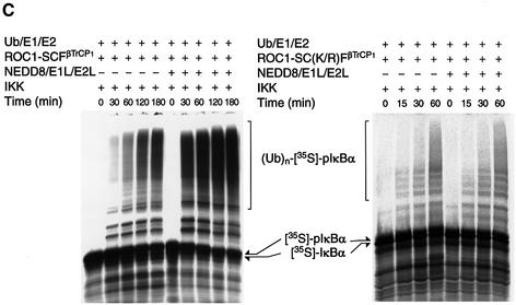
Fig. 1. NEDD8-ylation of Cul-1 stimulates ubiquitylation of IκBα by the ROC1–SCFβTrCP1 complex. (A) Electrophoretic patterns of recombinant proteins consisting of the IκBα-ubiquitylating [E1 (Uba1), E2 (Ubc4) and (E3) ROC1–SCFβTrCP1] and NEDD8-modifying [E1L (APP-BP1/Uba3) and E2L (Ubc12)] systems. A 5 µg aliquot of each affinity-purified protein, except ROC1–SCFβTrCP1 (10 µg), was subjected to SDS–PAGE followed by staining with Coomassie blue R-250. (B) Modification of Cul-1 by a reconstituted NEDD8 conjugation system. The ROC1–SCFβTrCP1 complex or ROC1–SC(K/R)FβTrCP1 was incubated at 25°C for 15–30 min in the presence or absence of the NEDD8 system containing His-NEDD8 or GST–NEDD8 as described in Materials and methods. After incubation, the reaction was terminated by the addition of 20 µl of SDS–PAGE sample buffer. One-quarter volume of the boiled supernatant was subjected to SDS–PAGE followed by western blotting with anti-HA antibodies to detect Cul-1. Arrows indicate Cul-1, His-NEDD8-Cul-1 and GST–NEDD8-Cul-1. (C) Time course of [35S]IκBα ubiquitylation by ROC1–SCFβTrCP1 (left) and ROC1–SC(K/R)FβTrCP1 (right). Ubiquitylation of [35S]IκBα by ROC1–SCFβTrCP1 was performed in the presence or absence of the NEDD8 system. IκBα pre-phosphorylated by IKK was used as a substrate, unless otherwise specified. The high molecular mass ubiquitylated [35S]IκBα is shown as (Ub)n-[35S]-pIκBα. (D) Effect of Ubc12(C/S) on the in vitro ubiquitylation of phosphorylated GST–IκBα. Experiments were similar to those of (B), except that GST–IκBα and Ubc12(C/S) were used for the assay. The high molecular mass ubiquitylated GST–IκBα detected by western blotting is shown as (Ub)n-GST–pIκBα.
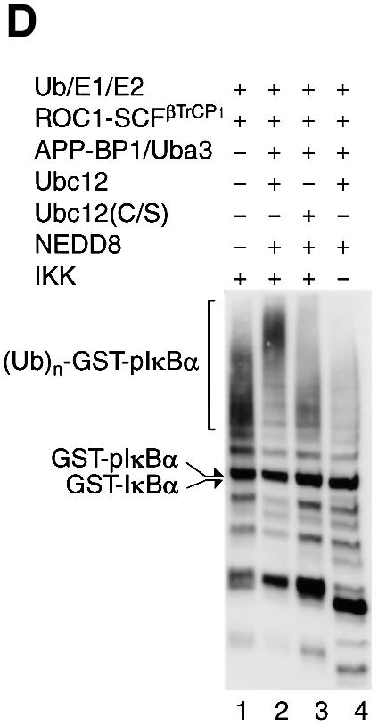
Fig. 1. NEDD8-ylation of Cul-1 stimulates ubiquitylation of IκBα by the ROC1–SCFβTrCP1 complex. (A) Electrophoretic patterns of recombinant proteins consisting of the IκBα-ubiquitylating [E1 (Uba1), E2 (Ubc4) and (E3) ROC1–SCFβTrCP1] and NEDD8-modifying [E1L (APP-BP1/Uba3) and E2L (Ubc12)] systems. A 5 µg aliquot of each affinity-purified protein, except ROC1–SCFβTrCP1 (10 µg), was subjected to SDS–PAGE followed by staining with Coomassie blue R-250. (B) Modification of Cul-1 by a reconstituted NEDD8 conjugation system. The ROC1–SCFβTrCP1 complex or ROC1–SC(K/R)FβTrCP1 was incubated at 25°C for 15–30 min in the presence or absence of the NEDD8 system containing His-NEDD8 or GST–NEDD8 as described in Materials and methods. After incubation, the reaction was terminated by the addition of 20 µl of SDS–PAGE sample buffer. One-quarter volume of the boiled supernatant was subjected to SDS–PAGE followed by western blotting with anti-HA antibodies to detect Cul-1. Arrows indicate Cul-1, His-NEDD8-Cul-1 and GST–NEDD8-Cul-1. (C) Time course of [35S]IκBα ubiquitylation by ROC1–SCFβTrCP1 (left) and ROC1–SC(K/R)FβTrCP1 (right). Ubiquitylation of [35S]IκBα by ROC1–SCFβTrCP1 was performed in the presence or absence of the NEDD8 system. IκBα pre-phosphorylated by IKK was used as a substrate, unless otherwise specified. The high molecular mass ubiquitylated [35S]IκBα is shown as (Ub)n-[35S]-pIκBα. (D) Effect of Ubc12(C/S) on the in vitro ubiquitylation of phosphorylated GST–IκBα. Experiments were similar to those of (B), except that GST–IκBα and Ubc12(C/S) were used for the assay. The high molecular mass ubiquitylated GST–IκBα detected by western blotting is shown as (Ub)n-GST–pIκBα.
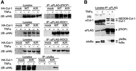
Fig. 2. Modification of Cul-1 by NEDD8 in vivo. (A) Association of Cul-1(K/R) with the SCF complex and IκBα in living cells. pcDNA3-HA-Cul-1 and HA-Cul-1(K/R) were co-expressed with FLAG-βTrCP1, FLAG-ROC1 or FLAG-ROC2 in 293 cells and either left untreated or treated with TNF-α. After immunoprecipitation (IP) with anti-FLAG (upper three rows) and anti-IκBα (lower row) antibodies, immunoblotting (IB) was carried out with anti-HA antibody for cell lysates and the immunoprecipitates. (B) Co-immunoprecipitation of NEDD8-modified Cul-1 with βTrCP1 in vivo. pcDNA3-βTrCP1-FLAG was transfected into 293 cells and treated or not with TNF-α. The extracts or anti-FLAG antibody immunoprecipitates were analysed by western blotting using anti-FLAG, anti-Cul-1 and anti-IκBα antibodies. (C) NEDD8 modification of Cul-1 associated with phosphoryl ated IκBα. HEK-293 cells were either left untreated or treated with TNF-α. The anti-IκBα antibody immunoprecipitates were analysed by western blotting using anti-Cul-1 and anti-NEDD8 antibodies. Experiments were conducted in duplicate. (D) βTrCP1 augments modification of Cul-1 by NEDD8 in reticulocyte lysates. Following the synthesis of [35S]Cul-1 in the presence or absence of [35S]βTrCP1 or GST–NEDD8 as indicated for 60 min at 30°C in a reticulocyte lysate transcription/translation system, samples of the resultant translational products were subjected directly to SDS–PAGE in the presence of dithiothreitol and then autoradiographed. Arrows indicate [35S]βTrCP1, [35S]-Cul1, endogenous NEDD8-modified [35S]Cul-1 and GST–NEDD8-modified [35S]Cul-1. (E) Co-immunoprecipitation of NEDD8-modified Cul-1 with βTrCP1 or βTrCP1 lacking the WD40-repeat region. Thirty-six hours after pcDNA3-FLAG-βTrCP1 or the cDNAs of FLAG-tagged βTrCP1 lacking the WD40-domains (designated FLAG-βTrCP1ΔW1–7) was transfected into 293 cells, crude extracts were prepared as described in Materials and methods. After immunopre cipitation by anti-FLAG antibody, the resulting immunoprecipitates were analysed by western blotting using anti-FLAG, anti-Skp1, anti-Cul-1 and anti-NEDD8 antibodies.

Fig. 2. Modification of Cul-1 by NEDD8 in vivo. (A) Association of Cul-1(K/R) with the SCF complex and IκBα in living cells. pcDNA3-HA-Cul-1 and HA-Cul-1(K/R) were co-expressed with FLAG-βTrCP1, FLAG-ROC1 or FLAG-ROC2 in 293 cells and either left untreated or treated with TNF-α. After immunoprecipitation (IP) with anti-FLAG (upper three rows) and anti-IκBα (lower row) antibodies, immunoblotting (IB) was carried out with anti-HA antibody for cell lysates and the immunoprecipitates. (B) Co-immunoprecipitation of NEDD8-modified Cul-1 with βTrCP1 in vivo. pcDNA3-βTrCP1-FLAG was transfected into 293 cells and treated or not with TNF-α. The extracts or anti-FLAG antibody immunoprecipitates were analysed by western blotting using anti-FLAG, anti-Cul-1 and anti-IκBα antibodies. (C) NEDD8 modification of Cul-1 associated with phosphoryl ated IκBα. HEK-293 cells were either left untreated or treated with TNF-α. The anti-IκBα antibody immunoprecipitates were analysed by western blotting using anti-Cul-1 and anti-NEDD8 antibodies. Experiments were conducted in duplicate. (D) βTrCP1 augments modification of Cul-1 by NEDD8 in reticulocyte lysates. Following the synthesis of [35S]Cul-1 in the presence or absence of [35S]βTrCP1 or GST–NEDD8 as indicated for 60 min at 30°C in a reticulocyte lysate transcription/translation system, samples of the resultant translational products were subjected directly to SDS–PAGE in the presence of dithiothreitol and then autoradiographed. Arrows indicate [35S]βTrCP1, [35S]-Cul1, endogenous NEDD8-modified [35S]Cul-1 and GST–NEDD8-modified [35S]Cul-1. (E) Co-immunoprecipitation of NEDD8-modified Cul-1 with βTrCP1 or βTrCP1 lacking the WD40-repeat region. Thirty-six hours after pcDNA3-FLAG-βTrCP1 or the cDNAs of FLAG-tagged βTrCP1 lacking the WD40-domains (designated FLAG-βTrCP1ΔW1–7) was transfected into 293 cells, crude extracts were prepared as described in Materials and methods. After immunopre cipitation by anti-FLAG antibody, the resulting immunoprecipitates were analysed by western blotting using anti-FLAG, anti-Skp1, anti-Cul-1 and anti-NEDD8 antibodies.
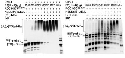
Fig. 3. Dose-dependent effect of E2 (Ubc4) on the ubiquitylation of phosphorylated IκBα. The ubiquitylated assays of [35S]IκBα (left) and GST–IκBα (right) were performed as in Figure 1C and D, except for the use of various amounts of E2 (Ubc4) as indicated.
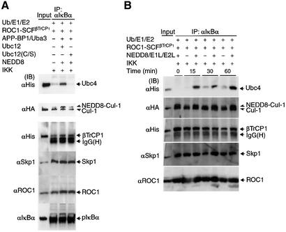
Fig. 4. Effect of the NEDD8 system on recruitment of E2 (Ubc4) onto ROC1–SCFβTrCP1 bound to phosphorylated IκBα. The ROC1–SCFβTrCP1 complex was incubated at 25°C for 30 min in the presence or absence of the NEDD8 system together with Ub, E1 (Uba1) and E2 (Ubc4) for ubiquitylation, IκBα and IKK at the indicated combinations. After incubation, the reaction mixtures were treated with anti-IκBα antibody and the resulting immunoprecipitates were used for western blotting with various antibodies as indicated. ‘Input’ denotes various input controls (1/10 the amounts added in the assay mixtures). The effects of Ubc12(C/S) (A) and time course (B) on the binding of Ubc4 to ROC1–SCFβTrCP1 are shown.
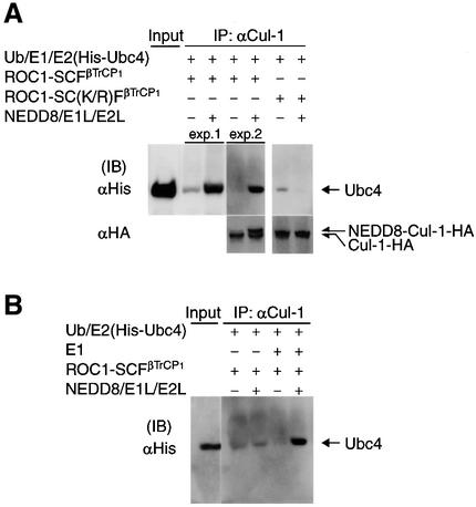
Fig. 5. Effect of the NEDD8 system on recruitment of E2 (Ubc4) to the ROC1–SCFβTrCP1 complex. (A) Effect of Ubc4 binding to ROC1–SCFβTrCP1 and ROC1–SC(K/R)FβTrCP1. Experiments and symbols are similar to those described in Figure 4, except that IκBα and IKK were not added and immunoprecipitation was carried out with anti-Cul-1 antibody. Some experiments were conducted in duplicate. (B) Only Ub-linked Ubc4 was recruited onto ROC1–SCFβTrCP1. The experiment was similar to that described in (A) except that E1 (Uba1) was omitted from the assay mixture. (C) The NEDD8 system does not support the binding of Ubc4(C/A) and Ubc4(C/S) to ROC1–SCFβTrCP1. The experiment was similar to that described in (A), except that Ubc4(C/A) and Ubc4(C/S) were used for wild-type Ubc4 (wt). (D) The NEDD8 system does not support the binding of Ubc4(C/A) and Ubc4(C/S) to pIκBα. The experiment was similar to that described in Figure 5 using Ubc4(C/A) and Ubc4(C/S). (E) Ubc4, but not Ubc4(C/A), co-immunoprecipitates Cul-1 in 293 cells. Thirty-six hours after pcDNA3-Cul-1-HA with unmodified FLAG-Ubc4(wt) or FLAG- Ubc4(C/A) were transfected into 293 cells, crude extracts were prepared as described in Materials and methods. After immuno precipitation by anti-FLAG antibody, the resulting immunoprecipitates were analysed by western blotting using anti-HA antibodies.
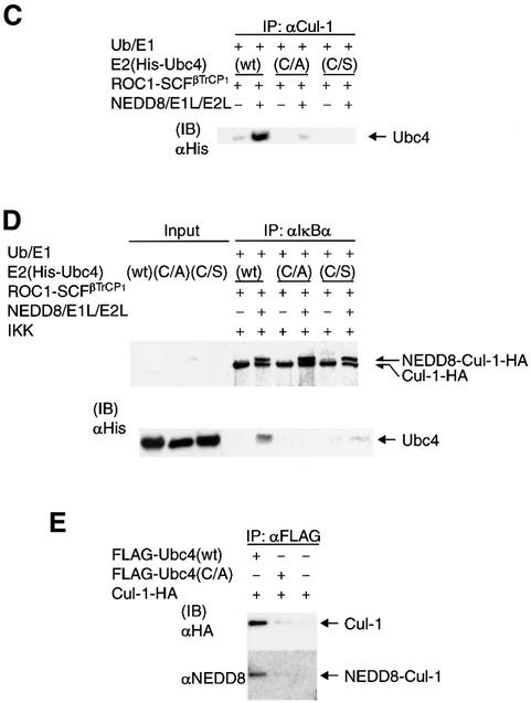
Fig. 5. Effect of the NEDD8 system on recruitment of E2 (Ubc4) to the ROC1–SCFβTrCP1 complex. (A) Effect of Ubc4 binding to ROC1–SCFβTrCP1 and ROC1–SC(K/R)FβTrCP1. Experiments and symbols are similar to those described in Figure 4, except that IκBα and IKK were not added and immunoprecipitation was carried out with anti-Cul-1 antibody. Some experiments were conducted in duplicate. (B) Only Ub-linked Ubc4 was recruited onto ROC1–SCFβTrCP1. The experiment was similar to that described in (A) except that E1 (Uba1) was omitted from the assay mixture. (C) The NEDD8 system does not support the binding of Ubc4(C/A) and Ubc4(C/S) to ROC1–SCFβTrCP1. The experiment was similar to that described in (A), except that Ubc4(C/A) and Ubc4(C/S) were used for wild-type Ubc4 (wt). (D) The NEDD8 system does not support the binding of Ubc4(C/A) and Ubc4(C/S) to pIκBα. The experiment was similar to that described in Figure 5 using Ubc4(C/A) and Ubc4(C/S). (E) Ubc4, but not Ubc4(C/A), co-immunoprecipitates Cul-1 in 293 cells. Thirty-six hours after pcDNA3-Cul-1-HA with unmodified FLAG-Ubc4(wt) or FLAG- Ubc4(C/A) were transfected into 293 cells, crude extracts were prepared as described in Materials and methods. After immuno precipitation by anti-FLAG antibody, the resulting immunoprecipitates were analysed by western blotting using anti-HA antibodies.
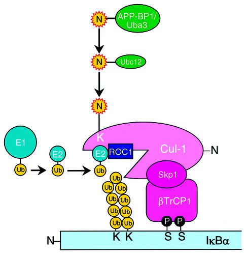
Fig. 6. Schematic model for the role of the NEDD8 system in IκBα ubiquitylation by a ROC1–SCFβTrCP1 Ub ligase complex. For details, see text. Ub, ubiquitin; E1 (Uba1) (Ub-activating); and E2 (Ubc4) (Ub-conjugating) enzymes; N, NEDD8; p, phosphorylation of serine. APP-BP1/Uba3 and Ubc12, as E1- and E2-like enzymes, respectively, for the NEDD8 system. S and K, serine and lysine residues, respectively.
References
-
- Baldwin A.S. (1996) The NF-κB and IκB proteins: new discoveries and insights. Annu. Rev. Immunol., 14, 649–681. - PubMed
-
- Chen Y., McPhie,D.L., Hirschberg,J. and Neve,R.L. (2000) The amyloid precursor protein-binding protein APP-BP1 drives the cell cycle through the S–M checkpoint and causes apoptosis in neurons. J. Biol. Chem., 275, 8929–8935. - PubMed
-
- Deshaies R.J. (1999) SCF and Cullin/RING-H2-based ubiquitin-ligases. Annu. Rev. Cell. Dev. Biol., 15, 435–467. - PubMed
-
- Finco T.S. and Baldwin,A.S. (1995) Mechanistic aspects of NF-κB regulation: the emerging role of phosphorylation and proteolysis. Immunity, 3, 253–272. - PubMed
Publication types
MeSH terms
Substances
LinkOut - more resources
Full Text Sources
Other Literature Sources
Miscellaneous
