. 2001 Aug 1;20(15):4173-82.
doi: 10.1093/emboj/20.15.4173.

Polymerization of the SAM domain of TEL in leukemogenesis and transcriptional repression

Affiliations

Polymerization of the SAM domain of TEL in leukemogenesis and transcriptional repression

C A Kim et al. EMBO J. .

Abstract

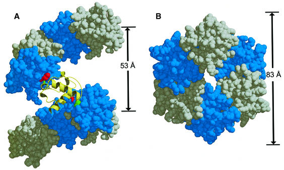
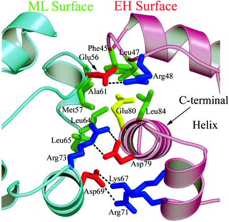
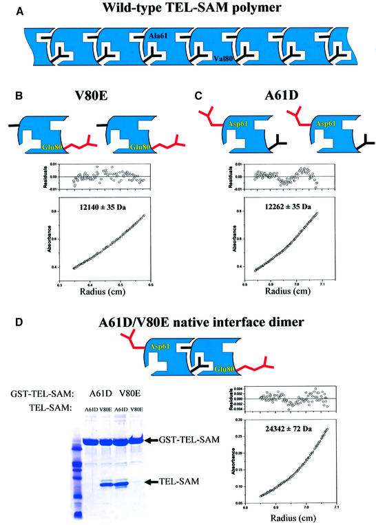
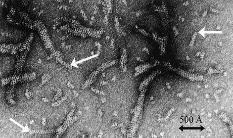
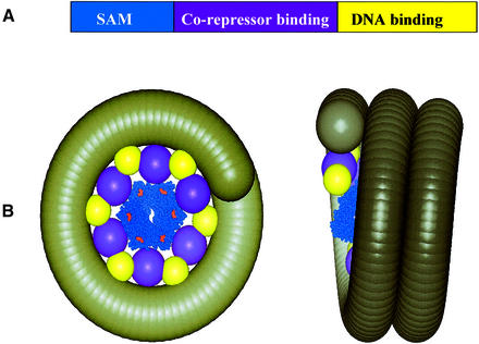
TEL is a transcriptional repressor that is a frequent target of chromosomal translocations in a large number of hematalogical malignancies. These rearrangements fuse a potent oligomerization module, the SAM domain of TEL, to a variety of tyrosine kinases or transcriptional regulatory proteins. The self-associating property of TEL-SAM is essential for cell transformation in many, if not all of these diseases. Here we show that the TEL-SAM domain forms a helical, head-to-tail polymeric structure held together by strong intermolecular contacts, providing the first clear demonstration that SAM domains can polymerize. Our results also suggest a mechanism by which SAM domains could mediate the spreading of transcriptional repression complexes along the chromosome.


Figures

None
Fig. 1. TEL–SAM polymer structure. (A) The V80E polymer structure viewed with the helix axis in the plane of the page, pointing up. The figure shows nine subunits of the polymer. The central subunit is shown in gold ribbon. V80E mutation on the EH surface is shown in red and ML surface apolar residue Ala61 is shown in green. The red and green coloring scheme representing the EH and ML surfaces, respectively, will be consistent for the other figures. (B) The V80E polymer structure viewed down the helix axis of the polymer.
None
Fig. 2. Detailed view of the TEL–SAM V80E polymer interface. Hydrophobic residues that make up the core of the interface are shown in green. The V80E mutation that renders the protein soluble is shown in yellow. In the wild-type sequence a hydrophobic Val side-chain would occupy this position. Salt-bridges in the interface that surround the core residues are also shown. Negatively charged residues are shown in red and positively charged residues are shown in blue.
None
Fig. 3. Test of the polymer model. (A) Schematic illustration of the putative wild-type polymeric structure highlighting two key residues, A61 and V80, on the different binding surfaces, ML and EH, respectively. (B) Equilibrium sedimentation results for the V80E mutant. The observed molecular weight was found to be 12 140 ± 35 Da and the calculated molecular weight for the monomer is 12 079 Da. (C) Equilibrium sedimentation results for the A61D mutant. The observed molecular weight was found to be 12 262 ± 35 Da and the calculated molecular weight for the monomer is 12 093 Da. (D) Testing mixed V80E–A61D dimer formation. The ability of V80E or A61D to bind to themselves or to each other was tested in GST pull-down experiments. As shown in the gel, A61D bound to V80E but not to itself, and V80E bound to A61D but not to itself. Equilibrium sedimentation results for an equimolar mixture of V80E and A61D are also shown. The observed molecular weight was found to be 24 342 ± 72 Da and the calculated molecular weight for the dimer is 24 172 Da.
None
Fig. 4. Electron microscopy of TEL–SAM. Electron microscopy photograph of the TEL–SAM polymer. Alhough most filaments appear tangled, isolated single filaments, indicated by arrows, have width dimensions similar to the width of the helical polymer observed in the crystal structure.
None
Fig. 5. Model of one possible TEL repression complex. (A) The domain structure of TEL. (B) A possible complex of TEL with chromatin. The TEL–SAM polymer structure from this work is shown as a space-filling model in blue. The C-termini are colored red and orient away from the axis of the polymer. This is the only known structure in the model shown. Spheres representing the co-repressor and DNA binding domains of TEL are placed on the outside of the polymer with the same helical pitch as the TEL–SAM polymer. The sphere volumes were determined assuming a partial specific volume of 0.7 ml/g. A coil representing chromatin is wrapped around the polymer, interacting with the DNA binding domains. The thickness of the coil is the same as the width of a nucleosome core particle (54 Å) (Luger et al., 1997). We note that this is a highly speculative model and serves only to illustrate one way that polymerization could lead to spreading of transcriptional repression. Many other models are possible.
None
Fig. 6. Sequence and structural alignment of the Ets family SAM domains. (A) The sequence of wild-type TEL–SAM aligned with SAM domains from members of the Ets family of transcription factors. The residues highlighted in red and green represent the EH and ML binding surfaces, respectively. Amino acids that have >75% of the residue buried in the core of the TEL–SAM structure and conserved apolar residues at that position are shaded in gray. The numbering scheme above the TEL–SAM sequence is of the construct used in this study. (B) Structural alignment of TEL–SAM with Ets1–SAM (Slupsky et al., 1998) showing the EH binding surface. TEL–SAM is shaded in blue while Ets1–SAM is shaded in gray. The apolar residues and the Val80Glu mutation that form the EH binding surface of TEL–SAM are colored red. The analogous Ets1–SAM residues from the sequence alignment in Figure 6A are colored pink. (C) The ML binding surface. The TEL–SAM hydrophobic residues that make up the core of the ML surface are green while the equivalent Ets1–SAM residues are colored yellow.

References

    1. Ahmad K.F., Engel,C.K. and Prive,G.G. (1998) Crystal structure of the BTB domain from PLZF. Proc. Natl Acad. Sci. USA, 95, 12123–12128. - PMC - PubMed
    1. Baker D.A., Mille-Baker,B., Wainwright,S.M., Ish-Horowicz,D. and Dibb,N.J. (2001) Mae mediates MAP kinase phosphorylation of Ets transcription factors in Drosophila. Nature, 411, 330–334. - PubMed
    1. Brunger A.T. et al. (1998) Crystallography & NMR system: a new software suite for macromolecular structure determination. Acta Crystallogr. D, 54, 905–921. - PubMed
    1. Buijs A. et al. (2000) The MN1-TEL fusion protein, encoded by the translocation (12;22)(p13;q11) in myeloid leukemia, is a transcription factor with transforming activity. Mol. Cell. Biol., 20, 9281–9293. - PMC - PubMed
    1. Carroll M., Tomasson,M.H., Barker,G.F., Golub,T.R. and Gilliland,D.G. (1996) The TEL/platelet-derived growth factor β receptor (PDGF β R) fusion in chronic myelomonocytic leukemia is a transforming protein that self-associates and activates PDGF β R kinase-dependent signaling pathways. Proc. Natl Acad. Sci. USA, 93, 14845–14850. - PMC - PubMed

Publication types

MeSH terms