. 2001 Aug 1;20(15):4214-21.
doi: 10.1093/emboj/20.15.4214.

The kink-turn: a new RNA secondary structure motif

Affiliations

The kink-turn: a new RNA secondary structure motif

D J Klein et al. EMBO J. .

Abstract

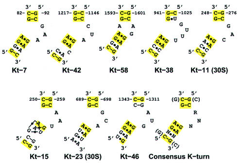
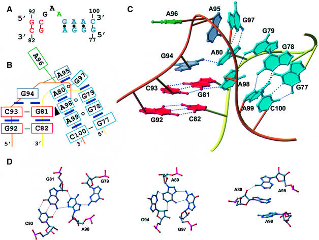
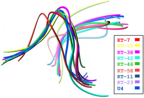
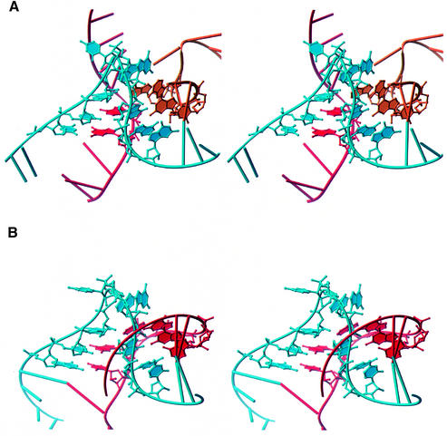
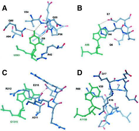
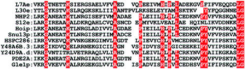
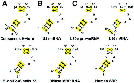
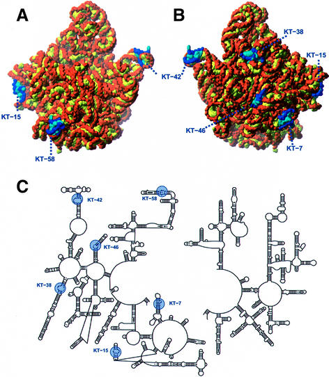
Analysis of the Haloarcula marismortui large ribosomal subunit has revealed a common RNA structure that we call the kink-turn, or K-turn. The six K-turns in H.marismortui 23S rRNA superimpose with an r.m.s.d. of 1.7 A. There are two K-turns in the structure of Thermus thermophilus 16S rRNA, and the structures of U4 snRNA and L30e mRNA fragments form K-turns. The structure has a kink in the phosphodiester backbone that causes a sharp turn in the RNA helix. Its asymmetric internal loop is flanked by C-G base pairs on one side and sheared G-A base pairs on the other, with an A-minor interaction between these two helical stems. A derived consensus secondary structure for the K-turn includes 10 consensus nucleotides out of 15, and predicts its presence in the 5'-UTR of L10 mRNA, helix 78 in Escherichia coli 23S rRNA and human RNase MRP. Five K-turns in 23S rRNA interact with nine proteins. While the observed K-turns interact with proteins of unrelated structures in different ways, they interact with L7Ae and two homologous proteins in the same way.


Figures

None
Fig. 1. Secondary structure diagrams of the eight K-turns found in the H.marismortui 50S and T.thermophilus 30S subunit structures and a derived consensus sequence. The names indicate which helix each example of the motif is found in, according to the helix numbering scheme of Leffers et al. (1987). Solid lines represent Watson–Crick pairings between bases, and black dots represent mismatched base pairings. Yellow shading indicates nucleotides that conform to the derived consensus sequence.
None
Fig. 2. Structure of KT-7. (A) Secondary structure with residues in the C-stem labeled red and the NC-stem blue. The bulged nucleotide is green. (B) Schematic representation of the relative base-stacking and pairing interactions. A black triangle represents an A-minor interaction. (C) Three-dimensional representation of KT-7 with the phosphate backbone of the kinked strand in orange and the unkinked strand in yellow. Hydrogen bonds are indicated by dashed lines. (D) Atomic details of individual base–base and base–backbone hydrogen bonds.
None
Fig. 3. Backbone representations of nine K-turn structures after superposition on KT-7. The average backbone r.m.s.d. of these structures to KT-7 is 1.7 Å.
None
Fig. 4. (A) Stereo view of the three-stranded K-turn located around nucleotide 46 in H.marismortui 23S rRNA. (B) Stereo image of KT-7 shown in the same relative orientation.
None
Fig. 5. Ribbon representations indicating the structural diversity of protein–K-turn complexes found in the structure of the H.marismortui 50S subunit. (A) L24 and L29 interact with opposite sides of KT-7. (B) Helical fragments of L10 bind to the bulged nucleotide in KT-42. (C) L19e and L37Ae make limited interactions with KT-58. (D) The β-extension of L4 interacts through the widened major groove of KT-46’s C-stem, while L32e recognizes its kinked backbone and protruded base. (E) L7Ae and L15e surround KT-15 with the majority of interactions involving hydrophobic packing made possible by the severe bend in the RNA helix.
None
Fig. 6. Recognition of the protruded nucleotide in ribosomal K-turns by specific amino acid contacts. Amino acids from (A) L7Ae and L15e, (B) L29, (C) L32e and (D) L10 interact with their respective K-turns by recognition of the protruded nucleotide (green).
None
Fig. 7. Multiple sequence alignment of proteins displaying homology to H.marismortui L7Ae. Red boxes indicate consensus residues. The structures of H.marismortui L7Ae, S.cerevisiae L30e and the human homolog of Snu13p (15.5 kDa spliceosomal protein) have been solved and show striking three-dimensional similarity.
None
Fig. 8. Secondary structure diagrams for the consensus and non-ribosomal K-turns. (A) Consensus derived from the eight K-turns found in the ribosome. (B) The secondary structure for the U4 snRNA K-turn adapted from Vidovic et al. (2000). (C) Predicted K-turn motifs in various non-ribosomal RNAs. The secondary structure we propose for L30e pre-mRNA is inconsistent with the NMR structure (Mao et al., 1999).
None
Fig. 9. Location of the K-turns in the H.marismortui 50S structure. (A) The 50S particle shown in the crown view. (B) The back side of the subunit, rotated 180° around the vertical axis from the crown view. K-turns are in blue, with sugars and phosphates in dark blue, and bases in light blue. The remaining RNA has sugars and phosphates in orange, and bases in yellow. (C) The positions of K-turns in the schematic secondary structure diagram of 23S rRNA. K-turns are highlighted in blue.

References

    1. Ban N., Nissen,P., Hansen,J., Moore,P.B. and Steitz,T.A. (2000) The complete atomic structure of the large ribosomal subunit at 2.4 Å resolution. Science, 289, 905–920. - PubMed
    1. Beckmann R., Bubeck,D., Grassucci,R., Penczek,P., Verschoor,A., Blobel,G. and Frank,J. (1997) Alignment of conduits for the nascent polypeptide chain in the ribosome–Sec61 complex. Science, 278, 2123–2126. - PubMed
    1. Brünger A.T. et al. (1998) Crystallography & NMR system: a new software suite for macromolecular structure determination. Acta Crystallogr. D, 54, 905–921. - PubMed
    1. Burd C.G. and Dreyfuss,G. (1994) Conserved structures and diversity of functions of RNA-binding proteins. Science, 265, 615–621. - PubMed
    1. Cate J.H., Yusupov,M.M., Yusupova,G.Z., Earnest,T.N. and Noller,H.F. (1999) X-ray crystal structures of 70S ribosome functional complexes. Science, 285, 2095–2104. - PubMed

Publication types

Associated data