. 2001 Aug 1;20(15):4222-32.
doi: 10.1093/emboj/20.15.4222.

Non-canonical mechanism for translational control in bacteria: synthesis of ribosomal protein S1

Affiliations

Non-canonical mechanism for translational control in bacteria: synthesis of ribosomal protein S1

I V Boni et al. EMBO J. .

Abstract

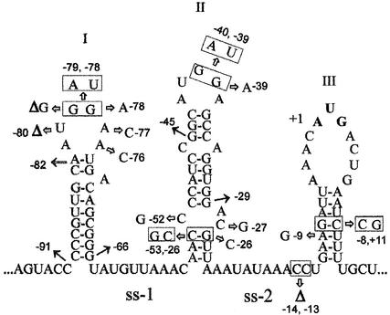
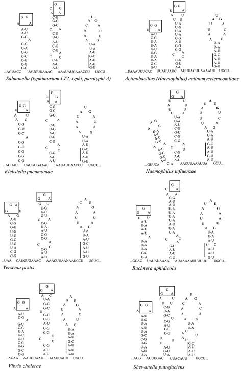
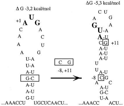
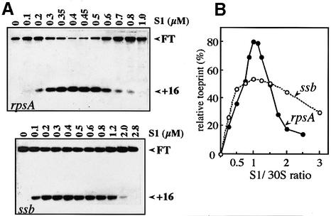
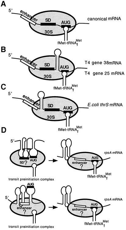
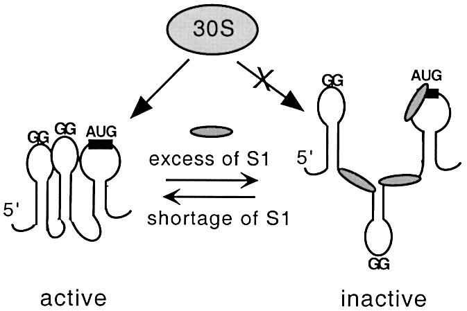
Translation initiation region (TIR) of the rpsA mRNA encoding ribosomal protein S1 is one of the most efficient in Escherichia coli despite the absence of a canonical Shine-Dalgarno-element. Its high efficiency is under strong negative autogenous control, a puzzling phenomenon as S1 has no strict sequence specificity. To define sequence and structural elements responsible for translational efficiency and autoregulation of the rpsA mRNA, a series of rpsA'-'lacZ chromosomal fusions bearing various mutations in the rpsA TIR was created and tested for beta-galactosidase activity in the absence and presence of excess S1. These in vivo results, as well as data obtained by in vitro techniques and phylogenetic comparison, allow us to propose a model for the structural and functional organization of the rpsA TIR specific for proteobacteria related to E.coli. According to the model, the high efficiency of translation initiation is provided by a specific fold of the rpsA leader forming a non-contiguous ribosome entry site, which is destroyed upon binding of free S1 when it acts as an autogenous repressor.


Figures

None
Fig. 1. Folding of the E.coli rpsA TIR predicted from in vitro enzymatic probing and computer modeling data. Single-stranded A/U-rich regions separating three consecutive hairpin-loop structures (I, II, III) are denoted ss-1 and ss-2. The sequence is numbered from the first base of the initiator codon (in bold). The location of the site-directed mutations and 5′ deletions described in the text are indicated by arrows. Double mutations are boxed, deletions of individual nucleotides are marked by ‘Δ’.
None
Fig. 2. Phylogenetic conservation of the rpsA TIR folding in proteobacteria from gamma subdivision. Conserved GGA-motifs in loops I and II are boxed, the degenerate SD-like sequences are indicated by vertical bars.
None
Fig. 3. Structural reorganization of the rpsA TIR hairpin III resulting from the double mutation –8G→C, +11C→G, as predicted by mfold algorithm. Mutated nucleotides are boxed, energy parameters are shown above each structure.
None
Fig. 4. S1 dependence of ternary initiation complex formation on the rpsA and ssb mRNAs (toeprint analysis). (A) Autoradiograms of 8% sequencing gels showing inhibition of the extension reactions (see Materials and methods). Concentration of individual components was: mRNA, 0.04 µM; 30S subunits lacking S1, 0.4 µM; uncharged initiator tRNA, 4 µM; free S1 as indicated over lanes. FT is the signal corresponding to full-length reverse transcript, +16 is the position of the toeprint signal. (B) Quantification of the toeprint results by densitometric scanning. Relative toeprint is a toeprint/(FT + toeprint) ratio, i.e. the percentage of the mRNA involved in initiation complex formation.
None
Fig. 5. Gel mobility shift assay of the S1-rpsA TIR complexes. Concentration of high specific activity RNA in each assay is 2–5 nM. (A) Results of band shifting (5% native gel) showing that all truncated rpsA leaders carry targets for S1 binding. The length of the rpsA leader is indicated below each lane. Formation of complexes with different stoichiometry in the case of 66-nt leader is clearly visible on the rightmost half of the gel. (B) Band shift assay for the 145- (left) and 45-nt (right) rpsA leaders at progressively decreased S1 concentration showing their different affinity for S1. Concentration of S1 (µM) in lanes: 1, 0.5; 2, 0.25; 3, 0.125; 4, 0.062; 5, 0.031.
None
Fig. 6. Footprint analysis of S1 binding to the rpsA TIR. (A) Diethyl pyrocarbonate (DEPC) modification of the A-residues within the rpsA mRNA in the absence (–) or presence (+) of S1 (2.4 µM). U, G, C, A lanes, dideoxy-sequencing of in vitro RNA transcript by primer extension. Structural elements (vertical bars) and certain positions of the rpsA TIR are indicated on the left and on the right of the panel. The A-residues that are modified only in the presence of S1 are indicated by arrows. (B) Partial digestion of the rpsA mRNA with RNase T1 in the absence (lane 1) or presence of increasing concentration of S1: lane 2, 0.5 µM; lane 3, 1.0 µM; lane 4, 2.0 µM. (C) The rpsA TIR structure with indication of positions protected by S1 against DEPC (black triangles), and positions with increased reactivity towards DEPC (gray triangles) or T1 digestion (gray circles) in the presence of S1.
None
Fig. 7. Models for ternary initiation complex formation on mRNAs with canonical (A) or non-contiguous ribosome binding sites (BD). Models (B)–(D) demonstrate that the ribosomal mRNA binding track can tolerate the presence of stable secondary structures which properly arrange essential sequence elements involved in ribosome binding (see text for comments). The enhancer (open boxes) is a single-stranded region found within 5′-leaders of many mRNAs that positively affects their translation efficiency (‘enhancing effect’). This enhancing effect can result from additional complementarity to the 16S RNA (as proposed by Olins and Rangwala, 1989) or from preferential binding by S1 within the 30S subunit (Boni et al., 1991; Zhang and Deutscher, 1992).
None
Fig. 8. Model for autogenous regulation of protein S1 synthesis. Active conformation of the rpsA TIR can be bound either by 30S subunit, thus providing efficient translation of the rpsA mRNA, or by free S1 when it is present in excess over ribosomes. Free S1 disturbs the active conformation of its cognate TIR, thereby acting as a specific translational repressor.

References

    1. Allen T., Shen,P., Samsel,L., Liu,R., Lindahl,L. and Zengel,J.M. (1999) Phylogenetic analysis of L4-mediated autogenous control of the S10 ribosomal protein operon. J. Bacteriol., 181, 6124–6132. - PMC - PubMed
    1. Boni I.V., Issaeva,D.M., Musychenko,M.L. and Tzareva,N.V. (1991) Ribosome-messenger recognition: mRNA target sites for ribosomal protein S1. Nucleic Acids Res., 19, 155–162. - PMC - PubMed
    1. Boni I.V., Artamonova,V.S. and Dreyfus,M. (2000) The last RNA-binding repeat of the Escherichia coli ribosomal protein S1 is specifically involved in autogenous control. J. Bacteriol., 182, 5872–5879. - PMC - PubMed
    1. Bycroft M., Hubbard,T.J., Proctor,M., Freund,S.M. and Murzin,A.G. (1997) The solution structure of the S1 RNA binding domain: a member of an ancient nucleic acid-binding fold. Cell, 88, 235–242. - PubMed
    1. Chen H., Bjerknes,M., Kumar,R. and Jay,E. (1994) Determination of the optimal aligned spacing between the Shine–Dalgarno sequence and the translation initiation codon of Escherichia coli mRNAs. Nucleic Acids Res., 22, 4953–4957. - PMC - PubMed

Publication types