. 2001 Aug 1;20(15):4233-42.
doi: 10.1093/emboj/20.15.4233.

Truncated initiation factor eIF4G lacking an eIF4E binding site can support capped mRNA translation

Affiliations

Truncated initiation factor eIF4G lacking an eIF4E binding site can support capped mRNA translation

I K Ali et al. EMBO J. .

Abstract

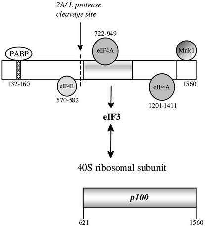
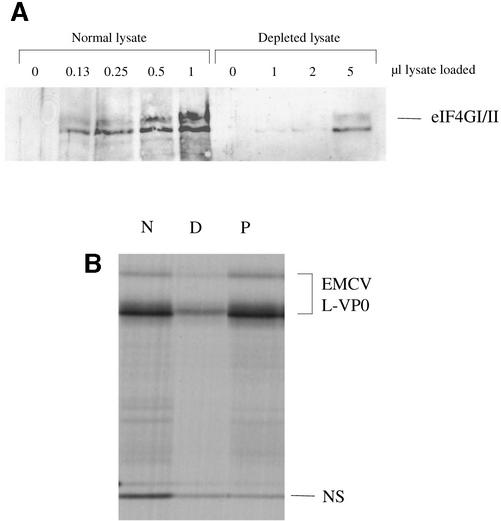
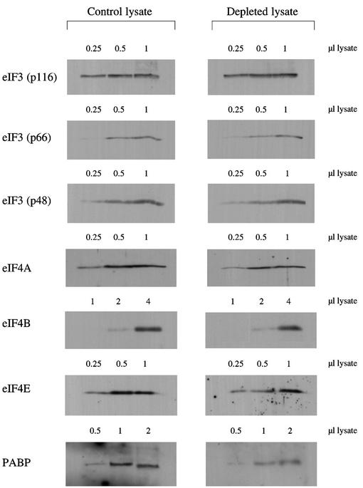
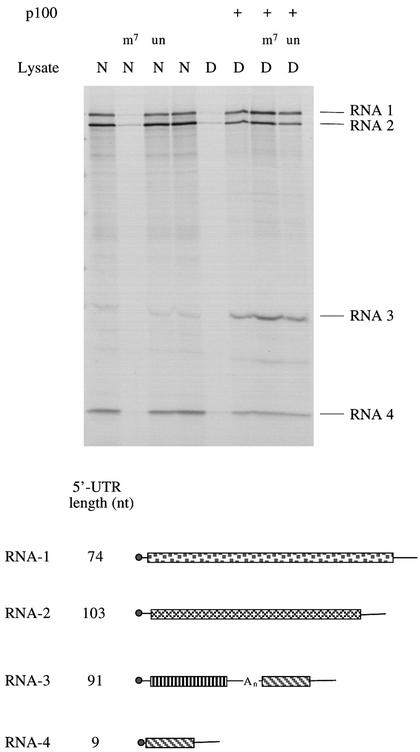
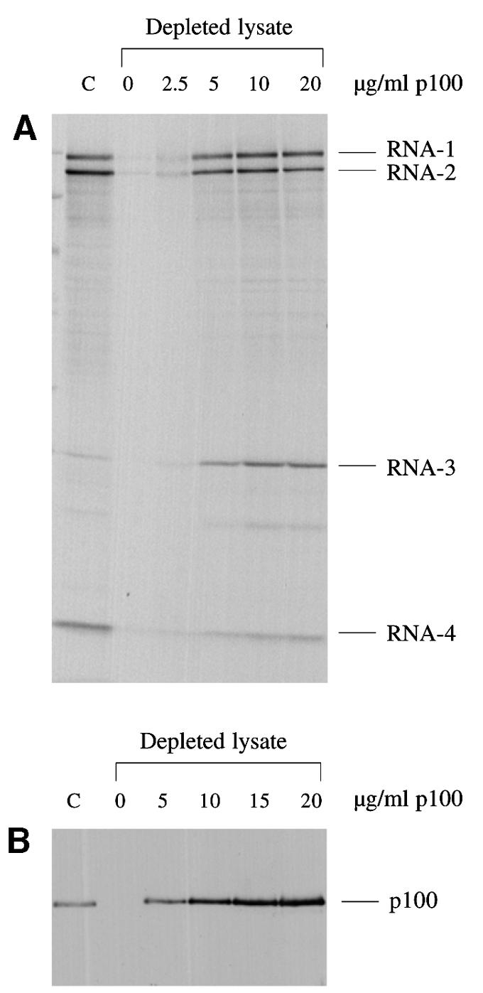
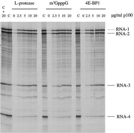
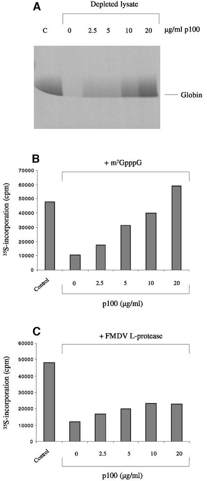
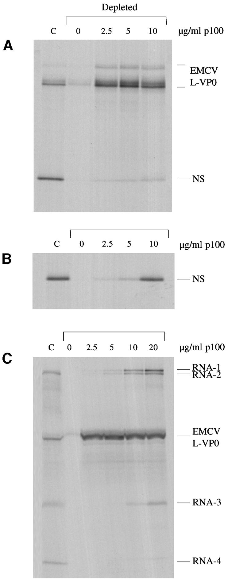
Picornavirus proteases cleave translation initiation factor eIF4G into a C-terminal two-thirds fragment (hereafter named p100) and an N-terminal one-third fragment, which interacts with the cap-binding factor eIF4E. As the timing of this cleavage correlates broadly with the shut-off of host cell protein synthesis in infected cells, a very widespread presumption has been that p100 cannot support capped mRNA translation. Through the use of an eIF4G-depleted reticulocyte lysate system, we show that this presumption is incorrect. Moreover, recombinant p100 can also reverse the inhibition of capped mRNA translation caused either by m7GpppG cap analogue, by 4E-BP1, which sequesters eIF4E and thus blocks its association with eIF4G, or by cleavage of endogenous eIF4G by picornavirus proteases. The concentration of p100 required for maximum translation of capped mRNAs is approximately 4-fold higher than the endogenous eIF4G concentration in reticulocyte lysates. Our results imply that picornavirus-induced shut-off is not due to an intrinsic inability of p100 to support capped mRNA translation, but to the viral RNA outcompeting host cell mRNA for the limiting concentration of p100.


Figures

None
Fig. 1. Domain structure of eIF4G(I) and sites of interaction of other proteins with eIF4G. eIF4GI is depicted as a rectangle. The amino acid numbering of the sites on eIF4G for interaction with PABP and eIF4E are from Gingras et al. (1999), for eIF4A binding to the central domain from Lomakin et al. (2000), and for eIF4A and Mnk-1 interaction near the eIF4G C-terminus from Morino et al. (2000). Note, however, that the upstream eIF4A binding site is defined as amino acids 672–970 by Morino et al. (2000) and 672–876 by Korneeva et al. (2000, 2001). The eIF3 interaction domain is shown as a stippled rectangle, and has been defined as amino acids 697–1076 by Lomakin et al. (2000), 672–1065 by Morino et al. (2000) but 975–1065 by Korneeva et al. (2000). A single arrow is used to denote the sites at which eIF4G is cleaved by enterovirus 2A protease and foot-and-mouth disease virus (FMDV) L-protease; the two cleavage sites are actually 7 amino acid residues apart. Below is depicted the p100 fragment of eIF4GI used in this study.
None
Fig. 2. Affinity depletion of eIF4G from reticulocyte lysates. (A) The indicated volumes of parent (normal) lysate and eIF4G-depleted lysate were subjected to SDS–PAGE and western blotting with anti-eIF4G antiserum and peroxidase-conjugated secondary antibody. Detection was by ECL. (B) Capped dicistronic mRNA with an upstream cistron coding for influenza virus NS1, an EMCV IRES, and EMCV coding sequences for L-VP0 (∼55 kDa) as downstream cistron, was translated at a final concentration of 25 µg/ml in the parent (non-depleted) lysate (N), in the depleted lysate (D) or in depleted lysate supplemented with 20 µg/ml recombinant p100 (P). Radiolabelled translation products were separated by gel electrophoresis, and the resulting autoradiograph is shown. Of the two L-VP0 products, the major (smaller) one results from translation of transcripts of the cDNA template linearized at the StuI site, and the minor (larger) protein is the translation product resulting from incomplete linearization: it is initiated at the same site as the smaller product, and is terminated at an in-frame termination codon that lies just within the vector sequences and beyond the StuI site.
None
Fig. 3. Western blotting assessment of degree of depletion of other factors. The indicated volumes (µl) of parent lysate and eIF4G-depleted lysate were resolved by SDS–PAGE, and after blotting the gels, the blots were probed with antisera against eIF3, eIF4A, eIF4B, eIF4E and PABP, as indicated. Horseradish peroxidase-conjugated secondary antibodies were used (against goat IgGs in the case of the eIF3 and eIF4B blots, and rabbit IgG in all other cases), and detection was by ECL.
None
Fig. 4. p100 can support the translation of capped BMV RNAs by a mechanism that is operationally cap independent. BMV RNA was translated at a final concentration of 20 µg/ml in either normal (parent) lysate (N) or eIF4G-depleted lysate (D), with the following additions where indicated: (m7), 0.4 mM m7GpppG cap analogue with 0.32 mM additional MgCl2; (un), 0.4 mM unmethylated GpppG with 0.32 mM additional MgCl2; (p100), 20 µg/ml (200 nM) recombinant p100. Radiolabelled translation products were separated by gel electrophoresis, and the resulting autoradiograph is shown. The products encoded by BMV RNA-1, RNA-2, RNA-3 and RNA-4 are indicated. The schematic diagram depicts the four BMV RNAs, approximately to scale, and the lengths of the 5′-UTRs (Ahlquist et al., 1981, 1984; Dasgupta and Kaesberg, 1982). Open reading frames are shown as rectangles, UTRs as lines. BMV RNA-3 is functionally monocistronic, but structurally dicistronic; the silent downstream open reading frame is identical to the ORF of RNA-4, and the intercistronic spacer has a 16–21 residue oligo(A) tract.
None
Fig. 5. The dependency of capped mRNA translation on p100 concentration, in relation to the concentration of endogenous eIF4G in the parent (non-depleted) lysate. (A) BMV RNA was translated at a final concentration of 20 µg/ml in either the control (non-depleted) lysate (C) or the eIF4G-depleted lysate supplemented with 0, 2.5, 5, 10 or 20 µg/ml recombinant p100, as indicated. Radiolabelled translation products were separated by gel electrophoresis, and the resulting autoradiograph is shown. (B) Western blot analysis. eIF4G-depleted lysate was supplemented with 0, 5, 10, 15 or 20 µg/ml recombinant p100, as indicated. Parent (non-depleted) lysate (C) was pre-incubated for 10 min at 30°C with in vitro expressed FMDV L-protease (see Materials and methods). Aliquots (equivalent to 1 µl lysate) were separated by gel electrophoresis, which was blotted and the blot probed with anti-eIF4G antiserum.
None
Fig. 6. p100 can reverse the inhibition of capped mRNA translation caused by either FMDV L-protease, m7GpppG cap analogue or 4E-BP1. BMV RNA was translated at a final concentration of 20 µg/ml in either control (untreated) lysate (C), control lysate supplemented with 20 µg/ml recombinant p100 (C + 20), or lysate subjected to one of the following regimes, and then supplemented with 0, 2.5, 5, 10 or 20 µg/ml recombinant p100 as indicated: pre-incubation for 10 min at 30°C with recombinant FMDV L-protease; supplementation with 0.4 mM m7GpppG (with 0.32 mM additional MgCl2); or pre-incubated for 10 min at 30°C with 10 µg/ml recombinant 4E-BP1. Radiolabelled translation products were separated by gel electrophoresis, and the resulting autoradiograph is shown.
None
Fig. 7. p100 can rescue the translation of capped polyadenylated globin mRNA. (A) Globin mRNA was translated at a concentration of 15 µg/ml in either the control (non-depleted) lysate (C) or the eIF4G-depleted lysate supplemented with 0, 2.5, 5, 10 or 20 µg/ml recombinant p100, as indicated. Radiolabelled translation products were separated by gel electrophoresis, and the resulting autoradiograph is shown. (B) Globin mRNA was translated at a final concentration of 15 µg/ml in either control (untreated) lysate or lysate supplemented with 0.4 mM m7GpppG (with 0.32 mM additional MgCl2) and either 0, 2.5, 5, 10 or 20 µg/ml recombinant p100, as indicated. The incorporation of [35S]methionine into acid-precipitable protein was determined by scintillation counting and is plotted. (C) Globin mRNA was translated at a final concentration of 15 µg/ml in either control (untreated) lysate or lysate pre-incubated for 10 min at 30°C with recombinant FMDV L-protease and then supplemented with 0, 2.5, 5, 10 or 20 µg/ml recombinant p100 as indicated.
None
Fig. 8. The EMCV IRES strongly outcompetes capped scanning-dependent mRNAs for functional interaction with p100. (A) Capped dicistronic mRNA with an upstream cistron coding for influenzavirus NS1, an EMCV IRES, and EMCV coding sequences for L-VP0 (∼55 kDa) as downstream cistron, was translated at a final concentration of 20 µg/ml in the control (non-depleted) lysate (C), and in the eIF4G-depleted lysate supplemented with 0, 2.5, 5 or 10 µg/ml p100, as indicated. Radiolabelled translation products were separated by gel electrophoresis, and the resulting autoradiograph is shown. (B) A capped monocistronic mRNA encoding NS, transcribed from the same cDNA construct as the dicistronic mRNA in (A) but with linearization in the 5′-proximal part of the IRES, was translated at a final concentration of 8 µg/ml (same molar concentration as the dicistronic mRNA assayed in A), under the same conditions as in (A). (C) An equimolar mixture of BMV RNAs (13 µg/ml final concentration) and an uncapped monocistronic mRNA with the EMCV IRES linked to viral L-VP0 coding sequences (7 µg/ml) were translated under the same conditions as for (A) and (B).
None
Fig. 9. FMDV L-protease can partially relieve inhibition of capped BMV RNA translation caused by m7GpppG cap analogue. BMV RNA was translated at a final concentration of 20 µg/ml in either control (untreated) lysate (C) or lysate supplemented with 0.4 mM m7GpppG cap analogue (with an additional 0.32 mM MgCl2), or supplemented with both recombinant FMDV L-protease and 0.4 mM m7GpppG cap analogue (with an additional 0.32 mM MgCl2), as indicated. Radiolabelled translation products were separated by gel electrophoresis, and the resulting autoradiograph is shown. The autoradiograph was exposed for longer than in the case of Figure 6 in order to facilitate visualization of the relative yield of products synthesized in the assay inhibited by cap analogue.

References

    1. Ahlquist P., Luckow,V. and Kaesberg,P. (1981) Complete nucleotide sequence of Brome Mosaic Virus RNA 3. J. Mol. Biol., 153, 23–38. - PubMed
    1. Ahlquist P., Dasgupta,R. and Kaesberg,P. (1984) Nucleotide sequence of the Brome Mosaic Virus genome and its implications for viral replication. J. Mol. Biol., 172, 369–383. - PubMed
    1. Ali I.K., McKendrick,L., Morley,S.J. and Jackson,R.J. (2001) The activity of the hepatitis A virus IRES requires association between the cap-binding translation initiation factor (eIF4E) and eIF4G. J. Virol., in press. - PMC - PubMed
    1. Bonneau A.-M. and Sonenberg,N. (1987) Proteolysis of the p220 component of the cap-binding protein complex is not sufficient for complete inhibition of host cell protein synthesis after poliovirus infection. J. Virol., 61, 986–991. - PMC - PubMed
    1. Dasgupta R. and Kaesberg,P. (1982) Complete nucleotide sequence of the coat protein messenger RNAs of Brome Mosaic Virus and Cowpea Chlorotic Mottle Virus. Nucleic Acids Res., 10, 703–713. - PMC - PubMed

Publication types

MeSH terms