. 2001 Aug 1;20(15):4278-86.
doi: 10.1093/emboj/20.15.4278.

Excision of deaminated cytosine from the vertebrate genome: role of the SMUG1 uracil-DNA glycosylase

Affiliations

Excision of deaminated cytosine from the vertebrate genome: role of the SMUG1 uracil-DNA glycosylase

H Nilsen et al. EMBO J. .

Abstract

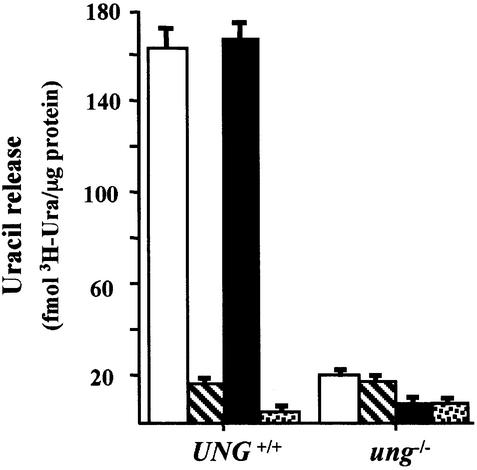
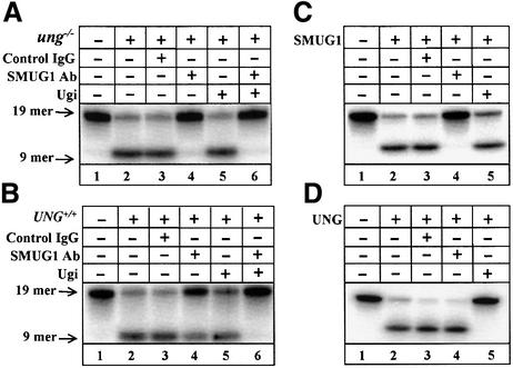
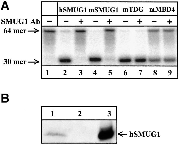
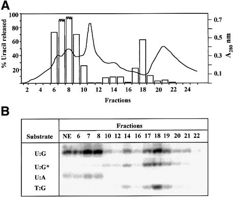
Gene-targeted mice deficient in the evolutionarily conserved uracil-DNA glycosylase encoded by the UNG gene surprisingly lack the mutator phenotype characteristic of bacterial and yeast ung(-) mutants. A complementary uracil-DNA glycosylase activity detected in ung(-/-) murine cells and tissues may be responsible for the repair of deaminated cytosine residues in vivo. Here, specific neutralizing antibodies were used to identify the SMUG1 enzyme as the major uracil-DNA glycosylase in UNG-deficient mice. SMUG1 is present at similar levels in cell nuclei of non-proliferating and proliferating tissues, indicating a replication- independent role in DNA repair. The SMUG1 enzyme is found in vertebrates and insects, whereas it is absent in nematodes, plants and fungi. We propose a model in which SMUG1 has evolved in higher eukaryotes as an anti-mutator distinct from the UNG enzyme, the latter being largely localized to replication foci in mammalian cells to counteract de novo dUMP incorporation into DNA.


Figures

None
Fig. 1. Uracil–DNA glycosylase activity in nuclear MEF extracts. Nuclear extracts from UNG+/+ and ung–/– MEF cell lines were incubated with a [3H]dUMP-containing double-stranded DNA substrate and release of acid soluble [3H]uracil was determined. Extracts were pre-incubated without additions (white bars), with Ugi (hatched bars), SMUG1 antibodies (black bars), or Ugi and SMUG1 antibodies together (dotted bars). Error bars show the SEM from three experiments.
None
Fig. 2. Uracil release from a double-stranded oligonucleotide substrate. Nuclear extracts from ung–/– (A) or UNG+/+ (B) MEF cell lines were incubated with a U:G-containing double-stranded oligonucleotide substrate with the uracil residue centrally placed in the 5′-32P-labelled strand (19mer; lane 1). Uracil release was determined following chemical cleavage of the abasic sites and resolution of the 9mer radio-labelled product by denaturing PAGE. Extracts were pre-incubated without additions (lane 2), with control IgG (lane 3), SMUG1 antibodies (lane 4), Ugi (lane 5) or SMUG1 antibodies and Ugi together (lane 6), as indicated. Release of uracil by purified recombinant human SMUG1 protein (C) or purified human UNG (UNGΔ84) protein (D) was similarly monitored.
None
Fig. 3. Specificity of SMUG1 antibodies. The specificity of the antibodies raised against recombinant hSMUG1 was investigated. (A) Uracil release from a UpG-containing, double-stranded 64mer oligonucleotide substrate (lane 1) was determined as in Figure 2. Uracil release was measured directly (–) or after pre-incubation with SMUG1 antibodies (+) using recombinant hSMUG1 (lanes 2 and 3), mSMUG1 (lanes 4 and 5), recombinant mTDG (lanes 6 and 7), and recombinant mMBD4 (lanes 8 and 9). (B) Western blot of coupled in vitro transcription–translation reaction mixtures. The SMUG1 antibodies specifically recognized mSMUG1 (lane 1) and there was no cross-reaction with reticulocyte lysate proteins when mSMUG1 cDNA was excluded from the reaction mixture (lane 2). Recombinant hSMUG1 (lane 3) is shown as a reference.
None
Fig. 4. Fractionation of ung–/– MEF nuclear extract by Mono S chromatography. Nuclear extract was prepared from ung–/– MEFs (108 cells) and the extract loaded onto a Mono S column. A linear NaCl gradient (50–500 mM) was applied and fractions collected (2–18), followed by elution in 1 M NaCl. (A) An aliquot of each fraction was assayed using the 19mer U:G-containing oligonucleotide substrate (as in Figure 2); uracil release (%) in each fraction is shown as open bars. (B) Nuclear extract (NE) and selected fractions were assayed for activity on oligonucleotide substrates containing U:G, U:A or T:G base pairs. The 9mer product is shown (see Figure 2 legend). Activity on the U:G substrate in fractions pre-incubated with SMUG1 antibodies is indicated by an asterisk.
None
Fig. 5. Stimulation of SMUG1 activity by APE1. Oligonucleotides containing uracil, as a single-stranded substrate (squares) or paired opposite G as a double-stranded substrate (triangles), were incubated with SMUG1 (open symbols) or SMUG1 plus APE1 (closed symbols). Following NaOH treatment to cleave AP sites and resolution of the product band by denaturing polyacrylamide gel electrophoresis, uracil release was quantitated by phosphoimager analysis.
None
Fig. 6. Tissue distribution of SMUG1 activity. Nuclear extracts were prepared of different organs from ung–/– mice. Uracil-excising activity was measured using the 19mer U:G containing double-stranded oligonucleotide substrate (as in Figure 2). Uracil release was measured directly (–) or after pre-incubating the extracts with SMUG1 antibodies (+).
None
Fig. 7. Alignment of putative SMUG1 orthologues. Complete cDNA sequences (Hs, Xl, Dm, Mm) and ESTs (Dr, Sp, Ag, Bm) were included in the alignment. Related sequences were not detected in plants, nematodes, or yeast and other fungi. Residues shaded magenta are proposed to be important for substrate recognition and catalysis (Haushalter et al., 1999; Aravind and Koonin, 2000). Conserved residues are highlighted yellow, and conservative substitutions are shaded grey. Gaps and missing residues are denoted by dashes and unassignable amino acid residues from the ESTs are denoted by the letter X. Species and DDBJ/EMBL/Genbank accession Nos are as follows: Hs, Homo sapiens (human), NP_055126.1; Mm, Mus musculus (mouse), BF467856; Xl, Xenopus laevis (African clawed toad), AAD17300.1; Dr, Danio rerio (zebrafish), compilation of AW419619, AI878196 and AW134258; Sp, Strongylocentrotus purpuratus (purple sea urchin), AF122749; Dm, Drosophila melanogaster (fruit fly), AAF55400; Ag, Anopheles gambiae (malaria mosquito), AJ282661; Bm, Bombyx mori (silk moth), AU004467.

References

    1. Aprelikova O.N. and Tomilin,N.V. (1982) Activity of uracil–DNA glycosylase in different rat tissues and in regenerating rat liver. FEBS Lett., 137, 193–195. - PubMed
    1. Aravind L. and Koonin,E.V. (2000) The α/β fold uracil-DNA glycosylases: a common origin with diverse fates. Genome Biol., 1, 0007.1–0007.8. - PMC - PubMed
    1. Brynolf K., Eliasson,R. and Reichard,P. (1978) Formation of Okazaki fragments in polyoma DNA synthesis caused by misincorporation of uracil. Cell, 13, 573–580. - PubMed
    1. Duncan B.K. and Miller,J.H. (1980) Mutagenic deamination of cytosine residues in DNA. Nature, 287, 560–561. - PubMed
    1. Duncan B.K. and Weiss,B. (1982) Specific mutator effects of ung (uracil–DNA glycosylase) mutations in Escherichia coli. J. Bacteriol., 151, 750–755. - PMC - PubMed

Publication types

MeSH terms