. 2001 Aug;11(8):1365-74.
doi: 10.1101/gr.181001.

Prokaryotic homologs of the eukaryotic DNA-end-binding protein Ku, novel domains in the Ku protein and prediction of a prokaryotic double-strand break repair system

Affiliations

Prokaryotic homologs of the eukaryotic DNA-end-binding protein Ku, novel domains in the Ku protein and prediction of a prokaryotic double-strand break repair system

L Aravind et al. Genome Res. 2001 Aug.

Abstract

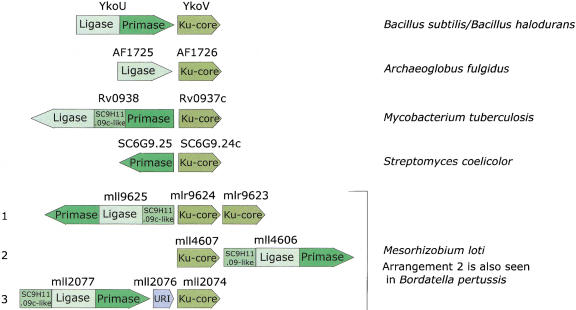
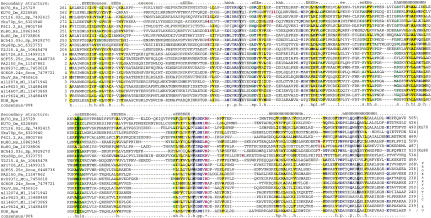
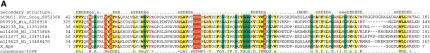
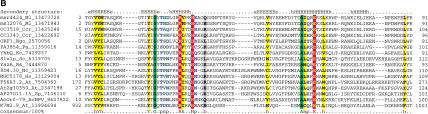
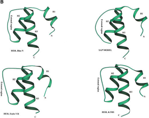
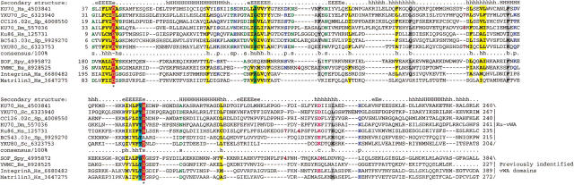
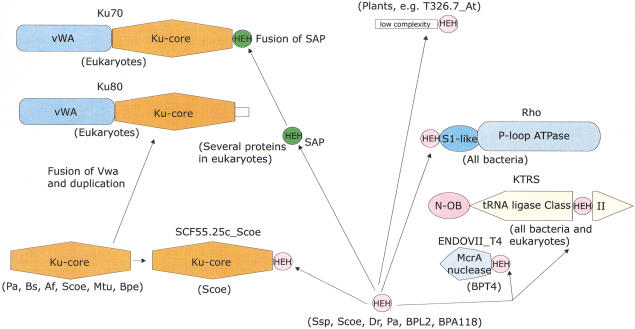
Homologs of the eukaryotic DNA-end-binding protein Ku were identified in several bacterial and one archeal genome using iterative database searches with sequence profiles. Identification of prokaryotic Ku homologs allowed the dissection of the Ku protein sequences into three distinct domains, the Ku core that is conserved in eukaryotes and prokaryotes, a derived von Willebrand A domain that is fused to the amino terminus of the core in eukaryotic Ku proteins, and the newly recognized helix-extension-helix (HEH) domain that is fused to the carboxyl terminus of the core in eukaryotes and in one of the Ku homologs from the Actinomycete Streptomyces coelicolor. The version of the HEH domain present in eukaryotic Ku proteins represents the previously described DNA-binding domain called SAP. The Ku homolog from S. coelicolor contains a distinct version of the HEH domain that belongs to a previously unnoticed family of nucleic-acid-binding domains, which also includes HEH domains from the bacterial transcription termination factor Rho, bacterial and eukaryotic lysyl-tRNA synthetases, bacteriophage T4 endonuclease VII, and several uncharacterized proteins. The distribution of the Ku homologs in bacteria coincides with that of the archeal-eukaryotic-type DNA primase and genes for prokaryotic Ku homologs form predicted operons with genes coding for an ATP-dependent DNA ligase and/or archeal-eukaryotic-type DNA primase. Some of these operons additionally encode an uncharacterized protein that may function as nuclease or an Slx1p-like predicted nuclease containing a URI domain. A hypothesis is proposed that the Ku homolog, together with the associated gene products, comprise a previously unrecognized prokaryotic system for repair of double-strand breaks in DNA.


Figures

Figure 1
Figure 1
Gene organization in the predicted operons encoding components of the postulated novel double-strand-break repair system. The direction of an arrow indicates the direction of transcription. Distinct regions of each gene encoding separate domains in the protein, such as primase and ligase, are indicated in different shades.
Figure 2
Figure 2
Multiple sequence alignment of the Ku-core domains. The secondary structure predicted using the PHD program is shown above the alignment. E indicates a β-strand and H indicates an α-helix, with the uppercase used to denote the most confident prediction (>82% accuracy). The 90% consensus shown below the alignment was derived using the following amino acid classes: polar (p: KRHEDQNST) colored blue; hydrophobic (h: ALICVMYFW) and the aliphatic subset of these are (l: ALIVMC) all shaded yellow; small (s: ACDGNPSTV) colored green, charged (c: DEHKR) colored pink, big (b: Q,E,R,K,Y,M,F,W,L,I) shaded gray. The limits of the domains are indicated by the position numbers on each side of the alignment. The subclasses of Ku-core domains are indicated to the right of the alignment. The sequences are denoted by their gene names followed by the species abbreviations and GenBank identifiers. Subsequent to the submission of this manuscript, the prokaryotic KU homologs were identified in the SMART database (Schultz et al. 1998). The species abbreviations are: At, Arabidopsis thaliana; Hs, Homo sapiens; Mm, Mus musculus; Dm, Drosophila melanogaster; Sc, Saccharomyces cerevisiae; Sp, Schizosaccharomyces pombe; Af,Archaeoglobus fulgidus; Pa, Pseudomonas aeruginosa; Bs, Bacillus subtilis; Scoe, Streptomyces coelicolor; Bpe, Bordatella pertussis; Mtu, Mycobacterium tuberculosis; Ml, Mesorhizobium loti.
Figure 3
Figure 3
Multiple sequence alignment of predicted nucleases associated with the hypothetical EP-ADDL-Ku repair system. The novel potential nuclease (A) and The URI-domain-containing nuclease (B). The potential metal chelating and active site residues are shown in reverse red shading. (B) The K7M2.9 from Arabidopsis is fused to a MutS domain at the amino terminus, whereas the other eukaryotic forms show carboxy-terminal fusion to a PHD fingers. The species abbreviations are as in Figure 2; the additional abbreviations not present in Figure 2 are: Nc, Neurospora crassa; AcNPV, Autographa californica Nuclear polyhedrosis virus; Ngo, Neisseria gonorrhea; Bs, Bacillus subtilis; Ec, Escherichia coli; Ccr, Caulobacter cresentus.
Figure 3
Figure 3
Multiple sequence alignment of predicted nucleases associated with the hypothetical EP-ADDL-Ku repair system. The novel potential nuclease (A) and The URI-domain-containing nuclease (B). The potential metal chelating and active site residues are shown in reverse red shading. (B) The K7M2.9 from Arabidopsis is fused to a MutS domain at the amino terminus, whereas the other eukaryotic forms show carboxy-terminal fusion to a PHD fingers. The species abbreviations are as in Figure 2; the additional abbreviations not present in Figure 2 are: Nc, Neurospora crassa; AcNPV, Autographa californica Nuclear polyhedrosis virus; Ngo, Neisseria gonorrhea; Bs, Bacillus subtilis; Ec, Escherichia coli; Ccr, Caulobacter cresentus.
Figure 4
Figure 4
(A) Multiple sequence alignment of different classes of HEH domains. Each of the alignments is colored according to a separate consensus using the rules described in the legend to Figure 2. The secondary structure shown above the alignment was derived from the structures of Rho, Endo-VII and K-TRS. For the SAP domains, the structure was predicted using the PHD program. The species abbreviations are the same as in Figure 2; those not present in Figure 2 are : Ec, Escherichia coli; Ssp, Synechocystis sp.; Tma, Thermotoga maritima; Dr, Deinococcus radiodurans; Aae, Aquifex aeolicus; BPL2, lactococcal Bacteriophage L2; BPA118, Listeria bacteriophage A118; T4, Bacteriophage T4; Miclu, Micrococcus luteus; Ce, Caenorhabditis elegans; Ct, Chlamydia trachomatis; Hp, Helicobacter pylori; Bst, Bacillus stearothermophilus. (B) Structures and models of different forms of the HEH domain shown in the alignment. The NH2 (N) and COOH (C) termini of the HEH domains are indicated.
Figure 4
Figure 4
(A) Multiple sequence alignment of different classes of HEH domains. Each of the alignments is colored according to a separate consensus using the rules described in the legend to Figure 2. The secondary structure shown above the alignment was derived from the structures of Rho, Endo-VII and K-TRS. For the SAP domains, the structure was predicted using the PHD program. The species abbreviations are the same as in Figure 2; those not present in Figure 2 are : Ec, Escherichia coli; Ssp, Synechocystis sp.; Tma, Thermotoga maritima; Dr, Deinococcus radiodurans; Aae, Aquifex aeolicus; BPL2, lactococcal Bacteriophage L2; BPA118, Listeria bacteriophage A118; T4, Bacteriophage T4; Miclu, Micrococcus luteus; Ce, Caenorhabditis elegans; Ct, Chlamydia trachomatis; Hp, Helicobacter pylori; Bst, Bacillus stearothermophilus. (B) Structures and models of different forms of the HEH domain shown in the alignment. The NH2 (N) and COOH (C) termini of the HEH domains are indicated.
Figure 5
Figure 5
Multiple sequence alignment of the vWA domains of Ku70 and Ku80. The secondary structure shown above the figure was based on the solved structures of vWA domains; the same consensus-based coloring scheme as in Figures 2 and 3 is used. The species abbreviation Spy is for Streptococcus pyogenes, whereas the rest are the same as in Figures 2 and 3.
Figure 6
Figure 6
Protein domain architectures and possible evolutionary trajectories for the constituent domains of the Ku proteins. The domains are indicated by different shapes; the two distinct forms of the HEH are indicated by differential coloring. For each domain architecture, the phyletic distribution is shown in parentheses; the species name abbreviations are as in Figures 2 and 3. The arrows indicate probable evolutionary events such as derivation of a new form of a particular domain (SAP from ancestral HEH) and domain fusion. The connection shown between prokaryotic and eukaryotic forms of the Ku protein does not differentiate between two evolutionary scenarios discussed in the text.

References

    1. Allison TJ, Wood TC, Briercheck DM, Rastinejad F, Richardson JP, Rule GS. Crystal structure of the RNA-binding domain from transcription termination factor rho [letter] Nat Struct Biol. 1998;5:352–356. - PubMed
    1. Altschul SF, Madden TL, Schaffer AA, Zhang J, Zhang Z, Miller W, Lipman DJ. Gapped BLAST and PSI-BLAST: A new generation of protein database search programs. Nucleic Acids Res. 1997;25:3389–3402. - PMC - PubMed
    1. Aravind L, Koonin EV. DNA-binding proteins and evolution of transcription regulation in the archaea. Nucleic Acids Res. 1999;27:4658–70. - PMC - PubMed
    1. ————— SAP — a putative DNA-binding motif involved in chromosomal organization. Trends Biochem Sci. 2000;25:112–114. - PubMed
    1. Aravind L, Walker DR, Koonin EV. Conserved domains in DNA repair proteins and evolution of repair systems. Nucleic Acids Res. 1999;27:1223–1242. - PMC - PubMed

MeSH terms

LinkOut - more resources