. 2001 Aug 1;534(Pt 3):691-700.
doi: 10.1111/j.1469-7793.2001.00691.x.

Expression and function of native potassium channel [K(V)alpha1] subunits in terminal arterioles of rabbit

Affiliations

Expression and function of native potassium channel [K(V)alpha1] subunits in terminal arterioles of rabbit

A Cheong et al. J Physiol. .

Abstract

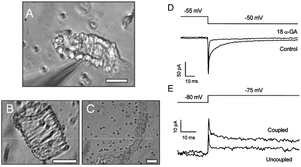
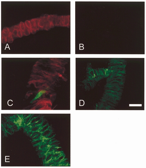
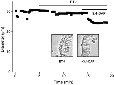
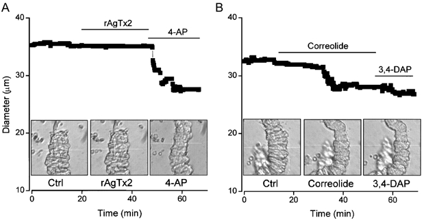
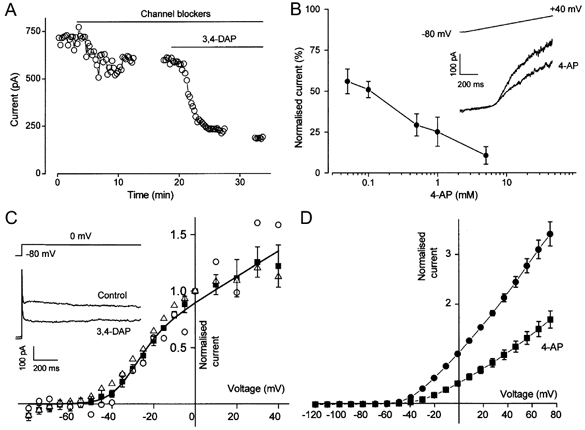
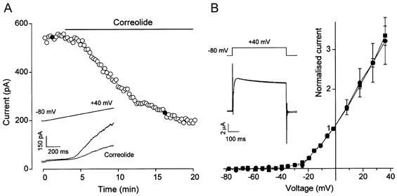
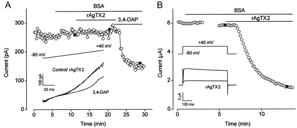
1. In this study we investigated the expression and function of the K(V)alpha1 subfamily of voltage-gated K(+) channels in terminal arterioles from rabbit cerebral circulation. 2. K(+) current was measured from smooth muscle cells within intact freshly isolated arteriolar fragments. Current activated on depolarisation positive of about -45 mV and a large fraction of this current was blocked by 3,4-diaminopyridine (3,4-DAP) or 4-aminopyridine (4-AP), inhibitors of K(V) channels. Expression of cRNA encoding K(V)1.6 in Xenopus oocytes also generated a 4-AP-sensitive K(+) current with a threshold for activation near -45 mV. 3. Immunofluorescence labelling revealed K(V)1.2 to be specifically localised to endothelial cells, and K(V)1.5 and K(V)1.6 to plasma membranes of smooth muscle cells. 4. K(V) channel current in arteriolar fragments was blocked by correolide (which is specific for the K(V)alpha1 family of K(V) channels) but was resistant to recombinant agitoxin-2 (rAgTX2; which inhibits K(V)1.6 but not K(V)1.5). Heterologously expressed K(V)2.1 was resistant to correolide, and K(V)1.6 was blocked by rAgTX2. 5. Arterioles that were mildly preconstricted and depolarised by 0.1-0.3 nM endothelin-1 constricted further in response to 3,4-DAP, 4-AP or correolide, but not to rAgTX2. 6. We suggest that K(V)alpha1 channels are expressed in smooth muscle cells of terminal arterioles, underlie a major part of the voltage-dependent K(+) current, and have a physiological function to oppose vasoconstriction. K(V)alpha1 complexes without K(V)1.5 appear to be uncommon.


Figures

Figure 2
Figure 2. Conventional whole-cell patch-clamp recordings from smooth muscle cells within intact arterioles
A, digital image of an arteriolar fragment with an attached patch pipette (out of focus object entering the image from the left corner). B, an isolated venule. C, a long isolated arteriole with exposed endothelial cells. Small circular cells are erythrocytes. The scale bars in A-C represent 30 μm. D and E, capacity currents recorded from arteriolar fragments in response to 5 mV steps in voltage. D, two currents from the same arteriole, one under control conditions, and the other in the presence of 50 μm 18α-glycyrrhetinic acid (18α-GA). E, two current traces from different arterioles, one showing electrical coupling between cells (Coupled) and the other in which the patched cell was spontaneously uncoupled from other cells in the arteriole (Uncoupled).
Figure 4
Figure 4. Immunofluorescence staining of native KV1.2, KV1.5 and KV1.6 in enzymatically isolated arterioles
A and B, control experiments showing an arteriole stained with anti-α-SMA-Cy3 (A) and secondary antibody conjugated to FITC but without primary (anti-KV) antibody (B). C, double staining with anti-α-SMA-Cy3 (red) and anti-KV1.2RK(461-480) (green). A fracture in the arteriole is evident, through which an endothelial cell (green) is exposed. D, staining with anti-Kv1.5R(578-598). E, staining with anti-Kv1.6RK(509-526). Scale bar, 30 μm.
Figure 1
Figure 1. Contraction in an arteriole exposed to 3,4-diaminopyridine, a blocker of KV channels
The filled squares in the plot indicate the external diameter of the arteriole. The bath solution was aCSF and a small level of preconstriction was induced with 0.3 nm endothelin-1 (ET-1) prior to application of 1 mm 3,4-diaminopyridine (3,4-DAP). Actual video images of the arteriole are shown inset for the two conditions. The white horizontal line in each image is the scan line used to detect the outer edge of the arteriole. It was moved slightly during the recording to ensure that edge-detection was maintained.
Figure 7
Figure 7. Effects of correolide and recombinant agitoxin-2 on arteriolar diameter
The filled squares in the plots indicate external diameter. The bath solution was aCSF and a small level of preconstriction was induced in both experiments with 0.3 nm ET-1. Actual video images of the arterioles are shown inset for the three conditions in each case. The white horizontal line in each image is the scan line used to detect the outer edge of the arteriole. It was moved slightly during recordings to maintain edge-detection. A, 1 nm rAgTX2 and 1 mm 4-AP. B, 1 μm correolide and 1 mm 3,4-DAP. Ctrl, control.
Figure 3
Figure 3. KV channel currents in arterioles and through heterologously expressed KV1.6
A, open circles show current amplitude at the end of 1 s ramp changes in voltage from -80 to +40 mV. ‘Channel blockers’ means replacement of extracellular Ca2+ by Mg2+, and application of 100 nm penitrem A, 1 μm glibenclamide and 50 μm niflumic acid. 3,4-DAP (1 mm) was applied as indicated. Gaps in the trace indicate the periods during which current-voltage relationships were constructed. B, plot of mean ±s.e.m. (n = 3-5) concentration-dependent block by 4-aminopyridine (4-AP) of outward current at 0 mV. Currents were normalised to the pre-4-AP amplitude. Inset, voltage protocol and example currents for control and in the presence of 50 μm 4-AP. C, voltage dependence of 1 mm 3,4-DAP-sensitive current measured at the end of 1 s square voltage steps and normalised to the amplitude at 0 mV. ▵, coupled cells under control conditions (s.e.m. bars not shown, n = 9); ▪, cells in the presence of 50 μm 18α-glycyrrhetinic acid (n = 4); ○, a spontaneously uncoupled cell. The curve is fitted to the 18α-glycyrrhetinic acid data and is the equation: gmax(V -Vrev)/{1 + exp((V - V1/2)/dV)}, in which V1/2 is the half-activation potential (-31.4 mV), Vrev is the reversal potential (-85 mV), dV is the slope (6.5 mV) and gmax is the maximum normalised conductance. Inset, example currents for one arteriole. The bath solution contained ‘channel blockers’. D, voltage dependence of current (means ±s.e.m.) through KV1.6 expressed as a homomultimer in Xenopus oocytes under control conditions (•, n = 7) and in 1 mm 4-AP (▪, n = 7). Currents were normalised to the amplitude at 0 mV.
Figure 5
Figure 5. Effect of correolide on K+ currents
A, plot of current amplitude at the end of 1 s ramp changes in voltage from -80 to +40 mV. Inset, two example current traces taken at the points marked by filled circles in the time-series plot. Correolide (1 μm) was bath applied. B, current-voltage relationships for KV2.1 homomultimers expressed in Xenopus oocytes before (▪) and after (•) application of 1 μm correolide (means ±s.e.m., n = 3). Currents were normalised to the amplitude at 0 mV. Inset, example current traces for control and correolide.
Figure 6
Figure 6. Effect of recombinant agitoxin-2 on K+ currents
A, plot of arteriolar current amplitude at the end of 1 s ramp changes in voltage from -80 to +40 mV. Inset, three example current traces taken at the points marked by filled circles in the time-series plot. BSA (0.01 %), recombinant agitoxin-2 (rAgTX2; 1 nm) and 3,4-DAP (1 mm) were bath applied. B, plot of current amplitude at +40 mV for KV1.6 homomultimers expressed in a Xenopus oocyte. BSA (0.01 %) and rAgTX2 (1 nm) were bath applied. The gap in the trace indicates the period during which a current-voltage relationship was constructed. Inset, example current traces taken at the points marked by filled circles in the time-series plot.

References

    1. Ahn DS, Hume JR. pH regulation of voltage-dependent K+ channels in canine pulmonary arterial smooth muscle cells. Pflügers Archiv. 1997;433:758–765. - PubMed
    1. Archer SL, Souil E, Dinh-Xuan AT, Schremmer B, Mercier JC, El Yaagoubi A, Nguyen-Huu L, Reeve HL, Hampl V. Molecular identification of the role of voltage-gated K+ channels, KV1. 5 and KV2.1, in hypoxic pulmonary vasoconstriction and control of resting membrane potential in rat pulmonary artery myocytes. Journal of Clinical Investigation. 1998;101:2319–2330. - PMC - PubMed
    1. Beech DJ. Actions of neurotransmitters and other messengers on Ca2+ channels and K+ channels in smooth muscle cells. Pharmacology and Therapeutics. 1997;73:91–119. - PubMed
    1. Beech DJ, Bolton TB. Two components of potassium current activated by depolarisation of single smooth muscle cells from the rabbit portal vein. Journal of Physiology. 1989;418:293–309. - PMC - PubMed
    1. Brayden JE, Nelson MT. Regulation of arterial tone by activation of calcium-dependent potassium channels. Science. 1992;256:532–535. - PubMed

Publication types

MeSH terms

LinkOut - more resources