. 2001 Sep;75(17):7828-39.
doi: 10.1128/jvi.75.17.7828-7839.2001.

Salicylates inhibit flavivirus replication independently of blocking nuclear factor kappa B activation

Affiliations

Salicylates inhibit flavivirus replication independently of blocking nuclear factor kappa B activation

C L Liao et al. J Virol. 2001 Sep.

Abstract

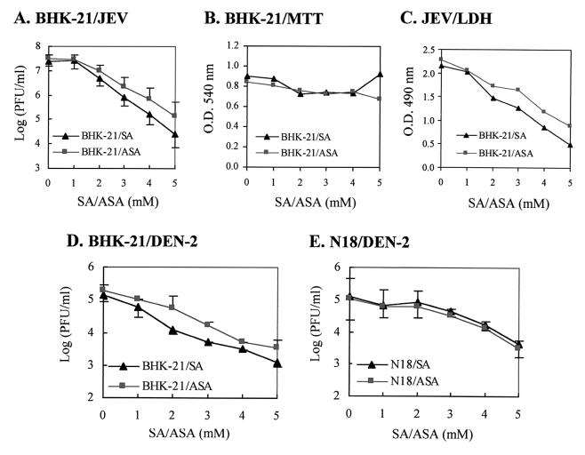
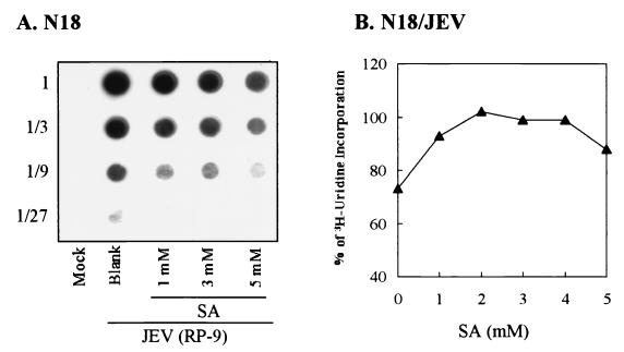
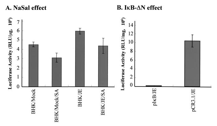
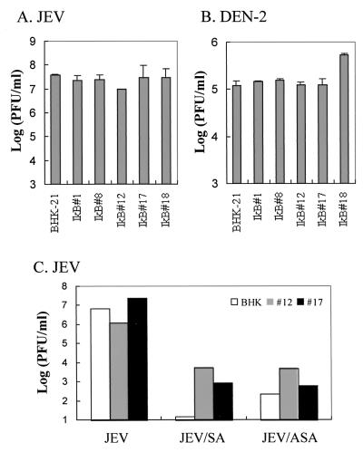
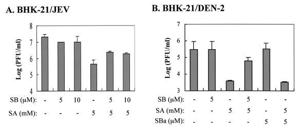
Flaviviruses comprise a positive-sense RNA genome that replicates exclusively in the cytoplasm of infected cells. Whether flaviviruses require an activated nuclear factor(s) to complete their life cycle and trigger apoptosis in infected cells remains elusive. Flavivirus infections quickly activate nuclear factor kappa B (NF-kappaB), and salicylates have been shown to inhibit NF-kappaB activation. In this study, we investigated whether salicylates suppress flavivirus replication and virus-induced apoptosis in cultured cells. In a dose-dependent inhibition, we found salicylates within a range of 1 to 5 mM not only restricted flavivirus replication but also abrogated flavivirus-triggered apoptosis. However, flavivirus replication was not affected by a specific NF-kappaB peptide inhibitor, SN50, and a proteosome inhibitor, lactacystin. Flaviviruses also replicated and triggered apoptosis in cells stably expressing IkappaBalpha-DeltaN, a dominant-negative mutant that antagonizes NF-kappaB activation, as readily as in wild-type BHK-21 cells, suggesting that NF-kappaB activation is not essential for either flavivirus replication or flavivirus-induced apoptosis. Salicylates still diminished flavivirus replication and blocked apoptosis in the same IkappaBalpha-DeltaN cells. This inhibition of flaviviruses by salicylates could be partially reversed by a specific p38 mitogen-activated protein (MAP) kinase inhibitor, SB203580. Together, these results show that the mechanism by which salicylates suppress flavivirus infection may involve p38 MAP kinase activity but is independent of blocking the NF-kappaB pathway.


Figures

FIG. 1
FIG. 1
NaSal and aspirin inhibit flavivirus replication in BHK-21 and N18 cells. (A) BHK-21 cells were infected by JEV at an MO1 of 5, and at 1 h postinfection, the infected cells were treated with varying concentrations of NaSal (SA) or aspirin (ASA). After a 48-h incubation at 37°C, the virus titers (expressed as PFU per milliliter) in the culture media were determined by a plaque forming assay as described in Materials and Methods. Values of virus titers are shown as means ± standard errors of the means for three independent experiments. (B) MTT assay of salicylate-treated BHK-21 cells. BHK-21 cells were treated with SA or ASA at the indicated concentrations for 48 h at 37°C, and the viabilities of the resulting cells were determined by an MTT assay. (C) LDH release assay. At 32 h postinfection, amounts of LDH released from the JEV-infected BHK-21 cells treated with varying amounts of salicylates were measured by an ELISA plate reader at 490 nm. Optical densities at 490 mm are shown as means for representative experiments performed in triplicate. (D) BHK-21 cells were infected by DEN at an MO1 of 5 and were treated with varying concentrations of SA or ASA as indicated. After a 48-h incubation at 37°C, the virus titers (in PFU per milliliter) in the culture media were determined as described for panel A). (E) N18 cells were infected by DEN and treated with SA or ASA. After a 48-h incubation at 37°C, the virus titers (in PFU per milliliter) in the culture media were determined as described for panel A.
FIG. 2
FIG. 2
Effects of NaSal on viral RNA synthesis in JEV-infected N18 cells. (A) RNA dot blotting. Total RNAs were isolated at 18 h postinfection from JEV-or mock-infected N18 cells treated with various doses of NaSa1 (SA) as indicated. Appropriate amounts of RNA from each sample were threefold serially diluted (marked as 1 to 1/27 on the left of the panel) and applied to a nylon membrane that was then hybridized with the DIG-labeled DNA probe specific for the JEV NS1 region. (B) [3H]uridine incorporation. JEV-infected N18 cells were treated with varying doses of SA and incubated for 17 h at 37°C. After the resulting cells were labeled with [3H]uridine for 1 h, the newly synthesized, actinomycin D-resistant RNA was isolated. One-tenth of the cell lysates from each sample was counted for incorporation of [3H]uridine into RNA as described in Materials and Methods.
FIG. 3
FIG. 3
Effects of NaSal on synthesis of JEV glycoproteins in infected BHK-21 and N18 cells. Immunoblot analysis of JEV E or NS1 protein in cell lysates (A) or in supernatants (B) from BHK-21 (lanes 1 to 6) or N18 cells (lanes 7 to 12) cells treated with doses of NaSa1 (SA) ranging from 0 to 5 mM as indicated. NS1′ is the longer version of the NS1 protein derived from an aberrant cleavage in the JEV-infected cells (36).
FIG. 4
FIG. 4
Salicylates suppress flavivirus-induced apoptosis in BHK-21 and N18 cells. (A) Agarose gel electrophoresis of DNA fragmentation. BHK-21 cells were either mock infected (lanes 2 and 6) or infected with JEV at an MOI of 5 (lanes 1, 3 to 5, and 7 to 9), and cells were then left untreated (lane 1) or treated with NaSal (SA) (lanes 2 to 5) or aspirin (ASA) (lanes 6 to 9) at the indicated concentrations. Low-molecular-weight DNA was isolated from cells at 32 h postinfection and analyzed by 2% agarose gel in the presence of EtBr. Lane M, 100-bp ladders as DNA markers. (B) Gel analysis of DNA ladders from JEV-infected N18 cells that were left untreated (lane 1) or treated with 5 mM SA (lane 2) at 32 h postinfection. (C) Kinetics of DNA fragmentation from JEV-infected N18 cells that were left untreated (filled squares) or treated (open circles) with 5 mM SA, determined by ELISA. Prior to virus infection, cells were labeled with BrdU overnight. At the indicated time points following infection, the cells were permeabilized to release the cytoplasmic DNA fragments into the supernatants. The amounts of BrdU-labeled DNA released were measured by ELISA as optical densities (O.D.) at 450 nm using antibodies against DNA and BrdU (see Materials and Methods).
FIG. 5
FIG. 5
Effect of NaSal or the dominant-negative mutant IκB-ΔN on JEV-induced NF-κB activation. (A) NaSal effect. A total of 8 × 10⁴ BHK-21 cells transfected with pNFκB-Luc were either left untreated or treated with 5 mM NaSa1 or for 18 h. The resulting cells were then either infected with JEV (at an MOI of 5) or mock infected, and at 6 h postinfection, cell lysates were prepared for determination of luciferase activity. (B) IκB-ΔN effect. A total of 8 × 10⁴ BHK-21 cells were transfected either with pNFκB-Luc together with plκB-ΔN or with pNFκB-Luc plus the pCR3.1 vector, and these cells were then infected with JEV at an MOI of 5 for another 6 h. The resulting cell lysates were prepared and assessed for luciferase activity. Values shown are representative of the results from three independent experiments. Luciferase activity is expressed as relative light units.
FIG. 6
FIG. 6
Role of NF-κB activation in JEV replication in BHK-21 and N18 cells. (A and B) Effect of the cell-permeating peptide inhibitor SN50 on JEV replication. At 1 h postinfection, JEV-infected BHK-21 (A) or N18 (B) cells were treated with varying doses of SN50, and virus yields from the cells were determined by a plaque assay after a 36-h incubation. As negative controls, the infected cells were either left untreated (Blank) or, treated with the mutant peptide SN50M; as a positive control, cells were treated with 5 mM NaSal (SA). (C and D) Effect of lactacystin (C) or indomethacin (D) on JEV reproduction in BHK-21 cells. At 1 h postinfection, JEV-infected BHK-21 cells were either left untreated (Blank) or treated with varying doses of lactacystin or indomethacin, and after 20 h of incubation, virus yields from the resulting cells were determined by plaque assay as described for Fig. 1A.
FIG. 7
FIG. 7
Effects of constitutive IκBα-ΔN expression on antiflavivirus capability of salicylates. The ability of JEV (A) or DEN (B) to replicate in IκBα-ΔN-expressing cell clones (IκB#1, #8, #12, #17, and #18) was compared to that in wild-type BHK-21 cells by measurement of titers of virus released into the culture medium. (C) The anti-JEV effects of salicylates were determined for the IκBα-ΔN-expressing cell clones (IκB#12 and #17) and the wild-type BHK-21 cells. After 1 h of JEV infection, cells were left untreated or treated with 5 mM NaSal (SA) or aspirin (ASA) and then incubated at 37°C for another 24 h. Virus production in the culture medium was determined by a plaque assay. Data are mean virus titers for two independent experiments.
FIG. 8
FIG. 8
Antiflavivirus capability of salicylates in cells treated with the p38 MAPK inhibitor SB203580. (A) BHK-21 cells were infected by JEV at an MOI of 5, and at 1 h postinfection SB203580 (SB) was added to the cells at varying micromolar concentrations in the presence or absence (−) of 5 mM NaSal (SA). After 36 h of incubation at 37°C, virus yields in the culture media were determined by a plaque assay. (B) BHK-21 cells were infected by DEN at an MOI of 5, and at 1 h postinfection varying doses (micromolar concentrations) of SB203580 or its ineffective analogue SB202474 (SBa) were added cells in the presence or absence (−) of 5 mM SA. After 36 h of incubation at 37°C, virus yields in the culture media were determined as described for Fig. 1A.

References

    1. Amano T, Richelson E, Nirenberg M. Neurotransmitter synthesis by neuroblastoma clones. Proc Natl Acad Sci USA. 1972;69:258–263. - PMC - PubMed
    1. Avirutnan P, Malasit P, Seliger B, Bhakdi S, Husmann M. Dengue virus infection of human endothelial cells leads to chemokine production, complement activation, and apoptosis. J Immunol. 1998;161:6338–6346. - PubMed
    1. Baeuerle P A, Baltimore D. NF-κB: ten years after. Cell. 1996;87:13–20.20. - PubMed
    1. Baeuerle P A, Henkel T. Function and activation of NF-κB in the immune system. Annu Rev Immunol. 1994;12:141–179. - PubMed
    1. Barkett M, Gilmore T D. Control of apoptosis by Rel/NF-κB transcription factors. Oncogene. 1999;18:6910–6924. - PubMed

Publication types

MeSH terms

LinkOut - more resources