. 2001 Sep;75(17):7854-63.
doi: 10.1128/jvi.75.17.7854-7863.2001.

Activity of the hepatitis A virus IRES requires association between the cap-binding translation initiation factor (eIF4E) and eIF4G

Affiliations

Activity of the hepatitis A virus IRES requires association between the cap-binding translation initiation factor (eIF4E) and eIF4G

I K Ali et al. J Virol. 2001 Sep.

Abstract

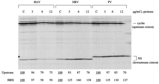
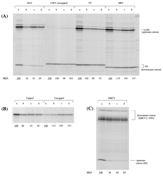
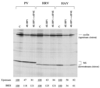
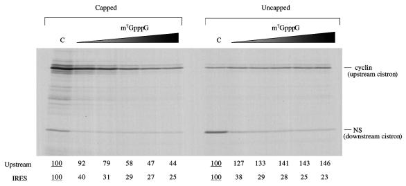
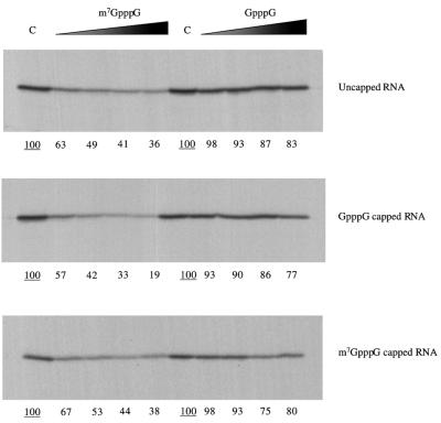
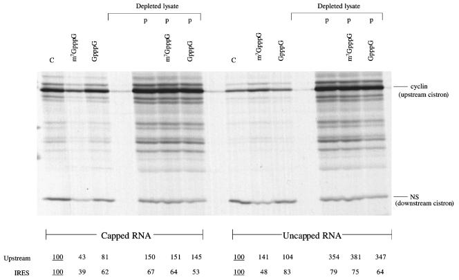
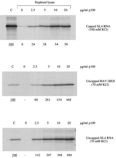
The question of whether translation initiation factor eIF4E and the complete eIF4G polypeptide are required for initiation dependent on the IRES (internal ribosome entry site) of hepatitis A virus (HAV) has been examined using in vitro translation in standard and eIF4G-depleted rabbit reticulocyte lysates. In agreement with previous publications, the HAV IRES is unique among all picornavirus IRESs in that it was inhibited if translation initiation factor eIF4G was cleaved by foot-and-mouth disease L-proteases. In addition, the HAV IRES was inhibited by addition of eIF4E-binding protein 1, which binds tightly to eIF4E and sequesters it, thus preventing its association with eIF4G. The HAV IRES was also inhibited by addition of m(7)GpppG cap analogue, irrespective of whether the RNA tested was capped or not. Thus, initiation on the HAV IRES requires that eIF4E be associated with eIF4G and that the cap-binding pocket of eIF4E be empty and unoccupied. This suggests two alternative models: (i) initiation requires a direct interaction between an internal site in the IRES and eIF4E/4G, an interaction which involves the cap-binding pocket of eIF4E in addition to any direct eIF4G-RNA interactions; or (ii) it requires eIF4G in a particular conformation which can be attained only if eIF4E is bound to it, with the cap-binding pocket of the eIF4E unoccupied.


Figures

FIG. 1
FIG. 1
Recombinant FMDV L-protease inhibits HAV IRES activity. Capped dicistronic mRNAs, each with the designated IRES, were translated at 25 μg/ml in the mixed reticulocyte lysate–HeLa cell HS-S100 system (see Materials and Methods), which had been preincubated for 10 min at 30°C with the indicated concentration of recombinant FMDV L-protease or with buffer (lanes C [control]). The concentration of added KCl in the assays was 70 mM. Translation was for 60 min, and the translation products were analyzed by SDS-PAGE followed by autoradiography. The positions of the upstream (cyclin) cistron product and downstream, IRES-dependent, NS-related product are shown. The NS-related product translated from the PV IRES is larger because this is full-length NS1 protein, rather than the slightly truncated form linked to the HAV and HRV IRESs, and because the NS coding sequences are joined to the authentic PV initiation codon via a short linker. The yields of radiolabeled translation products of the upstream and IRES-driven cistrons were determined by scanning densitometry and are expressed relative to the yield in the corresponding control assay, which was set at 100 (the underlined value).
FIG. 2
FIG. 2
The activity of the HAV IRES is inhibited by 4E-BP1 and by FMDV L-protease expressed in vitro. All RNAs were translated at 25 μg/ml in the mixed reticulocyte lysate–HeLa cell HS-S100 system (see Materials and Methods), which had been preincubated at 30°C as follows: lanes a, 15-min preincubation with buffer (control); lanes b, 10-min preincubation with 4E-BP1 (10 μg/ml); lanes c, 5-min preincubation with FMDV L-protease expressed in vitro; lanes d, 5-min preincubation with FMDV L-protease followed by 10-min preincubation with 4E-BP1 (10 μg/ml). The concentration of added KCl in all of the assays was 70 mM. (A) All dicistronic mRNAs have an upstream cistron coding for X. laevis cyclin B2 and a downstream cistron coding for an influenza virus NS1 derivative, and all except for the RNA with the CSFV IRES were capped. (B) Control assays carried out with monocistronic mRNAs coding for unr in both capped and uncapped forms. (C) The template was a capped dicistronic mRNA with a slightly truncated form of the influenza virus NS1 coding sequence as the upstream cistron, an EMCV IRES, and EMCV sequences coding for viral L-VP0 as the downstream IRES-dependent cistron. In all cases, translation was for 60 min and the translation products were analyzed by SDS-PAGE followed by autoradiography. The positions of the upstream cistron product and downstream, IRES-dependent product are shown. The different sizes of the IRES-dependent NS-related translation product are discussed in Results. The yields of radiolabeled translation products of the IRES-driven cistrons (A and C) and of the single product in the case of the monocistronic RNAs (B) were determined by scanning densitometry and are expressed relative to the yield in the corresponding control assay, which was set at 100 (the underlined value).
FIG. 3
FIG. 3
The inhibition of HAV IRES activity by 4E-BP1 can be reversed by addition of eIF4E. Mixed reticulocyte lysate–HeLa cell HS-S100 (see Materials and Methods) was preincubated for 10 min at 30°C with 4E-BP1 (10 μg/ml) or with buffer control (C); then KCl was added to 70 mM together with the other components of the translation assay, including capped dicistronic mRNAs, each with the designated IRES, at 25 μg/ml. Where indicated, eIF4E (purified from pig brain) was added at 25 μg/ml. Translation was for 60 min, and the translation products were analyzed by SDS-PAGE followed by autoradiography. The positions of the upstream (cyclin) cistron product and downstream, IRES-dependent, NS-related product are shown. The NS-related product translated from the PV IRES is larger because this is full-length NS1 protein, rather than the slightly truncated form linked to the HAV and HRV IRESs, and because the NS coding sequences are joined to the authentic PV initiation codon via a short linker. The yields of radiolabeled translation products of the upstream and IRES-driven cistrons were determined by scanning densitometry and are expressed relative to the yield in the corresponding control assay, which was set at 100 (the underlined value).
FIG. 4
FIG. 4
The activity of the HAV IRES is inhibited by m7GpppG cap analogue. Capped or uncapped dicistronic mRNAs with the HAV IRES were translated at 25 μg/ml in reticulocyte lysate, in the presence of added KCl at 70 mM. Cap analogue (m7GpppG) was added at 0 (lane C), 0.025, 0.05, 0.1, 0.2, and 0.4 mM, and additional MgCl2 was also added at 0.8 mol/mol of cap analogue. Translation was at 30°C for 60 min, and the translation products were analyzed by SDS-PAGE followed by autoradiography. The positions of the upstream (cyclin) cistron product and downstream, IRES-dependent, NS-related product are shown. The yields of radiolabeled translation products of the upstream and IRES-driven cistrons were determined by scanning densitometry and are expressed relative to the yield in the corresponding control assay, which was set at 100 (the underlined value).
FIG. 5
FIG. 5
The inhibitory effect of m7GpppG cap analogue on HAV IRES activity is independent of the nature of the 5′ end. Monocistronic mRNAs synthesized either as uncapped RNAs or with GpppG or m7GpppG capped 5′ ends, as indicated, were translated at 20 μg/ml in rabbit reticulocyte lysate, in the presence of added KCl at 70 mM. Cap analogues, either m7GpppG or GpppG, as indicated, were added at 0 (lane C), 0.025, 0.05, 0.1 or 0.2 mM, and additional MgCl2 was also added at 0.8 mol/mol of cap analogue. Translation was at 30°C for 60, min and the translation products were analyzed by SDS-PAGE followed by autoradiography. The yields of radiolabeled translation products were determined by scanning densitometry and are expressed relative to the yield in the corresponding control assay, which was set at 100 (the underlined value).
FIG. 6
FIG. 6
Translation driven by the HAV IRES can be supported by the p100 fragment (C-terminal two-thirds) of eIF4G. Capped and uncapped dicistronic mRNAs, as indicated, were translated at 25 μg/ml in either eIF4G-depleted reticulocyte lysate or parent (nondepleted lysate), in the presence of added KCl at 70 mM. Cap analogues, either m7GpppG or GpppG, were added at 0.4 mM, where indicated, together with 0.32 mM additional MgCl2. In lanes labeled “p,”, recombinant p100 was added at 20 μg/ml. Translation was at 30°C for 60 min, and the translation products were analyzed by SDS-PAGE followed by autoradiography. The positions of the upstream (cyclin) cistron product and downstream, IRES-dependent, NS-related product are shown. The yields of radiolabeled translation products of the upstream and IRES-driven cistrons were determined by scanning densitometry and are expressed relative to the yield in the corresponding control assay, which was set at 100 (the underlined value).
FIG. 7
FIG. 7
Dose response of p100 rescue of HAV IRES activity. Monocistronic XL4 mRNA (coding for X. laevis cyclin A), in either capped or uncapped form, and uncapped monocistronic HAV-NS RNA were translated at 20 μg/ml either in the parent (nondepleted) lysate (lanes C) or in eIF4G-depleted lysate supplemented with the designated concentrations of recombinant p100. Translation was at 30°C for 60 min, and the concentration of added KCl was either 70 mM (both uncapped RNAs) or 100 mM (capped XL4 mRNA), as indicated. The translation products were analyzed by SDS-PAGE followed by autoradiography. The yields of radiolabeled translation products were determined by scanning densitometry and are expressed relative to the yield in the corresponding control assay, which was set at 100 (the underlined value).

References

    1. Ali, I. K., L. McKendrick, S. J. Morley, and R. J. Jackson. Truncated initiation factor eIF4G lacking an eIF4E binding site can support capped mRNA translation. EMBO J., in press. - PMC - PubMed
    1. Belsham G J, Jackson R J. Translation initiation on picornavirus RNA. In: Sonenberg N, Hershey J W B, Mathews M B, editors. Translational control of gene expression. Cold Spring Harbor, N.Y: Cold Spring Harbor Laboratory Press; 2000. pp. 869–900.
    1. Borman A, Jackson R J. Initiation of translation of human rhinovirus RNA: mapping the internal ribosome entry site. Virology. 1992;188:685–696. - PubMed
    1. Borman A M, Kean K M. Intact eukaryotic initiation factor 4G is required for hepatitis A virus internal initiation of translation. Virology. 1997;237:129–136. - PubMed
    1. Borman A M, Bailly J-L, Girard M, Kean K M. Picornavirus internal ribosome entry segments—comparison of translation efficiency and the requirements for optimal internal initiation of translation in vitro. Nucleic Acids Res. 1995;23:3656–3663. - PMC - PubMed

Publication types

MeSH terms