. 2001 Sep;75(17):7893-903.
doi: 10.1128/jvi.75.17.7893-7903.2001.

The envelope glycoprotein of friend spleen focus-forming virus covalently interacts with and constitutively activates a truncated form of the receptor tyrosine kinase Stk

Affiliations

The envelope glycoprotein of friend spleen focus-forming virus covalently interacts with and constitutively activates a truncated form of the receptor tyrosine kinase Stk

K Nishigaki et al. J Virol. 2001 Sep.

Abstract

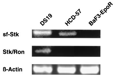
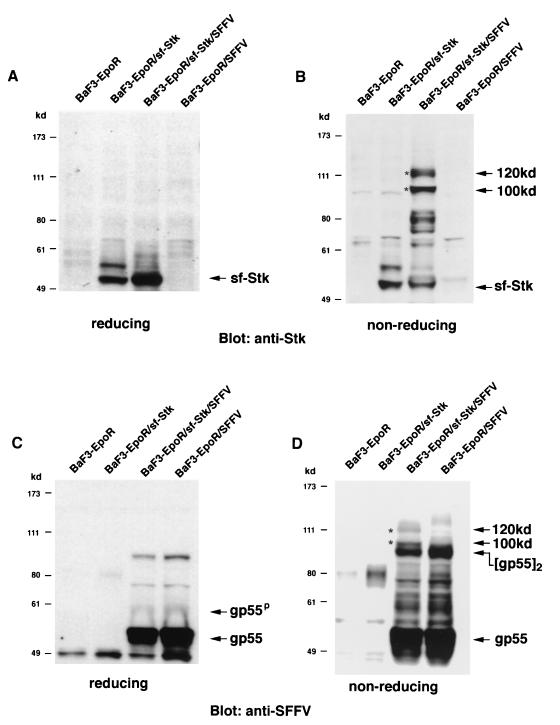
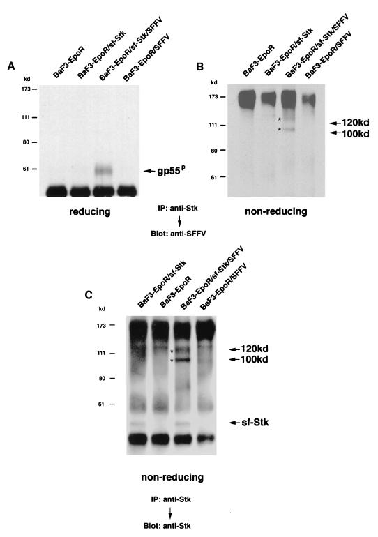
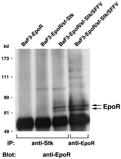
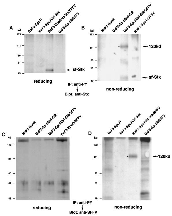
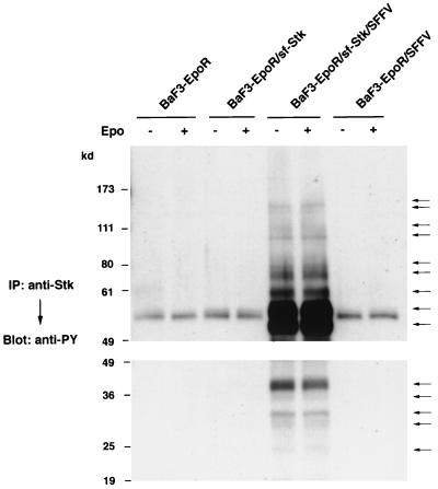
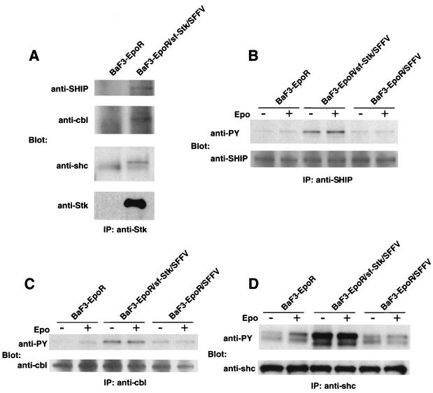
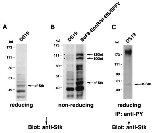
The Friend spleen focus-forming virus (SFFV) encodes a unique envelope glycoprotein, gp55, which allows erythroid cells to proliferate and differentiate in the absence of erythropoietin (Epo). SFFV gp55 has been shown to interact with the Epo receptor complex, causing constitutive activation of various signal-transducing molecules. When injected into adult mice, SFFV induces a rapid erythroleukemia, with susceptibility being determined by the host gene Fv-2, which was recently shown to be identical to the gene encoding the receptor tyrosine kinase Stk/Ron. Susceptible, but not resistant, mice encode not only full-length Stk but also a truncated form of the kinase, sf-Stk, which may mediate the biological effects of SFFV infection. To determine whether expression of SFFV gp55 leads to the activation of sf-Stk, we expressed sf-Stk, with or without SFFV gp55, in hematopoietic cells expressing the Epo receptor. Our data indicate that sf-Stk interacts with SFFV gp55 as well as gp55(P), the biologically active form of the viral glycoprotein, forming disulfide-linked complexes. This covalent interaction, as well as noncovalent interactions with SFFV gp55, results in constitutive tyrosine phosphorylation of sf-Stk and its association with multiple tyrosine-phosphorylated signal-transducing molecules. In contrast, neither Epo stimulation in the absence of SFFV gp55 expression nor expression of a mutant of SFFV that cannot interact with sf-Stk was able to induce tyrosine phosphorylation of sf-Stk or its association with any signal-transducing molecules. Covalent interaction of sf-Stk with SFFV gp55 and constitutive tyrosine phosphorylation of sf-Stk can also be detected in an erythroleukemia cell line derived from an SFFV-infected mouse. Our results suggest that SFFV gp55 may mediate its biological effects in vivo by interacting with and activating a truncated form of the receptor tyrosine kinase Stk.


Figures

FIG. 1
FIG. 1
Expression of full-length and truncated Stk in EpoR-expressing cell lines. Total RNA was obtained from DS19, an erythroleukemia cell line derived from an SFFV-infected mouse; HCD-57, an Epo-dependent mouse erythroleukemia cell line; and BaF3-EpoR, an IL-3-dependent pro-B-cell line engineered to express the EpoR. RT-PCR using specific primer pairs was then carried out to examine expression of full-length Stk (Stk/Ron) or truncated Stk (sf-Stk).
FIG. 2
FIG. 2
Analysis of BaF3-EpoR cells engineered to express sf-Stk and SFFV gp55. Cell lysates from BaF3-EpoR cells, BaF3-EpoR cells expressing sf-Stk, BaF3-EpoR cells coexpressing sf-Stk and SFFV gp55, and BaF3-EpoR cells expressing SFFV gp55 were separated by electrophoresis under reducing (A and C) or nonreducing (B and D) conditions. After transfer to nitrocellulose filters, the proteins were immunoblotted with an antiserum against the C-terminal region of Stk (A and B) or against 7C10, a monoclonal antibody specific for SFFV gp55 (C and D). Asterisks indicate 100- and 120-kDa complexes.
FIG. 3
FIG. 3
Interaction between SFFV gp55 and sf-Stk. Cell lysates from BaF3-EpoR cells, BaF3-EpoR cells expressing sf-Stk, BaF3-EpoR cells coexpressing sf-Stk and SFFV gp55, and BaF3-EpoR cells expressing SFFV gp55 were immunoprecipitated (IP) with an antiserum against the C-terminal region of Stk and then separated by electrophoresis under reducing (A) or nonreducing (B and C) conditions. After transfer to nitrocellulose filters, the proteins were immunoblotted with an antiserum against 7C10, a monoclonal antibody specific for SFFV gp55 (A and B), or with an anti-Stk antiserum (C). Asterisks indicate 100- and 120-kDa complexes.
FIG. 4
FIG. 4
Association of sf-Stk with the EpoR. Cell lysates from BaF3-EpoR cells, BaF3-EpoR cells expressing sf-Stk, and BaF3-EpoR cells coexpressing sf-Stk and SFFV gp55 were immunoprecipitated (IP) with an antiserum against the C-terminal region of Stk or an antiserum against the EpoR (lane 4) and then separated by electrophoresis under reducing conditions. After transfer to nitrocellulose filters, the proteins were immunoblotted with an antiserum against the EpoR.
FIG. 5
FIG. 5
Tyrosine phosphorylation of sf-Stk. Cell lysates from BaF3-EpoR cells, BaF3-EpoR cells expressing sf-Stk, BaF3-EpoR cells coexpressing sf-Stk and SFFV gp55, and BaF3-EpoR cells expressing SFFV gp55 were immunoprecipitated (IP) with the anti-phosphotyrosine antibody 4G10 (anti-PY) and then separated by electrophoresis under reducing (A and C) or nonreducing (B and D) conditions. After transfer to nitrocellulose filters, the proteins were immunoblotted with an antiserum against the C-terminal region of Stk (A and B) or 7C10, a monoclonal antibody specific for SFFV gp55 (C and D). Asterisks indicate the 120-kDa tyrosine-phosphorylated complex.
FIG. 6
FIG. 6
Association of tyrosine-phosphorylated sf-Stk with other tyrosine-phosphorylated proteins. BaF3-EpoR cells, BaF3-EpoR cells expressing sf-Stk, BaF3-EpoR cells coexpressing sf-Stk and SFFV gp55, and BaF3-EpoR cells expressing SFFV gp55 were left unstimulated or stimulated with Epo for 15 min. Cell lysates were then immunoprecipitated (IP) with an antiserum against the C-terminal region of Stk and separated by electrophoresis under reducing conditions on 8% (upper panel) and 10% (lower panel) polyacrylamide gels. After transfer to nitrocellulose filters, the proteins were immunoblotted with the anti-phosphotyrosine antiserum 4G10 (anti-PY). Tyrosine-phosphorylated proteins immunoprecipitated with the anti-Stk antiserum are indicated by arrows.
FIG. 7
FIG. 7
Association of phosphorylated sf-Stk with known signal-transducing molecules and their tyrosine phosphorylation. (A) Lysates from BaF3-EpoR cells and BaF3-EpoR cells coexpressing sf-Stk and SFFV gp55 were immunoprecipitated (IP) with an antiserum to the C-terminal region of Stk and separated by electrophoresis under reducing conditions. After transfer to nitrocellulose filters, the proteins were immunoblotted with antisera to SHIP, Cbl, Shc, or Stk. (B to D) BaF3-EpoR cells, BaF3-EpoR cells coexpressing sf-Stk and SFFV gp55, and BaF3-EpoR cells expressing SFFV gp55 were left unstimulated or stimulated with Epo for 15 min. Lysates were immunoprecipitated (IP) with an antiserum against SHIP (B), Cbl (C), or Shc (D) and then separated by electrophoresis under reducing conditions. After transfer to nitrocellulose filters, the proteins were immunoblotted with the anti-phosphotyrosine antiserum 4G10 (anti-PY) (B to D) or an antiserum to SHIP (B), Cbl (C), or Shc (D).
FIG. 8
FIG. 8
The mutant SFFV BB6 does not induce the tyrosine phosphorylation of sf-Stk. Cell lystates from BaF3-EpoR cells expressing sf-Stk, BaF3-EpoR cells expressing sf-Stk and then infected with the BB6 virus, and BaF3-EpoR cells infected with the BB6 virus were immunoprecipitated (IP) with an antiserum to the C-terminal region of Stk and separated by electrophoresis under reducing conditions. After transfer to nitrocellulose filters, the proteins were immunoblotted with the anti-phosphotyrosine antiserum 4G10 (anti-PY) or the anti-Stk antiserum.
FIG. 9
FIG. 9
Association of sf-Stk and SFFV gp55 in an SFFV-induced erythroleukemia cell line. (A and B) A cell lysate prepared from DS19 cells, an erythroleukemia cell line derived from an SFFV-infected mouse, was separated by electrophoresis under reducing (A) or nonreducing (B) conditions. After transfer to nitrocellulose filters, the proteins were immunoblotted with an antiserum against the C-terminal region of Stk. BaF3-EpoR cells coexpressing sf-Stk and SFFV gp55 are shown in panel B for comparison. Asterisks indicate 100- and 120-kDa complexes. (C) A lysate from DS19 cells was immunoprecipitated (IP) with the anti-phosphotyrosine antiserum 4G10 (anti-PY) and then separated by electrophoresis under reducing conditions. After transfer to nitrocellulose filters, the proteins were immunoblotted with an antiserum against the C-terminal region of Stk in order to detect the phosphorylated form of sf-Stk.

References

    1. Amanuma H, Katori A, Obata M, Sagata N, Ikawa Y. Complete nucleotide sequence of the gene for the specific glycoprotein (gp55) of Friend spleen focus-forming virus. Proc Natl Acad Sci USA. 1983;80:3913–3917. - PMC - PubMed
    1. Behringer R R, Dewey M J. Cellular site and mode of Fv-2 gene action. Cell. 1985;40:441–447. - PubMed
    1. Bondurant M C, Koury M J, Krantz S B. The Fv-2 gene controls induction of erythroid burst formation by Friend virus infection in vitro: studies of growth regulators and viral replication. J Gen Virol. 1985;66:83–93. - PubMed
    1. Casadevall N, Lacombe C, Muller O, Gisselbrecht S, Mayeux P. Multimeric structure of the membrane erythropoietin receptor of murine erythroleukemia cells (Friend cells) J Biol Chem. 1991;266:16015–16020. - PubMed
    1. Chung S, Wolff L, Ruscetti S K. Transmembrane domain of the envelope gene of a polycythemia-inducing retrovirus determines erythropoietin-independent growth. Proc Natl Acad Sci USA. 1989;86:7957–7960. - PMC - PubMed

MeSH terms

Substances

LinkOut - more resources