. 2001 Sep;75(17):7904-12.
doi: 10.1128/jvi.75.17.7904-7912.2001.

Posttranslational processing of infected cell proteins 0 and 4 of herpes simplex virus 1 is sequential and reflects the subcellular compartment in which the proteins localize

Affiliations

Posttranslational processing of infected cell proteins 0 and 4 of herpes simplex virus 1 is sequential and reflects the subcellular compartment in which the proteins localize

S J Advani et al. J Virol. 2001 Sep.

Abstract

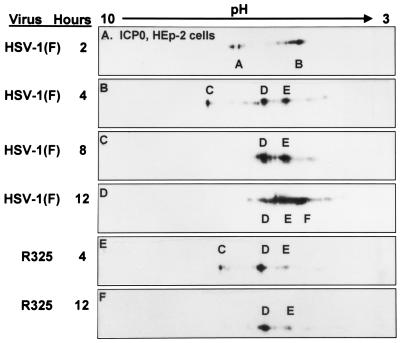
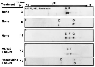
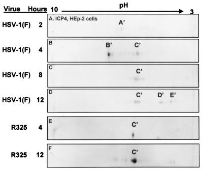
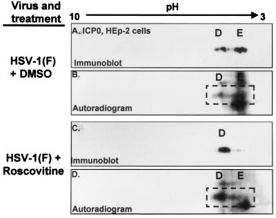
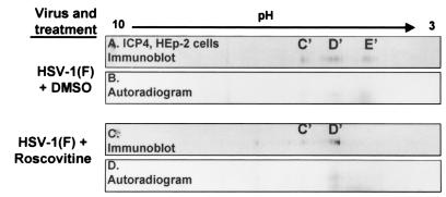
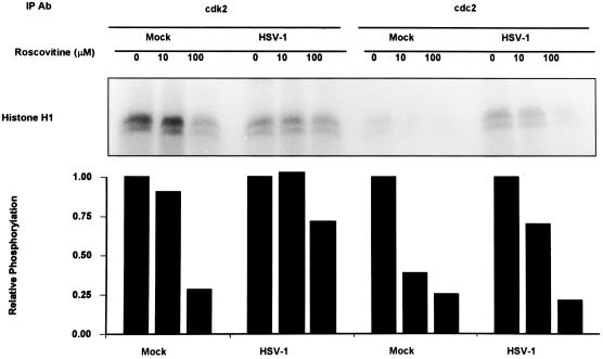
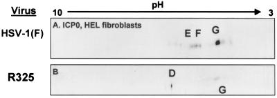
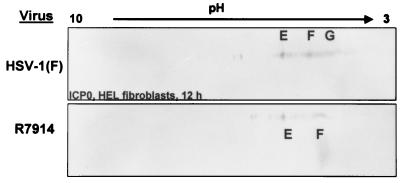
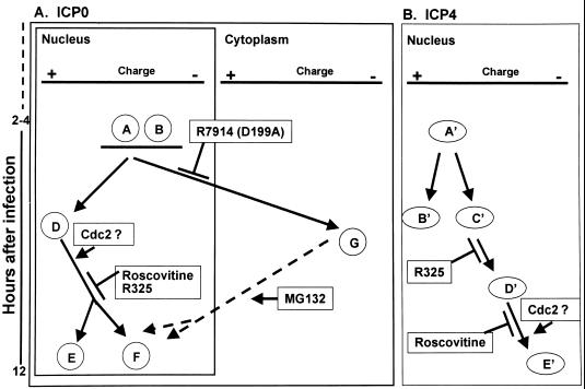
The herpes simplex virus 1 (HSV-1) infected cell proteins 0 and 4 (ICP0 and ICP4) are multifunctional proteins extensively posttranscriptionally processed by both cellular and viral enzymes. We examined by two-dimensional separations the posttranslational forms of ICP0 and ICP4 in HEp-2 cells and in human embryonic lung (HEL) fibroblasts infected with wild-type virus, mutant R325, lacking the sequences encoding the U(S)1.5 protein and the overlapping carboxyl-terminal domain of ICP22, or R7914, in which the aspartic acid 199 of ICP0 was replaced by alanine. We report the following (i) Both ICP0 and ICP4 were sequentially posttranslationally modified at least until 12 h after infection. In HEL fibroblasts, the processing of ICP0 shifted from A+B forms at 4 h to D+G forms at 8 h and finally to G, E, and F forms at 12 h. The ICP4 progression was from the A' form noted at 2 h to B' and C' forms noted at 4 h to the additional D' and E' forms noted at 12 h. The progression tended to be toward more highly charged forms of the proteins. (ii) Although the overall patterns were similar, the mobility of proteins made in HEp-2 cells differed from those made in HEL fibroblasts. (iii) The processing of ICP0 forms E and F was blocked in HEL fibroblasts infected with R325 or with wild-type virus and treated with roscovitine, a specific inhibitor of cell cycle-dependent kinases cdc2, cdk2, and cdk5. R325-infected HEp-2 cells lacked the D' form of ICP4, and roscovitine blocked the appearance of the most highly charged E' form of ICP4. (iv) A characteristic of ICP0 is that it is translocated into the cytoplasm of HEL fibroblasts between 5 and 9 h after infection. Addition of MG132 to the cultures late in infection resulted in rapid relocation of cytoplasmic ICP0 back into the nucleus. Exposure of HEL fibroblasts to MG132 late in infection resulted in the disappearance of the highly charged ICP0 G isoform. The G form of ICP0 was also absent in cells infected with R7914 mutant. In cells infected with this mutant, ICP0 is not translocated to the cytoplasm. (v) Last, cdc2 was active in infected cells, and this activity was inhibited by roscovitine. In contrast, the activity of cdk2 exhibited by immunoprecipitated protein was reduced and resistant to roscovitine and may represent a contaminating kinase activity. We conclude from these results that the ICP0 G isoform is the cytoplasmic form, that it may be phosphorylated by cdc2, consistent with evidence published earlier (S. J., Advani, R. R. Weichselbaum, and B. Roizman, Proc. Natl. Acad. Sci. USA 96:10996-11001, 2000), and that the processing is reversed upon relocation of the G isoform from the cytoplasm into the nucleus. The processing of ICP4 is also affected by R325 and roscovitine. The latter result suggests that ICP4 may also be a substrate of cdc2 late in infection. Last, additional modifications are superimposed by cell-type-specific enzymes.


Figures

FIG. 1
FIG. 1
Immunoblots of ICP0 in two-dimensionally separated HSV-1- or R325-infected HEp-2 cell lysates. Infected cells were harvested at the indicated times postinfection. Time zero was defined as the time the viral inoculum was added to the cells. Lysates were first separated by charge on linear pH 3 to 10 gradients and then separated by size on 6% bisacrylamide gels as described in Materials and Methods. Blots were reacted with a monoclonal antibody to ICP0. The major isoforms that accumulated are given letter designations.
FIG. 2
FIG. 2
Immunoblots of ICP0 in two-dimensionally separated HSV-1-infected HEL fibroblast cell lysates. HSV-1-infected cells were harvested at 4, 8, and 12 h postinfection (P.I.). In addition cells were also treated with MG132 or roscovitine. MG132 was given to HSV-1-infected cells 8 h after infection and harvested at 12 h. Roscovitine was added to HSV-1-infected cells at 5 h after infection, and cells were harvested at 12 h postinfection. Immunoblots were reacted with ICPO monoclonal antibody.
FIG. 3
FIG. 3
Immunoblots of ICP4 in two-dimensionally separated HSV-1-+ and R325-infected HEp-2 cell lysates. Immunoblots were prepared and reacted with the anti-ICP4 monoclonal antibody as described in the legend to Fig. 1.
FIG. 4
FIG. 4
Immunoblots (A and C) and corresponding autoradiograms (B and D) of ICPO in two-dimensionally separated 32P-orthophosphate-labeled HSV-1-infected HEp-2 cell lysates to determine roscovitine-sensitive ICP0 phosphorylation. HSV-1-infected HEp-2 cells were treated with roscovitine or an equivalent volume of DMSO at 5 h after infection in phosphate-free medium. Infected cells were labeled with 32P-orthophosphate from 6 to 10 h postinfection in the presence of roscovitine. Cells were harvested at 10 h postinfection, and whole-cell lysates were subjected to two-dimensional electrophoresis. Membranes were developed by autoradiography and immunoblotted with an ICP0 antibody. Isoform designations are identical to those shown in Fig. 1.
FIG. 5
FIG. 5
Immunoblots (A and C) and corresponding autoradiograms (B and D) of ICP4 in two-dimensionally separated 32P-orthophosphate-labeled HSV-1-infected HEp-2 cell lysates to determine roscovitine-sensitive ICP4 phosphorylation. HSV-1-infected HEp-2 cells were treated with roscovitine or an equivalent volume of DMSO at 5 h after infection in phosphate-free medium. Infected cells were labeled with 32P-orthophosphate from 6 to 10 h after infection in the presence of roscovitine. Cells were harvested at 10 h postinfection, and whole-cell lysates were subjected to two-dimensional electrophoresis. Membranes were developed by autoradiography and immunoblotted with an ICP4 antibody. Isoform designations are idential to those shown in Fig. 3.
FIG. 6
FIG. 6
Autoradiograms of histone H1 phosphorylated by in vitro kinase assays using immunoprecipitated cdk2 or cdc2 from HSV-1- or mock-infected cells. HEp-2 cells were HSV-1 or mock infected; 5 h after infection, the medium was replaced with medium containing 0, 10, or 100 μM roscovitine. Cells were harvested 12 h after infection, and cdk2 or cdc2 was immunoprecipitated (IP) with antibodie (Ab) as described in Materials and Methods. Kinase assays were done using histone H1 as the substrate. Phosphorylated histone H1 was quantitated by a PhosphorImager. Relative reduction in the phosphorylation of histone H1 due to roscovitine was determined relative to untreated samples.
FIG. 7
FIG. 7
Immunoblots of ICP0 in two-dimensionally separated lysates of HSV-1- or R325-infected HEL fibroblasts. Cells were harvested at 12 h after infection and separated by two-dimensional electrophoresis. Immunoblots were reacted with ICP0 monoclonal antibody. Isoform designations are the same as in Fig. 2.
FIG. 8
FIG. 8
Immunoblots of ICP0 in two-dimensionally separated lysates of HSV-1 or R7914 (alanine substituted for aspartic acid of ICP0)-infected HEL fibroblasts. The cells were harvested at 12 h after infection, lysed, and subjected to two-dimensional electrophoresis. Immunoblots were reacted with anti ICP0 antibody. Isoform designations are the same as in Fig. 2.
FIG. 9
FIG. 9
Schematic representation of ICP0 and ICP4 isoforms observed after two-dimensional separation of lysates of HSV-1-infected HEL fibroblasts. (A) Absence of specific isoforms from lysates of HEL fibroblasts infected with HSV-1 mutants or with wild-type virus and treated with roscovitine. Isoforms E and F failed to accumulate in lysates of R325-infected or roscovitine-treated, HSV-1-infected cells. These isoforms are putative nuclear forms, and their processing is dependent on cdc2 kinase. HEL fibroblasts infected with R7914 or wild-type virus and treated with MG132 failed to accumulate the G isoform, and this isoform appears to be associated with ICP0 translocated into the cytoplasm. (B) Isoforms D′ and E′ of ICP4 failed to accumulate in HEL fibroblasts infected with R325 mutant. The most negatively charged, E′ isoform of ICP4 was not detected in significant amounts in cells infected with wild-type virus and treated with roscovitine. The accumulation of the ICP4 isoform E′ may also be dependent on cdc2 kinase.

References

    1. Ackermann M, Braun D K, Pereira L, Roizman B. Characterization of herpes simplex virus 1 alpha proteins 0, 4, and 27 with monoclonal antibodies. J Virol. 1984;52:108–118. - PMC - PubMed
    1. Advani S J, Brandimarti R, Weichselbaum R R, Roizman B. The disappearance of cyclins A and B and the increase in activity of the G2/M phase cellular kinase cdc2 in herpes simplex virus 1-infected cells requires the expression of α22/US1.5 and UL13 viral genes. J Virol. 2000;74:8–15. - PMC - PubMed
    1. Advani S J, Weichselbaum R R, Roizman B. The role of cdc2 in the expression of herpes simplex virus genes. Proc Natl Acad Sci USA. 2000;97:10996–11001. - PMC - PubMed
    1. Blaho J A, Roizman B. ICP4, the major regulatory protein of herpes simplex virus, shares features common to GTP-binding proteins and is adenylated and guanylated. J Virol. 1991;65:3759–3769. - PMC - PubMed
    1. Blaho J A, Mitchell C, Roizman B. Guanylylation and adenylylation of the alpha regulatory proteins of herpes simplex virus require a viral beta or gamma function. J Virol. 1993;67:3891–3900. - PMC - PubMed

Publication types

MeSH terms

LinkOut - more resources