. 2001 Sep;75(17):7995-8007.
doi: 10.1128/jvi.75.17.7995-8007.2001.

Specific interaction of a novel foamy virus Env leader protein with the N-terminal Gag domain

Affiliations

Specific interaction of a novel foamy virus Env leader protein with the N-terminal Gag domain

T Wilk et al. J Virol. 2001 Sep.

Abstract

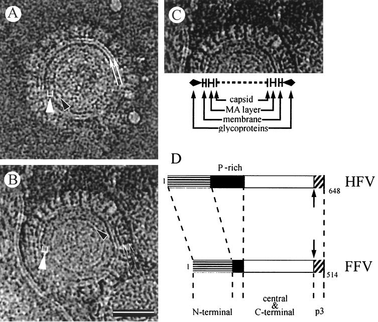
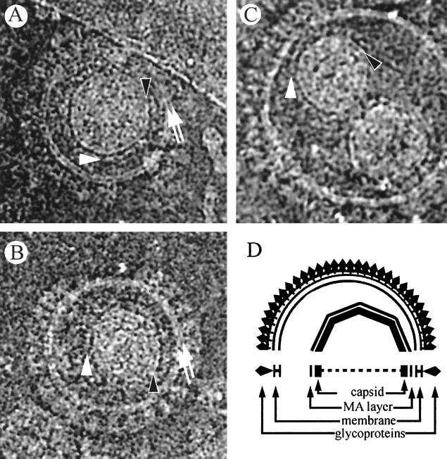
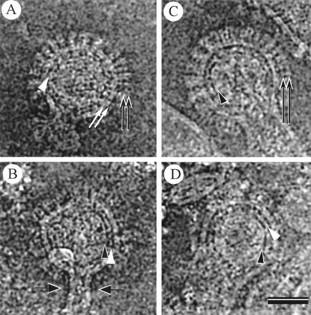
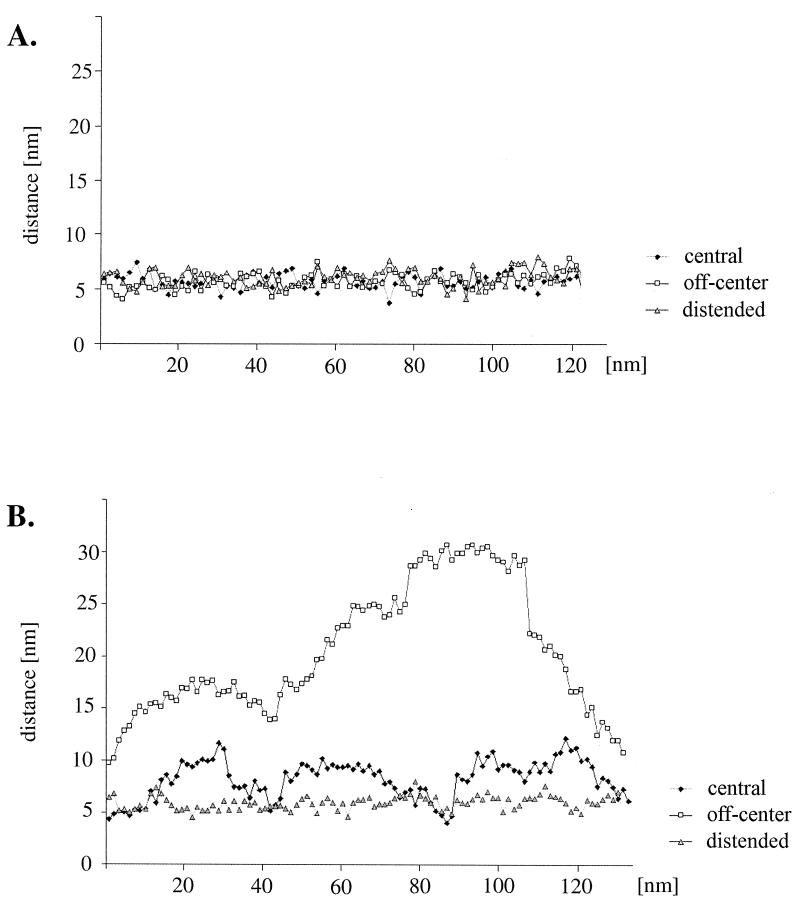
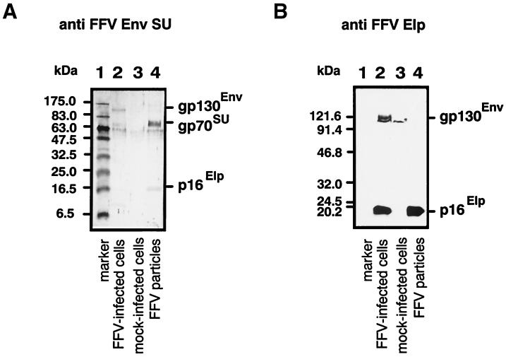
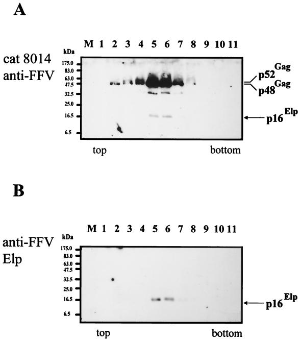
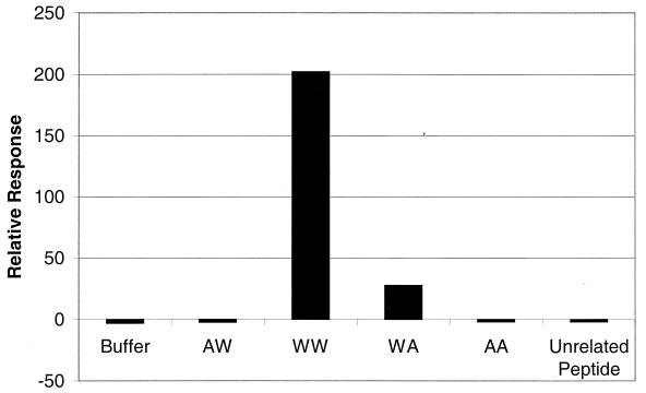
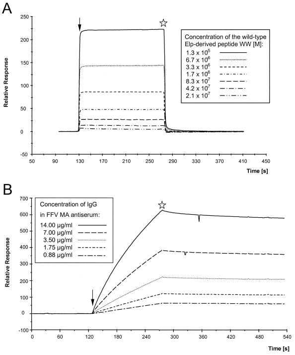
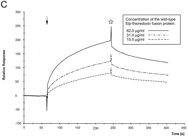
Cryoelectron micrographs of purified human foamy virus (HFV) and feline foamy virus (FFV) particles revealed distinct radial arrangements of Gag proteins. The capsids were surrounded by an internal Gag layer that in turn was surrounded by, and separated from, the viral membrane. The width of this layer was about 8 nm for HFV and 3.8 nm for FFV. This difference in width is assumed to reflect the different sizes of the HFV and FFV MA domains: the HFV MA domain is about 130 residues longer than that of FFV. The distances between the MA layer and the edge of the capsid were identical in different particle classes. In contrast, only particles with a distended envelope displayed an invariant, close spacing between the MA layer and the Env membrane which was absent in the majority of particles. This indicates a specific interaction between MA and Env at an unknown step of morphogenesis. This observation was supported by surface plasmon resonance studies. The purified N-terminal domain of FFV Gag specifically interacted with synthetic peptides and a defined protein domain derived from the N-terminal Env leader protein. The specificity of this interaction was demonstrated by using peptides varying in the conserved Trp residues that are known to be required for HFV budding. The interaction with Gag required residues within the novel virion-associated FFV Env leader protein of about 16.5 kDa.


Figures

FIG. 1
FIG. 1
Ultrastructure of released HFV particles (A to C) and schematic alignment of HFV and FFV Gag proteins (D). cEM of HFV reveals closely packed envelope proteins with distinct layers of density within the glycoproteins (pair of black arrows in B) covering the surface of the budded virion. A separation between the viral membrane (pair of white arrows) and the broad MA layer (white bracket and white arrowhead) is clearly visible and characteristic for budded FV particles. The MA layer width of about 8 nm is characteristic for HFV. The prominent margin of the angular HFV capsid is marked with a black arrowhead. The capsid in panel A has a central position in contrast to the off-center position of the capsid shown in panel B. The scale bar in panel B represents 50 nm. (C) Schematic presentation of structural features of HFV particles analyzed by cEM. (D) Schematic alignment of HFV and FFV Gag proteins. The presence of the C-terminal p3 cleavage site (arrow), the p3 protein, and the N-terminal, Pro-rich (P-rich), and central and C-terminal subdomains of FV Gag proteins is shown. For details, see the text.
FIG. 2
FIG. 2
Ultrastructure of released FFV particles (A to C) and schematic diagram of FFV particles (D). The MA layer (white arrowheads) follows the shape of the capsids in particles with central (A) and off-center (B) capsids as schematically shown in panel D. Many particles in the population show the internal angular capsid (the edge of the capsid is marked by black arrowheads) displaced from the center of the particle (B). Views in which the capsid appeared to be almost in the center of the particle were also observed (A). Occasionally, two capsids were surrounded by an almost perfect spherical viral membrane (pair of white arrows), resulting in particles with a greater diameter (B and C).
FIG. 3
FIG. 3
Features of FFV particles with a distended morphology. Particles with a distended morphology (A to D) show tight interaction of the MA layer (white arrowheads) with the viral membrane (pair of white arrows). The two opposed arrows mark the stalk of cellular membrane which is still attached to the particle (B). The pairs of black arrows mark spike proteins, and the black arrowheads point to the margin of the capsid. The scale bar in panel D represents 50 nm.
FIG. 4
FIG. 4
Relative localization of the MA layer in FFV particles. Arrangement of the MA layer in FFV particles with central capsids (diamonds), off-center capsids (squares), and distended morphology (triangles). The plots show the distribution of distances between the MA layer and the edge of the capsid structure (A) and between the MA layer and the inner leaflet of the viral membrane (B). The distances in nanometers are plotted against the position of the measurement on the circumference of the MA layer and were measured at 1-nm intervals. A typical result is shown for each particle type. No significant difference is seen between the mean distances from the capsid to the MA layer of the three particle types (A). In contrast, only the distended FFV particles showed a constant and close juxtaposition between the MA layer and the membrane, with a distance of 6.0 ± 0.8 nm (mean ± standard deviation) (n = 83) (B). The mean distances from the membrane to the MA layer were 8.8 ± 1.4 nm (n = 85) for central capsids and 18.3 ± 11.4 nm (n = 133) for off-center capsids.
FIG. 5
FIG. 5
Detection of FFV Elp in released FFV particles and FFV-infected cells. FFV particles were enriched from the cell culture supernatant from FFV-infected CRFK cells by centrifugation through sucrose as described in Materials and Methods, lysed, separated on a denaturing gel, and analyzed by immunoblotting (lanes 4). In parallel, proteins from FFV-infected (lanes 2) and mock-infected CRFK cells (lanes 3) were analyzed. The blots were reacted with the FFV SU antiserum (A) (56) and the FFV Elp antiserum (B) and specific proteins were detected by diamino-benzidine staining (A) and enhanced chemiluminescence (B). The positions of Elp, Env-SU, and the gp130Env precursor are marked. Two different prestained molecular mass markers were used in lanes 1. The gel in panel A contained 16% polyacrylamide, and that in panel B contained 14% polyacrylamide.
FIG. 6
FIG. 6
Cosedimentation of FFV Elp with FFV particles. FFV particles enriched by centrifugation through 20% sucrose were analyzed on preformed 10 to 32% iodixanol gradients as described in Materials and Methods. Regular aliquots of the gradient fractions (as indicated) were directly analyzed by immunoblotting using the FFV-specific cat antiserum 8014 (A) and the FFV Elp antiserum (B). Proteins were detected by enhanced chemiluminescence. The positions of the p52 and p48 Gag and the FFV Elp proteins are marked. In lanes M, prestained molecular mass markers were separated in parallel.
FIG. 7
FIG. 7
Interaction of FFV Elp-derived peptides with FFV Gag 1–154. SPR analysis of the binding of FFV Elp-derived peptides to the recombinant N-terminal FFV Gag 1–154 domain bound to the sensor surface. Each peptide solution (50 μg/ml) was passed over the FFV Gag sensor surface at a flow rate of 10 μl/min for 3 min. The peptide representing the wt FFV (Elp residues 1 to 30) is designated WW, AW represents the single W12A, WA represents the W15A exchange, and both Trp residues have been replaced by Ala in peptide AA. A human collagen-derived peptide (unrelated peptide) served as an additional control. The signals for binding were automatically recorded after the binding reaction reached equilibrium and are presented as relative response units.
FIG. 8
FIG. 8
(A) Kinetic analyses of the interaction of the authentic Elp-derived peptide with FFV Gag 1–154 coupled to the CM5 sensor. The peptide WW was passed over the sensor chip at a flow rate of 10 μl/min for 2.5 min at concentrations ranging from 2.08 × 10⁻⁷ to 1.33 × 10⁻⁵ M as shown in the inset. The fast associations and dissociations are graphically expressed as relative response over time in minutes. Regeneration of the sensor surface was complete and achieved by changing to HBS buffer. Arrow, start of the binding reaction; asterisk, beginning of the washing with HBS buffer. (B) Kinetic analyses of the interaction of the FFV MA antiserum with FFV Gag 1–154 coupled to the CM5 sensor. Defined dilutions of the FFV MA antisera ranging from 1:500 to 1:8,000 were passed over the sensor chip at a flow rate of 10 μl/min for 2.5 min corresponding to immunoglobulin G concentrations from 14 to 0.88 μg/ml as shown in the inset. The slow association of the polyclonal antiserum at concentrations reaching saturation and the very low dissociation of the bound antibodies are graphically expressed as relative response over time in minutes. (C) Kinetic analyses of the interaction of the FFV Gag 1–154 protein coupled to the CM5 sensor with the purified thioredoxin-Elp 1–65 fusion protein. Defined concentrations of the recombinant thioredoxin-Elp 1–65 fusion protein ranging from 62 to 15.5 μg/ml were passed over the sensor chip at a flow of 15 μl/min for 3 min. The slow and specific association of the thioredoxin-Elp 1–65 and the slow dissociation of the bound Elp protein are graphically expressed as relative response over time in minutes. The thioredoxin protein without the FFV Elp domain did not show any specific binding. Arrow, start of the binding reaction; asterisk, beginning of the washing with HBS buffer.
FIG. 8
FIG. 8
(A) Kinetic analyses of the interaction of the authentic Elp-derived peptide with FFV Gag 1–154 coupled to the CM5 sensor. The peptide WW was passed over the sensor chip at a flow rate of 10 μl/min for 2.5 min at concentrations ranging from 2.08 × 10⁻⁷ to 1.33 × 10⁻⁵ M as shown in the inset. The fast associations and dissociations are graphically expressed as relative response over time in minutes. Regeneration of the sensor surface was complete and achieved by changing to HBS buffer. Arrow, start of the binding reaction; asterisk, beginning of the washing with HBS buffer. (B) Kinetic analyses of the interaction of the FFV MA antiserum with FFV Gag 1–154 coupled to the CM5 sensor. Defined dilutions of the FFV MA antisera ranging from 1:500 to 1:8,000 were passed over the sensor chip at a flow rate of 10 μl/min for 2.5 min corresponding to immunoglobulin G concentrations from 14 to 0.88 μg/ml as shown in the inset. The slow association of the polyclonal antiserum at concentrations reaching saturation and the very low dissociation of the bound antibodies are graphically expressed as relative response over time in minutes. (C) Kinetic analyses of the interaction of the FFV Gag 1–154 protein coupled to the CM5 sensor with the purified thioredoxin-Elp 1–65 fusion protein. Defined concentrations of the recombinant thioredoxin-Elp 1–65 fusion protein ranging from 62 to 15.5 μg/ml were passed over the sensor chip at a flow of 15 μl/min for 3 min. The slow and specific association of the thioredoxin-Elp 1–65 and the slow dissociation of the bound Elp protein are graphically expressed as relative response over time in minutes. The thioredoxin protein without the FFV Elp domain did not show any specific binding. Arrow, start of the binding reaction; asterisk, beginning of the washing with HBS buffer.

Similar articles

Cited by

References

    1. Alke A, Schwantes A, Zemba M, Flügel R M, Löchelt M. Characterization of the humoral immune response and virus replication in cats experimentally infected with feline foamy virus. Virology. 2000;275:170–176. - PubMed
    1. Baker T S, Olson N H, Fuller S D. Adding the third dimension to virus life cycles: three-dimensional reconstruction of icosahedral viruses from cryo-electron micrographs. Microbiol Mol Biol Rev. 1999;63:862–922. - PMC - PubMed
    1. Baldwin D N, Linial M L. Proteolytic activity, the carboxy terminus of Gag, and the primer binding site are not required for Pol incorporation into foamy virus particles. J Virol. 1999;73:63687–63693. - PMC - PubMed
    1. Baldwin D N, Linial M L. The roles of Pol and Env in the assembly pathway of human foamy virus. J Virol. 1998;72:3658–3665. - PMC - PubMed
    1. Bodem J, Zemba M, Flügel R M. Nuclear localization of the functional Bel 1 transactivator but not of the gag proteins of the feline foamy virus. Virology. 1998;251:22–27. - PubMed

Publication types

LinkOut - more resources