. 2001 Aug;3(8):730-9.
doi: 10.1038/35087045.

The role of Drosophila CID in kinetochore formation, cell-cycle progression and heterochromatin interactions

Affiliations

The role of Drosophila CID in kinetochore formation, cell-cycle progression and heterochromatin interactions

M D Blower et al. Nat Cell Biol. 2001 Aug.

Abstract

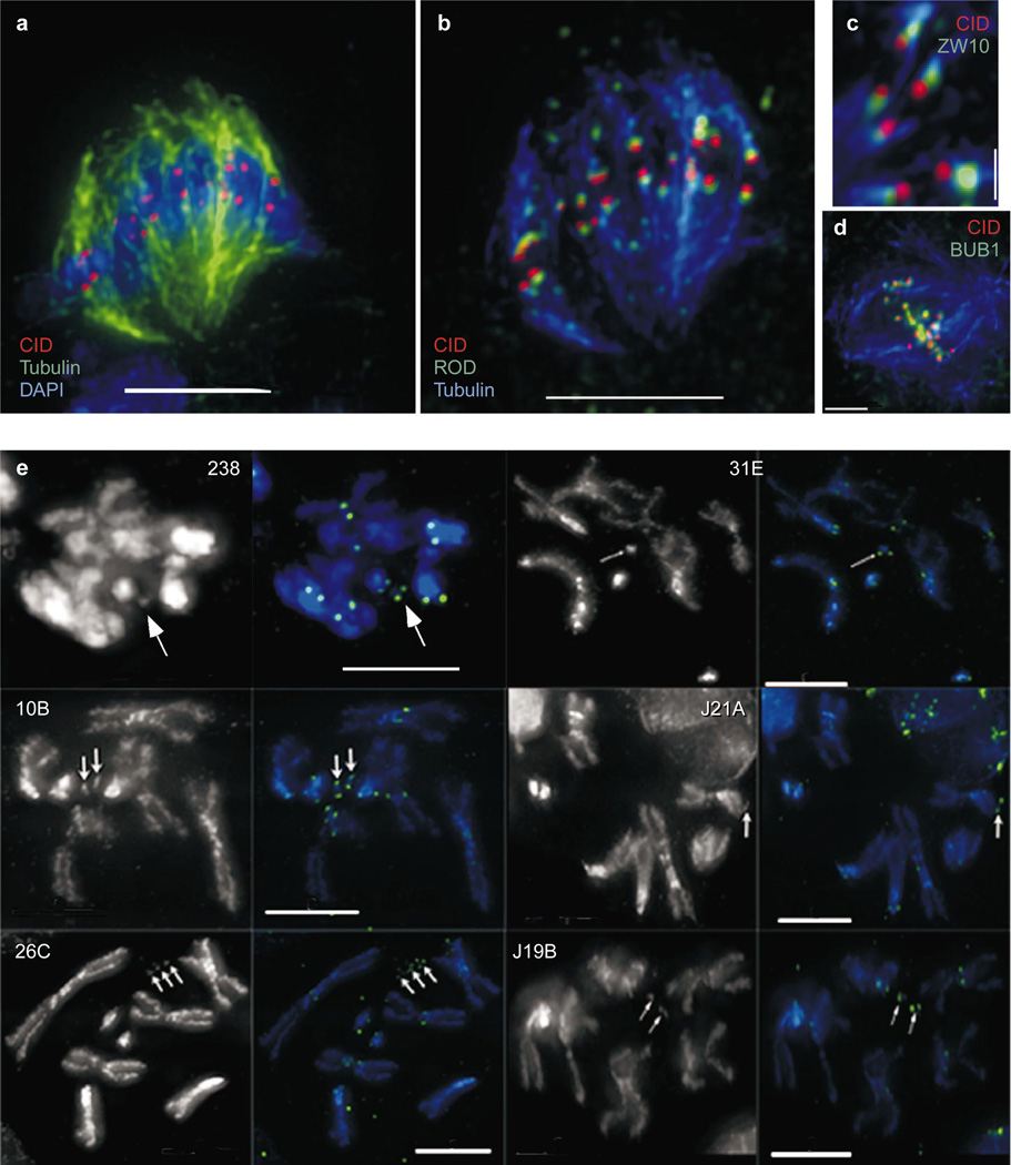
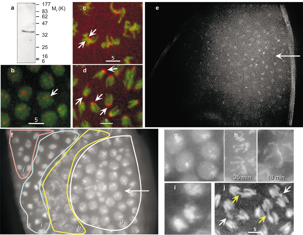
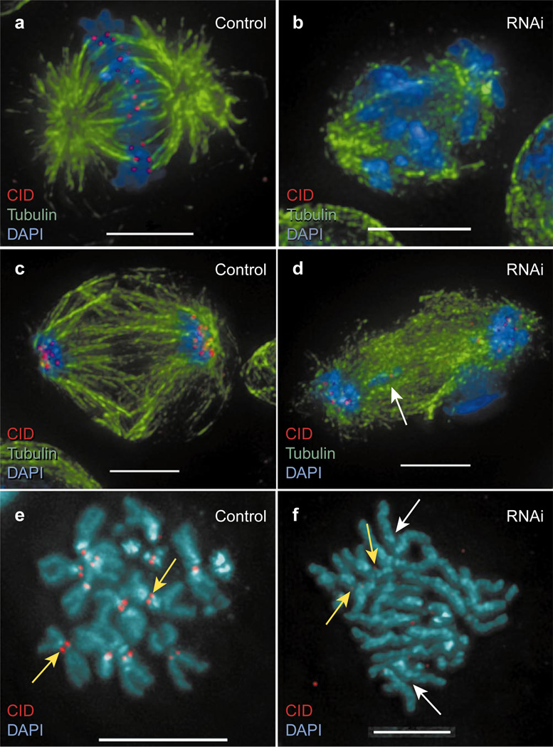
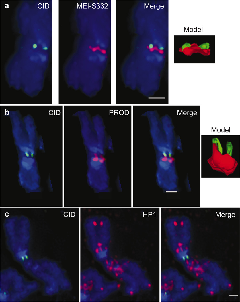
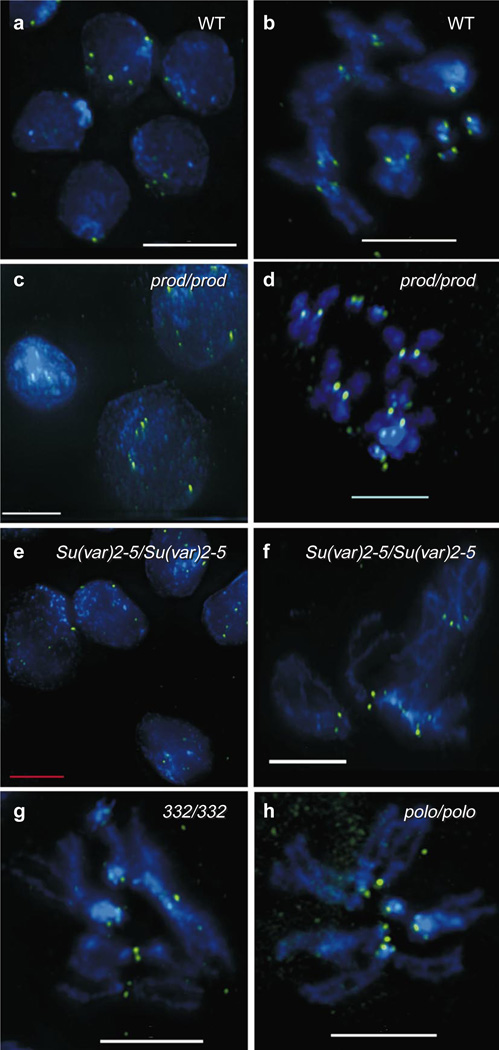
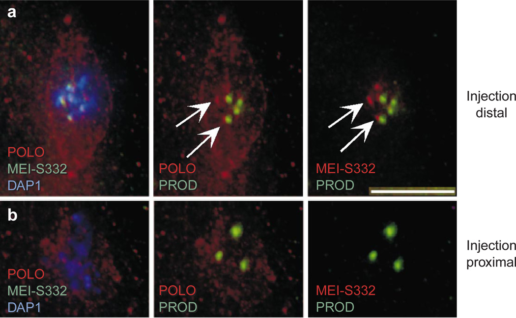
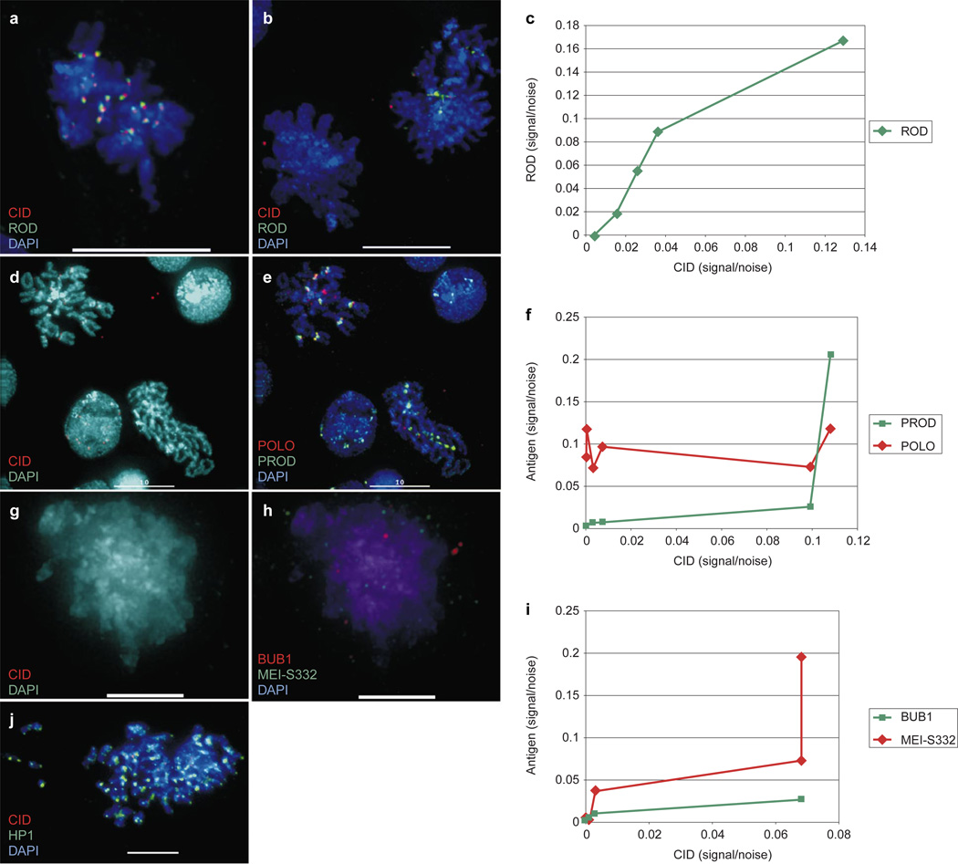
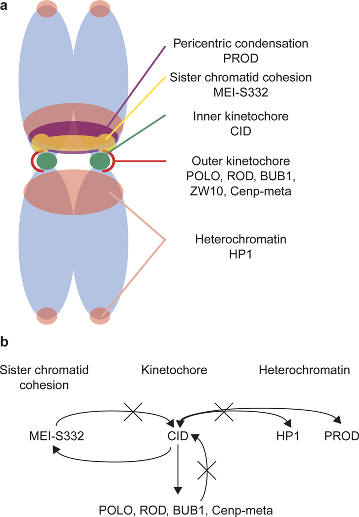
Centromere function requires the coordination of many processes including kinetochore assembly, sister chromatid cohesion, spindle attachment and chromosome movement. Here we show that CID, the Drosophila homologue of the CENP-A centromere-specific H3-like proteins, colocalizes with molecular-genetically defined functional centromeres in minichromosomes. Injection of CID antibodies into early embryos, as well as RNA interference in tissue-culture cells, showed that CID is required for several mitotic processes. Deconvolution fluorescence microscopy showed that CID chromatin is physically separate from proteins involved in sister cohesion (MEI-S332), centric condensation (PROD), kinetochore function (ROD, ZW10 and BUB1) and heterochromatin structure (HP1). CID localization is unaffected by mutations in mei-S332, Su(var)2-5 (HP1), prod or polo. Furthermore, the localization of POLO, CENP-meta, ROD, BUB1 and MEI-S332, but not PROD or HP1, depends on the presence of functional CID. We conclude that the centromere and flanking heterochromatin are physically and functionally separable protein domains that are required for different inheritance functions, and that CID is required for normal kinetochore formation and function, as well as cell-cycle progression.


Figures

Figure 1
Figure 1. CID is localized to the inner kinetochore and the functional centromere
CID was simultaneously localized with BUB1, ZW10, ROD and spindle microtubules in mitotic figures from Kc cells. a, CID is localized in paired spots along the spindle equator at metaphase. b, CID is localized closer to the chromosomes and further from kinetochore microtubules than ROD; the same cell as in a is shown. c, High-magnification view showing that CID is located further from kinetochore microtubules than ZW10. d, CID and BUB1 are offset but show significant colocalization at unattached kinetochores. e, Indirect immunofluorescence with anti- CID antibody was performed in larval neuroblasts from animals carrying one or more copies of each of the indicated derivatives. CID (green) is present on 100% of all derivatives (arrows) in staining intensities comparable to endogenous chromosomes. In all spreads minichromosomes are present as paired sister chromatids, and CID staining appears as double dots, as observed for endogenous chromosomes. See Supplementary Information for Dp derivative structures and transmission rates to progeny. Scale bars, 2 µm (d); 1 µm (c); and 5 µm (a, b, e).
Figure 2
Figure 2. Affinity-purified chicken anti-CID binds centromeres at all stages of the cell cycle in vivo, and induces several mitotic and cell-cycle defects
a, Western blot shows that affinity-purified chicken anti-CID antibody recognizes a single protein of relative molecular mass (Mr) ~32,000 (~32K) in total nuclear protein prepared from embryos, consistent with the predicted size of CID and with no cross-reactivity to histone H3 (16K). bd, Rhodamine-labelled chicken anti-CID (red) binds the centromeres (arrows) of all chromosomes (green) in vivo in all stages of the cell cycle. e, Injection of chicken anti-CID results in a gradient of antibody binding in the embryo, centred at the injection site (arrow). f, Antibody injection results in a gradient of phenotypes in the embryo. Italic lettering refers to regions that contain the phenotypes represented by the higher magnification images in gj. g, Interphase arrest phenotype prevalent nearest the site of antibody injection. h, Chromosomes that have begun condensation, reversed condensation and arrested (compare 35 min to 48 min). i, Chromosomes arrested in metaphase. j, Chromosomes exhibiting anaphase defects, such as lagging chromosomes (yellow arrow) and chromosomes left at the metaphase plate (white arrow). See Supplementary Information for movies. Scale bars, 5 µm.
Figure 3
Figure 3. CID RNAi results in several mitotic phenotypes in tissue culture cells
Kc cells were treated with dsRNA from the full CID transcript and observed for mitotic defects by comparison with control cells. a, Untreated control cell showing chromosome alignment at the metaphase plate and kinetochore microtubule attachment. b, RNAi cell exhibiting chromosome misalignment, failure to capture spindle microtubules, and spindle disorganization. Note the absence of CID staining. c, Untreated control anaphase showing all chromosomes present near the poles and a well-organized spindle. d, RNAi anaphase showing a lagging chromosome (white arrow) and spindle disorganization. Note the greatly reduced amount of CID staining compared with controls. e, Untreated control metaphase spread showing prominent CID staining at the primary constrictions of all chromosomes (yellow arrow). f, RNAi metaphase spread showing precocious sister chromatid separation (white arrows). Faint CID staining is visible at a reduced constriction on two autosomes that retain sister chromatid cohesion (yellow arrow). Scale bars, 5 µm (ad, f); 10 µm (e).
Figure 4
Figure 4. The centromere region comprises several, spatially separable domains
CID was simultaneously localized with MEI-S332, HP1 and PROD on metaphase chromosomes of S2 tissue culture cells. a, MEI-S332 (red) is offset from CID (green) to one side of the chromosome and seems to form a bridge between the paired sister chromatids. b, PROD (red) is displaced towards the arms, always in the same direction as MEI-S332. c, HP1 (red) is present near but not in centromere chromatin. In all panels, DNA was counterstained with DAPI (blue). All models were created directly from the raw data. All measurement bars are 1 µM.
Figure 5
Figure 5. CID localization is unaffected by mutations in other centromere components and proteins involved in heterochromatin structure
CID (green) was localized by indirect immunofluorescence in larval neuroblasts from wild-type (WT), prod, Su(var)2–5, mei-S332 and polo homozygous mutant animals. a, b, CID localization in wild-type interphase and metaphase figures from a line containing the minichromosome derivative Dp31E. c, d, CID localization is unaffected by mutations in prod, even in the presence of visible centric decondensation. e, f, CID localization is unaffected in interphase cells and metaphase chromosomes of Su(var)2–5 mutants. g, CID localization is unaffected by mutations in mei-S332. h, A circular metaphase spread from a polo mutant shows that CID localization is unaffected by mutations in polo kinase. Scale bars, 5 µm.
Figure 6
Figure 6. CID disruption results in mislocalization of transient kinetochore components and a sister cohesion protein
Embryos injected with anti-CID antibody were fixed after injection and processed for immunofluorescence to determine whether localizations of other centromere region components were disrupted. a, Nucleus distal to the site of injection from the same embryo as the nucleus shown in b. Chromosomes aligned at the metaphase plate show concentrated PROD (on chromosomes 2 and 3 only), POLO and MEI-S332 localization to the kinetochore or pericentric heterochromatin (arrows), as well as POLO localization to the centrosomes, and a well-organized spindle (faint POLO staining). b, Chromosomes proximal to the injection site display chromosome misalignment and a disorganized spindle, normal PROD staining, and no MEI-S332 and POLO concentration in the centromere region. Scale bar, 5 µm.
Figure 7
Figure 7. CID RNAi results in mislocalization of many transient kinetochore components and a sister cohesion protein
Kc cells treated with CID dsRNA were processed for immunofluorescence to determine whether the localizations of other centromere components were disrupted. a, Control cell showing normal CID and ROD localization. b, Metaphases from RNAi-treated cells. The metaphase on the right shows a small but detectable amount of CID staining, accompanied by a decreased and delocalized amount of ROD staining; the metaphase on the left shows no detectable CID staining and no detectable ROD staining. c, Quantitative immunofluorescence shows that ROD localization depends on the amount of CID present at the kinetochore. d, Metaphase spreads exhibiting varying degrees of CID inhibition. The spread on the left has a small but detectable amount of CID, whereas the spread on the left has no detectable CID. e, Same spreads as in d; the spread with CID has detectable POLO and PROD; the spread on the right has no detectable POLO, but does have detectable, normally localized PROD. f, Quantitative immunofluorescence shows that POLO localization, but not PROD, is dependent on the amount of CID present at the kinetochore. g, h, Mitotic figure lacking detectable CID, also lacks detectable localized BUB1 and MEI-S332. i, Quantitative immunofluorescence shows that both BUB1 and MEI-S332 localization are dependent on the amount of CID present at the kinetochore. j, Mitotic figure with very low CID levels shows no disruption of telomeric or diffuse pericentric HP1 localization. Scale bars, 5 µm (g, h); 10 µm (a, b, d, e, j).
Figure 8
Figure 8. Structural and functional relationships within the Drosophila centromere region
a, Summary of the spatial relationships of various centromere region and kinetochore components on metaphase chromosomes. b, Epistasis diagram depicting the functional relationships of the components shown in a. Three separate domains and pathways, which all affect chromosome inheritance, are shown: kinetochore, centric heterochromatin, and sister chromatid cohesion. Epistasis analyses show that CID is essential for recruiting all outer kinetochore proteins tested here, as well as a sister cohesion protein (MEI-S332), and that CID and two flanking heterochromatin proteins are functionally independent. Thus, CID is at or near the top of the kinetochore assembly and the MEI-S332-mediated cohesion pathways.

References

    1. Dobie KW, Hari KL, Maggert KA, Karpen GH. Centromere proteins and chromosome inheritance: a complex affair. Curr. Opin. Genet. Dev. 1999;9:206–217. - PubMed
    1. Palmer DK, O’Day K, Trong HL, Charbonneau H, Margolis RL. Purification of the centromere-specific protein CENP-A and demonstration that it is a distinctive histone. Proc. Natl Acad. Sci. USA. 1991;88:3734–3738. - PMC - PubMed
    1. Meluh PB, Yang P, Glowczewski L, Koshland D, Smith MM. Cse4p is a component of the core centromere of Saccharomyces cerevisiae. Cell. 1998;94:607–613. - PubMed
    1. Takahashi K, Chen ES, Yanagida M. Requirement of Mis6 centromere connector for localizing a CENP-A-like protein in fission yeast. Science. 2000;288:2215–2219. - PubMed
    1. Buchwitz BJ, Ahmad K, Moore LL, Roth MB, Henikoff S. A histone-H3-like protein in C. elegans. Nature. 1999;401:547–548. - PubMed

Publication types

MeSH terms

Substances