. 2001 Sep;69(3):471-80.
doi: 10.1086/323265. Epub 2001 Jul 31.

Identification and functional consequences of a new mutation (E155G) in the gene for GCAP1 that causes autosomal dominant cone dystrophy

Affiliations

Identification and functional consequences of a new mutation (E155G) in the gene for GCAP1 that causes autosomal dominant cone dystrophy

S E Wilkie et al. Am J Hum Genet. 2001 Sep.

Abstract

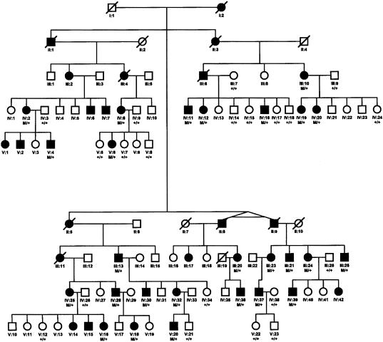
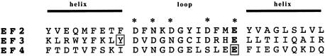
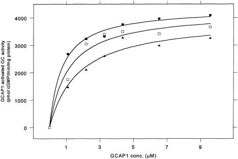
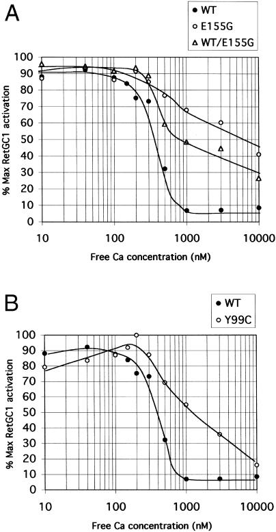
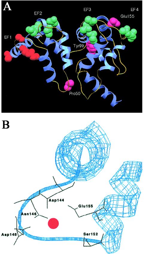
Mutations in the gene for guanylate cyclase-activating protein-1 (GCAP1) (GUCA1A) have been associated with autosomal dominant cone dystrophy (COD3). In the present study, a severe disease phenotype in a large white family was initially shown to map to chromosome 6p21.1, the location of GUCA1A. Subsequent single-stranded conformation polymorphism analysis and direct sequencing revealed an A464G transition, causing an E155G substitution within the EF4 domain of GCAP1. Modeling of the protein structure shows that the mutation eliminates a bidentate amino acid side chain essential for Ca2+ binding. This represents the first disease-associated mutation in GCAP1, or any neuron-specific calcium-binding protein within an EF-hand domain, that directly coordinates Ca2+. The functional consequences of this substitution were investigated in an in vitro assay of retinal guanylate cyclase activation. The mutant protein activates the cyclase at low Ca2+ concentrations but fails to inactivate at high Ca2+ concentrations. The overall effect of this would be the constitutive activation of guanylate cyclase in photoreceptors, even at the high Ca2+ concentrations of the dark-adapted state, which may explain the dominant disease phenotype.


Figures

Figure  1
Figure 1
Autosomal dominant inheritance of cone dystrophy. Individuals are identified by generation and pedigree number; squares indicate males, and circles indicate females. Blackened symbols indicate affected individuals; open symbols, indicate unaffected individuals.
Figure  2
Figure 2
Clinical evaluation. A, Fundus photograph from a patient (IV:37) with cone dystrophy, aged 26 years, with visual acuity 20/200, showing central macular pigmentary changes. B, ERGs recorded from the left (OS) and right (OD) eye of patient V:6 (aged 43 years), showing responses to ganzfeld stimuli (times of flash indicated by vertical bars) presented to the dark-adapted eye with a low-intensity strobe flash (top row) and with a high-intensity strobe flash (middle row) and to the light-adapted eye with a high-intensity strobe flash (bottom row). Two successive responses are shown for each stimulus condition. Note that the ERG of the light-adapted eye (cone) is diminished below the normal range, whereas responses of the dark-adapted eye (rod-dominated) fall within normal limits.
Figure  3
Figure 3
Alignment of the three functional EF-hand domains in GCAP1. The α-helix and loop regions of the domain are identified above the sequences. Residues in the loop that coordinate the Ca2+ ion are marked with an asterisk. The invariant Glu residues at position 12 of each Ca2+-binding loop (E75, E111, and E155) are highlighted in bold. The positions of the E155G mutation in EF4 present in our pedigree and of the previously reported Y99C mutation in EF3 are boxed.
Figure  4
Figure 4
GCAP1 titrations of RetGC1 activity with wild-type GCAP1 (blackened circles), E155G mutant (blackened triangles) and an equimolar mixture of wild-type and E155G mutant (open squares). Curves were fitted to the data points on the basis of the relation v=[GCAP1]Vmax/(Kmapp[GCAP1]+[GCAP1]), where Km app[GCAP1] is the GCAP1 concentration required for half maximal activation and where Vmax is the maximum specific activation. Values of Vmax and Km app[GCAP1] are derived from the fitted curves are given in table 1.
Figure  5
Figure 5
Ca2+ sensitivity of activation of wild-type RetGC1 by wild-type and mutant GCAP1. A, Activation by wild-type (blackened circles) and E155G mutant (open circles) GCAP1 and by an equimolar mixture of wild-type and E155G mutant (open triangles). B, Activation by wild-type (blackened circles) and Y99C mutant (open circles) GCAP1. Guanylate cyclase activity was determined in membrane preparations expressing RetGC1 activity stimulated with 8 μM GCAP1 in all cases. Results are presented as percentage of maximum RetGC1 activation (maximum RetGC1 activity minus basal activity in absence of GCAP1).
Figure  6
Figure 6
Model structure of GCAP1 showing the overall fold of the molecule. A, Complete structure, showing helices (blue) and loops (yellow). The key residues determining the calcium-binding ability of each EF hand are shown in space-filling mode and are labeled. The EF1 hand, which is predicted to be incapable of binding calcium, is shown in orange, to distinguish it from the calcium-binding EF hands 2–4 (green). Amino acid residues 50, 99, and 155, which correspond to the detected mutations in GCAP1, are shown in pink. The figure was produced using SETOR (Evans 1993). B, Enlargement of the EF4 domain, showing the polypeptide backbone in ribbon form (blue) and the five amino acid side chains, including E155, that assist in calcium coordination (black). The Ca2+ ion is shown as a red circle. The figure was produced using Swiss-Model (Peitsch 1996).

Similar articles

Cited by

References

Electronic-Database Information

    1. Online Mendelian Inheritance in Man (OMIM), http://www.ncbi.nlm.nih.gov/Omim (for autosomal dominant retinal cone dystrophy, COD3, and GCAP1 [OMIM 180020, 602093, and 600364])
    1. Swiss-Model, http://www.expasy.ch/swissmod/SWISS-MODEL.html (for protein modeling)

References

    1. Balciuniene J, Johanssen K, Sandgren O, Wachmeister L, Homgren G, Forsman G (1995) A gene for autosomal dominant progressive cone dystrophy (CORD5) maps to chromosome 17p12-p13. Genomics 30:281–286 - PubMed
    1. Baylor D (1996) How photons start vision. Proc Natl Acad Sci USA 93:560–565 - PMC - PubMed
    1. Cuenca N, Lopez S, Howes K, Kolb H (1998) The localization of guanylyl cyclase-activating proteins in the mammalian retina. Invest Ophthalmol Vis Sci 39:1243–1250 - PubMed
    1. Dizhoor AM, Boikov SG, Olshevskaya EV (1998) Constitutive activation of photoreceptor guanylate cyclase by Y99C mutant of GCAP-1. J Biol Chem 273:17311–17314 - PubMed
    1. Dizhoor AM, Hurley JB (1999) Regulation of photoreceptor membrane guanylate cyclases by guanylate cyclase activator proteins. Methods 19:521–531 - PubMed

Publication types

MeSH terms

Associated data