. 2001 Aug;159(2):501-12.
doi: 10.1016/S0002-9440(10)61722-1.

The perifollicular and marginal zones of the human splenic white pulp : do fibroblasts guide lymphocyte immigration?

Affiliations

The perifollicular and marginal zones of the human splenic white pulp : do fibroblasts guide lymphocyte immigration?

B Steiniger et al. Am J Pathol. 2001 Aug.

Abstract

We investigate the white pulp compartments of 73 human spleens and demonstrate that there are several microanatomical peculiarities in man that do not occur in rats or mice. Humans lack a marginal sinus separating the marginal zone (MZ) from the follicles or the follicular mantle zone. The MZ is divided into an inner and an outer compartment by a special type of fibroblasts. An additional compartment, termed the perifollicular zone, is present between the follicular MZ and the red pulp. The perifollicular zone contains sheathed capillaries and blood-filled spaces without endothelial lining. In the perifollicular zone, in the outer MZ, and in the T cell zone fibroblasts of an unusual phenotype occur. These cells stain for the adhesion molecules MAdCAM-1, VCAM-1 (CD106), and VAP-1; the Thy-1 (CD90) molecule; smooth muscle alpha-actin and smooth muscle myosin; cytokeratin 18; and thrombomodulin (CD141). They are, however, negative for the peripheral node addressin, the cutaneous lymphocyte antigen, CD34, PECAM-1 (CD31), and P- and E-selectin (CD62P and CD62E). In the MZ the fibroblasts are often tightly associated with CD4-positive T lymphocytes, whereas CD8-positive cells are almost absent. Our findings lead to the hypothesis, that recirculating CD4-positive T lymphocytes enter the human splenic white pulp from the open circulation of the perifollicular zone without crossing an endothelium. Specialized fibroblasts may attract these T cells and guide them into the periarteriolar T cell area.


Figures
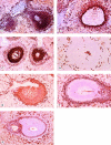
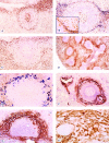
Figure 1.
Figure 1.
A–C: Variable distribution of surface IgD-positive B cells in individual specimens (ABC technique on autoclaved paraffin sections using polyclonal anti-human IgD). A: Strongly IgD-positive recirculating B cells occupy the mantle zones of two tangentially sectioned secondary follicles. In this case strongly IgD-positive cells are densely distributed in the inner MZ, obscuring the memory B cells that are only weakly positive for surface IgD. Strongly IgD-positive B cells occur in the outer MZ and extend along the PALS (top left) as a small stripe. Final magnification, ×55. B: Strongly IgD-positive recirculating B cells form a small mantle zone in the secondary follicle. The inner MZ is occupied by weakly IgD-positive memory B cells mixed with few strongly IgD-positive cells. The outer MZ is hardly recognizable. There are only few strongly IgD-positive B cells at the periphery of the PALS (left). Final magnification, ×88. C: A broad IgD-positive mantle zone surrounds the unstained germinal centers of two secondary follicles. The memory B cells of the inner MZ are IgD-negative in this specimen. A prominent ring-like accumulation of strongly IgD-positive B cells is seen in the outer MZ, whereas fewer of these cells are scattered in the inner MZ. Strongly IgD-positive B cells are almost absent at the periphery of the PALS. Final magnification, ×55. D: Distribution of CD34 in a secondary follicle. Few endothelia and perivascular fibroblasts are stained in the follicle interior. CD34 is not detected between the mantle zone and the MZ, indicating absence of a marginal sinus. A few capillary endothelia are reactive in the perifollicular zone. Weakly CD34-positive sinus endothelia occur only in this region. ABC technique on paraffin section using mAb QBend10. Final magnification, ×110. E: B lymphocytes stained for CD79a. All compartments of the secondary follicle are positive. The reactivity is most pronounced in the outer MZ. ABC technique on autoclaved paraffin section using mAb JCB117. Final magnification, ×69. F: Smooth muscle α-actin in a secondary follicle: A network of strongly actin-positive branched cells divides the inner MZ from the outer MZ. In this specimen a relatively large part of the outer MZ contains actin-positive cells. More weakly stained cells are present in the perifollicular zone. The actin-positive branched cells continue into a network filling the entire PALS (right). The red pulp also harbors some actin-positive cells. ABC technique on autoclaved paraffin section using mAb asm-1. Final magnification, ×88. G: Thrombomodulin in a primary (or tangentially sectioned secondary) follicle. Thrombomodulin-positive branched cells occur at the border between inner MZ and outer MZ and continue into the PALS (left). Red pulp sinus endothelia are also thrombomodulin-positive, especially in the perifollicular region. ABC technique on paraffin section using mAb TM 1009. Final magnification, ×69.
Figure 2.
Figure 2.
A: CD4-positive T lymphocytes in a secondary follicle. Top: A longitudinal and partially tangential section of the PALS. Many CD4-positive T cells are present in the germinal center, whereas only few CD4-positive T cells occur in the mantle zone and in the inner MZ. A prominent ring-like accumulation of CD4-positive T cells occurs at the border between inner MZ and outer MZ. ABC technique on autoclaved paraffin section using mAb 1F6. Final magnification, ×69. B: Double staining for smooth muscle α-actin (dark blue) and CD3 (brown) in a degenerating secondary follicle. Inset shows a 1.4-fold magnification of the top right part of the figure. CD3-positive T cells are closely associated with the most strongly smooth muscle α-actin-positive branched cells in the MZ. The PALS is situated on the right and left side of the follicle. mAb asm-1 is highly diluted to permit better visualization of the apposed CD3-positive cells. APAAP technique with nitro blue tetrazolium/BCIP for mAb asm-1 and ABC with DAB for CD3 antiserum. Final magnification, ×110. C: CD8-positive cells in the same secondary follicle as shown in A. CD8-positive cells do not accumulate in the follicle. Many small round cells and sinus endothelia are stained in the red pulp. ABC technique on autoclaved paraffin section using mAb 4B11. Final magnification, ×69. D: Erythrocytes in the perifollicular zone demonstrated by DAB. A similar pattern is revealed by staining for CD14 or CD15. Final magnification, ×28. E: Strongly sialoadhesin-positive macrophages (blue) scattered in the perifollicular zone. The brown background staining is because of residual peroxidatic activity of erythrocytes outlining the perifollicular zone. Cryosection without nuclear counterstain. mAb HSN-1 revealed by an APAAP technique with fast blue, erythrocytes are stained by DAB. Final magnification, ×110. F: MAdCAM-1-positive cells in a secondary follicle and PALS. Rows of strongly MAdCAM-1-positive branched cells occur at the border of the inner and outer MZ. More weakly staining cells are located in the outer MZ and perifollicular zone. MAdCAM-1-positive cells also form a network in the PALS (right). ABC technique on cryosection using mAb 10A6. Final magnification, ×69. G: Primary (or tangentially sectioned secondary) follicle stained for MAdCAM-1. In this specimen more branched cells are positive for MAdCAM-1 than in F. There are strongly positive elongated cells in the outer MZ and more weakly staining cells with finer and more elaborate projections in the perifollicular zone. The PALS is located at the bottom. ABC technique on cryosection using mAb 10A6. Final magnification, ×110. H: Higher magnification of MAdCAM-1-positive cells in the PALS. A central arteriole is located to the left. The MAdCAM-1-positive branched cells ensheath bundles of fibers that appear as pale stripes within the brown envelope of immunoreactive cells. No nuclear counterstain. ABC technique on cryosection using mAb 10A6. Final magnification, ×277.

Similar articles

Cited by

References

    1. Timens W, Poppema S: Lymphocyte compartments in human spleen. An immunohistologic study in normal spleens and noninvolved spleens in Hodgkin’s disease. Am J Pathol 1985, 120:443-454 - PMC - PubMed
    1. Van Krieken JHJM, te Velde J: Immunohistology of the human spleen: an inventory of the localization of lymphocyte subpopulations. Histopathology 1986, 10:285-294 - PubMed
    1. Grogan TM, Jolley CS, Rangel CS: Immunoarchitecture of the human spleen. Lymphology 1983, 16:72-82 - PubMed
    1. Grogan TM, Rangel CS, Richter LC, Wirt DP, Villar HV: Further delineation of the immuno-architecture of the human spleen. Lymphology 1984, 17:61-68 - PubMed
    1. Buckley PJ, Smith MR, Braverman MF, Dickson SA: Human spleen contains phenotypic subsets of macrophages and dendritic cells that occupy discrete microanatomic locations. Am J Pathol 1987, 128:505-520 - PMC - PubMed