. 2001 Aug;159(2):591-7.
doi: 10.1016/s0002-9440(10)61730-0.

Role of peroxisome proliferator-activated receptor gamma and its ligands in non-neoplastic and neoplastic human urothelial cells

Affiliations

Role of peroxisome proliferator-activated receptor gamma and its ligands in non-neoplastic and neoplastic human urothelial cells

K I Nakashiro et al. Am J Pathol. 2001 Aug.

Abstract

Peroxisome proliferator-activated receptor gamma (PPARgamma) is a member of the nuclear receptor superfamily of ligand-activated transcription factors and is expressed in several types of tissue. Although PPARgamma reportedly is expressed in normal urothelium, its function is unknown. We examined the expression of PPARgamma in normal urothelium and bladder cancer in an attempt to assess its functional role. Immunohistochemical staining revealed normal urothelium to express PPARgamma uniformly. All low-grade carcinomas were positive either diffusely or focally, whereas staining was primarily focal or absent in high-grade carcinomas. A nonneoplastic urothelial cell line (1T-1), a low-grade (RT4) carcinoma cell line, and two high-grade (T24 and 253J) carcinoma cell lines in culture expressed PPARgamma mRNA and protein. Luciferase assay indicated that PPARgamma was functional. PPARgamma ligands (15-deoxy-Delta(12,14)-prostaglandin J(2), troglitazone and pioglitazone) suppressed the growth of nonneoplastic and neoplastic urothelial cells in a dose-dependent manner. However, neoplastic cells were more resistant than were nonneoplastic cells. Failure to express PPARgamma or ineffective transcriptional activity may be some of the mechanisms responsible for resistance to the inhibitory action of PPARgamma ligands.


Figures

Figure 1.
Figure 1.
Immunohistochemical staining for PPARγ protein in normal human ureter (A) and low-grade (B) and high-grade (C and D) bladder carcinomas. All nuclei are uniformly stained in ureteral urothelium [original magnification, ×100 (A)] and grade 1 transitional cell carcinoma [original magnification, ×100 (B)]. In grade 3 transitional cell carcinoma, nuclear staining is focal [original magnification, ×40 (C)] or completely absent [original magnification, ×100 (D)].
Figure 2.
Figure 2.
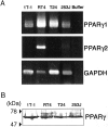
Expression of PPARγ by nonneoplastic and neoplastic human urothelial cells. A: RT-PCR for expression of PPARγ1 and -2 mRNA. All samples demonstrate PPARγ1 message whereas PPARγ2 was expressed only in RT4 cells. Primers specific for PPARγ1 and -2 and glyceraldehyde-3-phosphate dehydrogenase cDNAs generated fragments of 790 bp, 877 bp, and 782 bp, respectively. B: Western blot analysis for PPARγ protein. Cell lysates were isolated and electrophoresed on 10% sodium dodecyl sulfate-polyacrylamide gel (50 μg of protein/lane). Proteins from gels were transferred to polyvinylidene difluoride membrane, and PPARγ was detected with mouse monoclonal anti-PPARγ antibody and an enhanced chemiluminescence kit. All samples demonstrate PPARγ protein (∼58 kd).
Figure 3.
Figure 3.
Effect of 15d-PGJ2 (A), TRO (B), and PIO (C) on growth of nonneoplastic and neoplastic urothelial cells. Cells were seeded on a 96-well or 6-well plate in the medium with 5% fetal bovine serum appropriate for each cell type. Twenty-four hours later, medium was changed to the same medium containing 15d-PGJ2 (0 to 10 μmol/L), TRO (0 to 50 μmol/L), or PIO (0 to 50 μmol/L). After incubation for 24 hours, cell proliferation was assessed by cell proliferation enzyme-linked immunosorbent assay, BrdU kit. After 3 days, the number of cells was counted with a hemocytometer after cells were recovered by trypsinization. Results are expressed as ratio to the respective control culture. Bars denote SD of triplicate samples. The ligand concentrations in the abscissa are on an arbitrary scale. *, P < 0.0001 compared with the respective control culture.
Figure 4.
Figure 4.
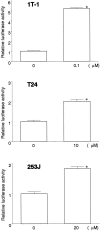
Ligand-induced transcriptional activation of PPARγ. Cells (1T1, T24, and 253J, 3 × 10 per well) were seeded on a 6-well plate in the medium appropriate for each cell type. Twenty-four hours later, transient transfection was done by using Effectene transfection reagent with PPRE-TK-LUC. The transfection mix was replaced by complete medium with or without TRO (0.1, 10, or 20 μmol/L) and incubated for an additional 24 hours. The cells were lysed with cell culture lysis reagent. Luciferase activity was measured by using luciferase assay reagent in a scintillation counter. Results are expressed as ratio to the respective control culture. Bars denote SD of triplicate samples. *, P < 0.0001 compared with the respective control culture.
Figure 5.
Figure 5.
Expression of PBP and RXRα protein by nonneoplastic and neoplastic human urothelial cells. Cell lysates were isolated and electrophoresed on 10% sodium dodecyl sulfate-polyacrylamide gel (50 μg of protein/lane). Proteins from gels were transferred to polyvinylidene difluoride membrane, and PBP and RXRα were detected with rabbit anti-PBP and -RXRα antibody and an enhanced chemiluminescence kit. Only RT4 cells demonstrate PBP protein. RXRα expression is at much higher levels in 1T-1 and low-grade carcinoma cell line RT4 than in high-grade carcinoma cell lines T24 and 253J.

References

    1. Zhu Y, Alvares K, Huang Q, Rao MS, Reddy JK: Cloning of a new member of the peroxisome proliferator-activated receptor gene family from mouse liver. J Biol Chem 1993, 268:26817-26820 - PubMed
    1. Tontonoz P, Hu E, Spiegelman BM: Stimulation of adipogenesis in fibroblasts by PPARγ2, a lipid-activated transcription factor. Cell 1994, 79:1147-1156 - PubMed
    1. Kliewer SA, Forman BM, Blumberg B, Ong ES, Borgmeyer U, Mangelsdorf DJ, Umesono K, Evans RM: Differential expression and activation of a family of murine peroxisome proliferator-activated receptors. Proc Natl Acad Sci USA 1994, 91:7355-7359 - PMC - PubMed
    1. Braissant O, Foufelle F, Scotto C, Dauca M, Wahli W: Differential expression of peroxisome proliferator-activated receptors (PPARs): tissue distribution of PPAR-alpha, -beta, and -gamma in the adult rats. Endocrinology 1996, 137:354-366 - PubMed
    1. Guan Y, Zhang Y, Davis L, Breyer MD: Expression of peroxisome proliferator-activated receptors in urinary tract of rabbits and humans. Am J Physiol 1997, 273:F1013-F1022 - PubMed

Publication types

MeSH terms