. 2001 Sep;21(17):5753-66.
doi: 10.1128/MCB.21.17.5753-5766.2001.

In vitro reconstitution of the end replication problem

Affiliations

In vitro reconstitution of the end replication problem

R Ohki et al. Mol Cell Biol. 2001 Sep.

Abstract

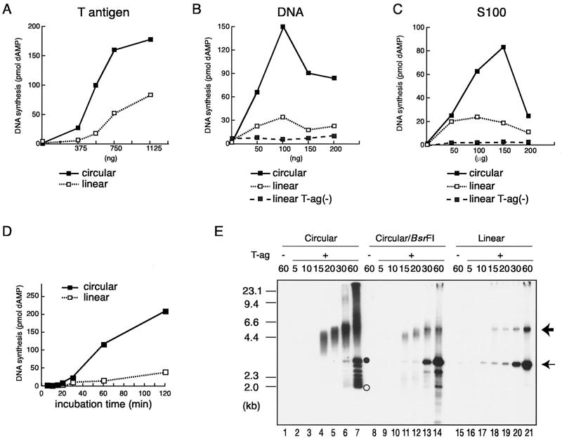
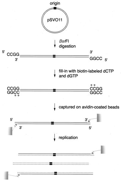
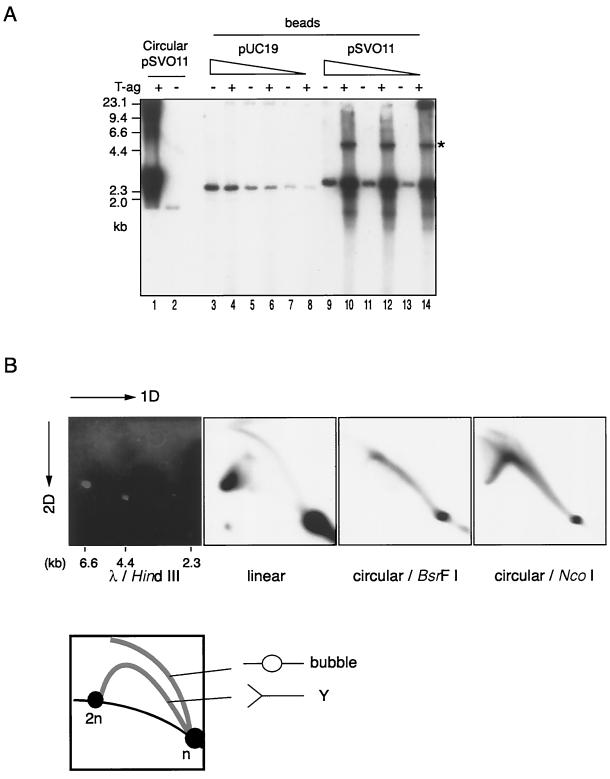
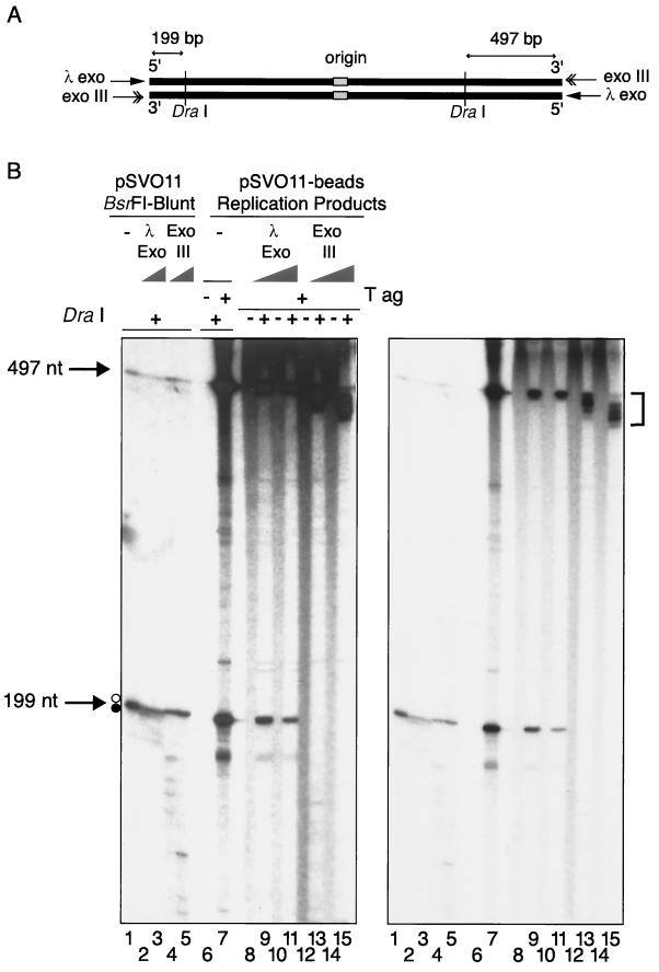
The end replication problem hypothesis proposes that the ends of linear DNA cannot be replicated completely during lagging strand DNA synthesis. Although the idea has been widely accepted for explaining telomere attrition during cell proliferation, it has never been directly demonstrated. In order to take a biochemical approach to understand how linear DNA ends are replicated, we have established a novel in vitro linear simian virus 40 DNA replication system. In this system, terminally biotin-labeled linear DNAs are conjugated to avidin-coated beads and subjected to replication reactions. Linear DNA was efficiently replicated under optimized conditions, and replication products that had replicated using the original DNA templates were specifically analyzed by purifying bead-bound replication products. By exploiting this system, we showed that while the leading strand is completely synthesized to the end, lagging strand synthesis is gradually halted in the terminal approximately 500-bp region, leaving 3' overhangs. This result is consistent with observations in telomerase-negative mammalian cells and formally demonstrates the end replication problem. This study provides a basis for studying the details of telomere replication.


Figures

FIG. 1
FIG. 1
In vitro replication of circular and linear pSVO11 DNAs. (A to C) Titrations of the amounts of SV40 T-ag, template DNA, and cytosol S100 extracts derived from 293 cells in DNA synthesis from circular and linear pSVO11 DNA templates. Reactions were conducted using varied amounts of the immunoaffinity-purified SV40 T-ag (A), pSVO11 template (B), and 293 cell S100 (C) extracts, as indicated in the figure. Other components in the reaction mixture (25 μl each) were 50 ng of DNA template and 80 μg of S100 extracts (A), 600 ng of T-ag and 80 μg of S100 extracts (B), and 50 ng of DNA template and 600 ng of T-ag (C). The samples were incubated at 37°C for 1.5 h. The average incorporation of [32P]dAMP (in picomoles) under each condition was measured. (D) Kinetics of DNA synthesis. Reaction mixtures (25 μl each) containing 50 ng of either circular or linearized pSVO11 DNA, 750 ng of T-ag, and 100 μg of cytosol S100 extracts were incubated for the times indicated, and the average incorporation of [32P]dAMP (in picomoles) was measured. (E) Gel electrophoresis analysis of the DNA products obtained from circular and linear pSVO11 template DNAs. One-fifth (for 20 to 60 min) or 1/2.5 (for 5 to 15 min) of each product obtained in the reactions containing the linear pSVO11 templates and 1/10 (20 to 60 min) or 1/5 (5 to 15 min) of each product obtained in the reactions containing the circular pSVO11 templates were run in a 0.8% agarose gel. For circular DNA reactions, the products were analyzed either with (Circular/BsrFI) or without (Circular) BsrFI restriction digestion prior to gel loading. Following electrophoresis, the gel was dried and autoradiographed. λ DNA digested with HindIII was used as a size marker. The closed and open circles indicate the positions of relaxed and supercoiled circular DNAs, respectively. The thin arrow marks the position of full-length linear DNA products. The linear dimer DNA products potentially produced by the endogenous DNA ligase activity present in S100 extracts are indicated by the thick arrow.
FIG. 2
FIG. 2
Method to analyze DNA products that have undergone a single round of replication reaction on an original DNA strand template. Plasmid DNA is linearized by BsrFI producing two ends with 5′-protruding 5′-CCGG-3′ sequences. The 3′-recessive ends are subsequently filled in with biotinylated dCTPs and dGTPs by the Klenow enzyme. The resultant DNA molecules possess two biotinylated, blunt ends. Both ends are conjugated to avidin beads. When these bead-captured linear DNAs are used as templates for in vitro DNA replication, their daughter DNA molecules remain bound to the beads, whereas daughter DNA molecules that have been replicated using nascent DNA strands as templates are liberated to the aqueous phase. By simply collecting the DNA molecules bound to the beads after replication, it is possible to analyze the products of a single round of DNA replication on the original DNA strand. Labeled biotin is represented by the small filled circles. Nascent DNA is shown by the dotted lines.
FIG. 3
FIG. 3
DNA replication of linear DNA molecules bound to beads. (A) One-dimensional gel electrophoresis of DNA products. Circular pSVO11, linear bead-captured pUC19 (pUC19-beads) and linear bead-captured pSVO11 (pSVO11-beads) were subjected to the replication reaction. Circular DNA was replicated in solution as described in the legend to Fig. 1. For pUC19-beads and pSVO11-beads, three batches of beads that had been incubated with three different amounts of DNA (60, 20, and 6.6 ng, indicated as the graded triangles) were used as templates. In a separate experiment, it was confirmed that the volume of beads used in these experiments had the capacity to bind these amounts of DNA. In the pSVO11-bead reactions, DNA products liberated from beads were discarded, while the bound DNA fractions (bound fraction) were collected and further analyzed. Aliquots of the DNA products were run on an agarose gel as described in the legend to Fig. 1. Reactions were assembled with (+) or without (−) T-ag. (B) 2D gel electrophoresis of DNA products. The labeled DNA obtained from circular pSVO11 and pSVO11-beads (bound fraction) was assayed by neutral-neutral 2D gel electrophoresis assay (4). λ DNA digested with HindIII was run in parallel and is shown in the leftmost panel to serve as a marker for the linear DNA species. The bound fraction obtained from the pSVO11-bead reaction was run in the second panel. DNA products obtained from circular pSVO11 were digested with either BsrFI or NcoI prior to gel loading. Both enzymes digest the circular DNA products at a single site. The BsrFI site is positioned approximately opposite the replication origin, whereas the NcoI site neighbors the origin. Typical Y arcs were observed for both BsrFI-digested and NcoI-digested circular pSVO11 products (the third and fourth panels). Although bubble-form intermediates, and not Y-form intermediates, were expected to comprise the majority of DNA species in the BsrFI-digested circular pSVO11 products, a bubble arc was not observed in the autoradiograph (the third panel). It has been reported that for an unknown reason, restriction enzymes disrupt bubble structures (3). Typical Y and bubble arcs were observed for the bound fraction of the pSVO11-bead reaction (the second panel). A schematic representation of the relative positions of linear DNA, bubble arcs, and Y arcs is shown below the electrophoresis data. n and 2n represent the positions of full-sized and double-sized linear DNA molecules. The strong signal found at the 2n position of linear species in the pSVO11-bead products probably represents the linear dimer, which is also denoted by an asterisk in panel A, in addition to replication intermediates.
FIG. 4
FIG. 4
Analysis of terminal restriction fragments from replicated linear DNAs. (A and B) The bound fraction of pSVO11-bead replication products was purified and treated with either λ exonuclease or exonuclease III. To know the exonuclease digestion rates, we treated a separately prepared 199-bp terminal fragment with these exonucleases and found that under the employed conditions, approximately 100 nt is digested from the ends, albeit relatively asymmetrically (data not shown). After the digestion, a half aliquot of the DNA was further treated with DraI, which produces 199- and 497-bp fragments from the left and right arms of the DNA, respectively (A). Samples were run in a 6% denaturing acrylamide gel, dried, and autoradiographed. Heavily and lightly exposed autoradiographs of the same gel are shown. Control pSVO11 DNA was digested with BsrFI, and the two ends were filled-in with dNTPs. The resultant blunt-ended linear pSVO11 was first treated with either λ exonuclease or exonuclease III, followed by DraI digestion. The products were first dephosphorylated by alkaline phosphatase at their 5′ ends and then labeled by T4 polynucleotide kinase and [γ-32P]ATP. DraI digests DNA at a TTT/AAA site, leaving blunt ends. Therefore, the two 199-nt and 497-nt fragment strands have the same nucleotide lengths (arrows). However, because of the effect of different base compositions on migration rates, two distinct 199-nt single-stranded DNA bands are visible in lane 1. The upper and lower bands (marked by open and filled circles, respectively) of the 199-nt doublet were completely digested by λ exonuclease and exonuclease III, respectively (lanes 2 to 5). The 199- and 197-nt bands were detected in pSV011-band replication products. These two bands were resistant to λ exonuclease (lanes 9 and 11). In contrast, the 199-nt band was completely digested, and the 497-nt band was significantly trimmed by exonuclease III (lanes 13 and 15; shorter-sized 497-nt bands are indicated by a bracket). These results indicate that the observed 497- and 199-nt bands were derived solely from a strand whose 3′ ends correspond to nascent radiolabeled DNA ends. Several extra bands were observed in lane 7. We do not know the precise origin of these signals. However, because they are both λ exonuclease and exonuclease III sensitive, it is likely they represent unligated lagging strand DNA molecules derived from internal template regions. It seemed that λ exonuclease had reached the DraI site on the template (cold) strand of some molecules, because the signal intensity of the 199-nt band decreased after the λ exonuclease treatment.
FIG. 5
FIG. 5
Relative efficiencies of local leading and lagging strand syntheses in the linear DNA replication system. (A) Labeled replication products from circular (lanes C), and linear (lanes F) pSVO11 in solution, and from pSVO11-beads (lanes B), were digested with the combinations of restriction enzymes indicated. The samples were run in 6% denaturing acrylamide gels and autoradiographed. Signals deriving from leading strand syntheses are shown by open circles, and those from lagging strand syntheses are shown by filled circles. Fragments are presented in the same order as their restriction sites are located along the linear pSVO11 DNA. The replication origin is shown by an arrow. Since linear DNA was digested with BsrFI and subsequently filled in by the Klenow enzyme, whereas circular DNA was only digested with BsrFI, there is a small difference in fragment size at both ends, as seen in panel MseI-BsrFI. (B) Relative efficiencies of lagging versus leading strand synthesis were calculated for different regions on the linear pSVO11 molecule. Experiments similar to those in panel A were done extensively, thus covering many regions of the replication products derived from pSVO11-beads and circular pSVO11. Nascent lagging and leading strand fragments for each restriction fragment were inferred from their expected DNA lengths. Then, the lagging strand intensity was divided by the leading strand intensity (Ilag/Ilead) for each restriction fragment. Because the circular pSVO11 products are completely replicated, the corresponding values serve as a reference for the variation in labeling efficiencies caused by nucleotide compositions. Ilag/Ilead was divided by this factor to compensate for the labeling efficiencies before plotting. The position of the SV40 replication origin is shown by an arrow. Most values are means of at least two replicate experiments. (C) Similar analyses were done for the pSVO10 molecule, which is a 7,933-bp SV40 origin-containing plasmid. Most values are means of at least two replicate experiments.
FIG. 5
FIG. 5
Relative efficiencies of local leading and lagging strand syntheses in the linear DNA replication system. (A) Labeled replication products from circular (lanes C), and linear (lanes F) pSVO11 in solution, and from pSVO11-beads (lanes B), were digested with the combinations of restriction enzymes indicated. The samples were run in 6% denaturing acrylamide gels and autoradiographed. Signals deriving from leading strand syntheses are shown by open circles, and those from lagging strand syntheses are shown by filled circles. Fragments are presented in the same order as their restriction sites are located along the linear pSVO11 DNA. The replication origin is shown by an arrow. Since linear DNA was digested with BsrFI and subsequently filled in by the Klenow enzyme, whereas circular DNA was only digested with BsrFI, there is a small difference in fragment size at both ends, as seen in panel MseI-BsrFI. (B) Relative efficiencies of lagging versus leading strand synthesis were calculated for different regions on the linear pSVO11 molecule. Experiments similar to those in panel A were done extensively, thus covering many regions of the replication products derived from pSVO11-beads and circular pSVO11. Nascent lagging and leading strand fragments for each restriction fragment were inferred from their expected DNA lengths. Then, the lagging strand intensity was divided by the leading strand intensity (Ilag/Ilead) for each restriction fragment. Because the circular pSVO11 products are completely replicated, the corresponding values serve as a reference for the variation in labeling efficiencies caused by nucleotide compositions. Ilag/Ilead was divided by this factor to compensate for the labeling efficiencies before plotting. The position of the SV40 replication origin is shown by an arrow. Most values are means of at least two replicate experiments. (C) Similar analyses were done for the pSVO10 molecule, which is a 7,933-bp SV40 origin-containing plasmid. Most values are means of at least two replicate experiments.
FIG. 6
FIG. 6
Analysis of the end structure of replicated DNAs. (A) Nondenaturing hybridization analysis of the replicated products. The circular pSVO11 (Circular) and pSVO11-bead (Linear) replication reactions were carried out in the absence (−) or presence (+) of SV40 T-ag with cold dNTPs. The replication products were treated (+) or not treated (−) with exonuclease I. pSVO11 products were subsequently digested with BsrFI (used to prepare pSVO11-beads) and HindIII, while pSVO11-bead products were digested with HindIII only. The approximate positions of the HindIII site on the linearized pSVO11 are shown. The restriction digests were run in an agarose gel and then subjected to in-gel hybridization with strand-specific probes. A mixture of two strand-specific probes, corresponding to the upper strands of the regions from nt 1 to 197 and nt 2681 to 2884, was used. Control DNA BsrFI-HindIII fragments possessing 5′-protruding (5′ overhang), 3′-protruding (3′ overhang), or blunt BsrFI sites were run in parallel. The 5′-protruding and 3′-protruding DNAs were prepared by treating the end-filled DNA with exonuclease III and λ exonuclease, respectively. (B) The ends containing the terminal nascent leading strand do not have 3′ overhangs. In vitro replication was performed with circular pSVO11 and pSVO11-beads in the presence of [α-32P]dATP. The products were treated with (+) or without (−) exonuclease I, followed by restriction enzyme digestion. The circular pSVO11 products were digested with BsrFI and MseI. The pSVO11-bead products were digested with MseI only. By MseI digestion, 147- and 94-nt signals deriving from both ends are expected as a result of leading strand synthesis for pSVO11-bead products without exonuclease I treatment. The digests were run in a 6% denaturing acrylamide gel and then autoradiographed. The reason for the observed difference in size between circular and pSVO11-bead replication products is described in the legend to Fig. 5A.

Similar articles

Cited by

References

    1. Blasco M A, Lee H W, Hande M P, Samper E, Lansdorp P M, DePinho R A, Greider C W. Telomere shortening and tumor formation by mouse cells lacking telomerase RNA. Cell. 1997;91:25–34. - PubMed
    1. Bodnar A G, Ouellette M, Frolkis M, Holt S E, Chiu C P, Morin G B, Harley C B, Shay J W, Lichtsteiner S, Wright W E. Extension of life-span by introduction of telomerase into normal human cells. Science. 1998;279:349–352. - PubMed
    1. Braguglia D, Heun P, Pasero P, Duncker B P, Gasser S M. Semi-conservative replication in yeast nuclear extracts requires Dna2 helicase and supercoiled template. J Mol Biol. 1998;281:631–649. - PubMed
    1. Brewer B J, Fangman W L. The localization of replication origins on ARS plasmids in S. cerevisiae. Cell. 1987;51:463–471. - PubMed
    1. Brush G S, Kelly T J, Stillman B. Identification of eukaryotic DNA replication proteins using simian virus 40 in vitro replication system. Methods Enzymol. 1995;262:522–548. - PubMed

Publication types

LinkOut - more resources