. 2001 Sep;21(17):5925-34.
doi: 10.1128/MCB.21.17.5925-5934.2001.

Induction of cyclin D1 transcription and CDK2 activity by Notch(ic): implication for cell cycle disruption in transformation by Notch(ic)

Affiliations

Induction of cyclin D1 transcription and CDK2 activity by Notch(ic): implication for cell cycle disruption in transformation by Notch(ic)

C Ronchini et al. Mol Cell Biol. 2001 Sep.

Abstract

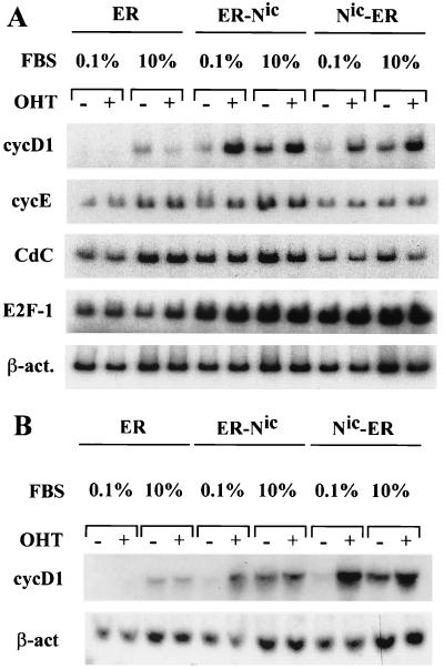
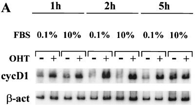
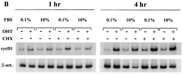
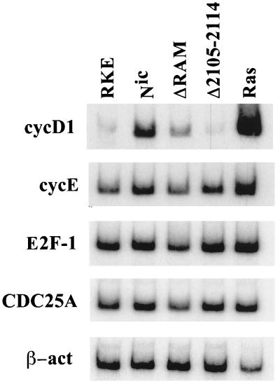
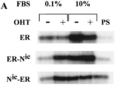
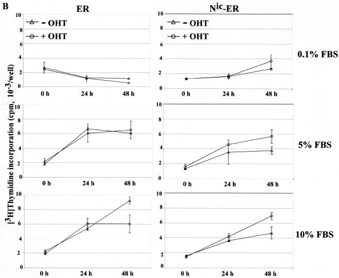
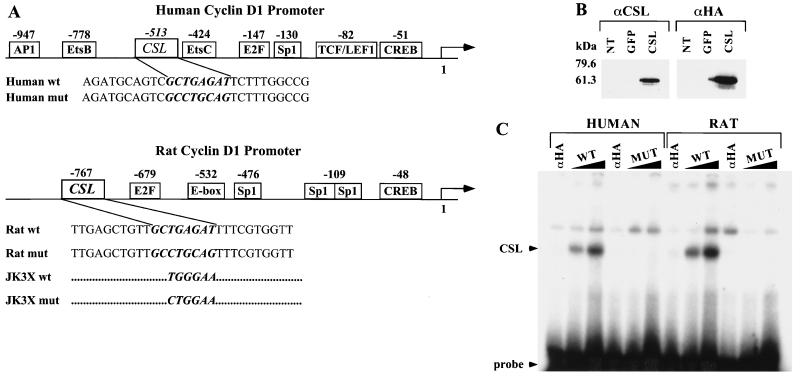
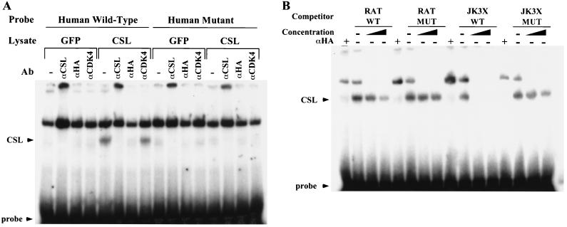
Notch genes encode a family of transmembrane proteins that are involved in many cellular processes such as differentiation, proliferation, and apoptosis. Although it is well established that all four Notch genes can act as oncogenes, the mechanism by which Notch proteins transform cells remains unknown. Previously, we have shown that transformation of RKE cells can be conditionally induced by hormone activation of Notch(ic)-estrogen receptor (ER) chimeras. Using this inducible system, we show that Notch(ic) activates transcription of the cyclin D1 gene with rapid kinetics. Transcriptional activation of cyclin D1 is independent from serum-derived growth factors and de novo synthesis of secondary transcriptional activators. Moreover, hormone activation of Notch(ic)-ER proteins induces CDK2 activity in the absence of serum. Upregulation of cyclin D1 and activation of CDK2 by Notch(ic) result in the promotion of S-phase entry. These data demonstrate the first evidence that Notch(ic) proteins can directly regulate factors involved in cell cycle control and affect cellular proliferation. Furthermore, nontransforming Notch(ic) proteins do not induce cyclin D1 expression, indicating that the mechanism of transformation involves cell cycle deregulation through constitutive expression of cyclin D1. Finally, we have identified a CSL [stands for CBF1, Su(H), and Lag-1] binding site within the human and rat cyclin D1 promoters, suggesting that Notch(ic) proteins activate cyclin D1 transcription through a CSL-dependent pathway.


Figures

FIG. 1
FIG. 1
Hormone-dependent upregulation of cyclin D1 mRNA level by Notchic-ER chimeras. (A) Following starvation for 48 h in medium containing 0.1% serum, cells were cultured for 5 h in low-concentration serum (FBS at 0.1%) or full-concentration serum (FBS at 10%) in the presence (+) or absence (−) of 1 μM OHT. Total RNA was extracted from the clonal RKE cell line expressing the ER hormone binding domain (lanes ER), the clonal RKE cell line expressing the ER-Nic chimera (lanes ER-Nic), and the clonal RKE cell line expressing the Nic-ER chimera (lanes Nic-ER). The levels of cyclin D1 mRNA (cycD1), cyclin E (cycE), Cdc25A (CdC), and E2F-1 were examined by RT-PCR as described in Materials and Methods. The level of β-actin mRNA (β-act.) is shown for normalization. (B) Northern blot analysis was performed on total RNA extracted from the clonal RKE cell lines treated as described for panel A.
FIG. 2
FIG. 2
The induction of cyclin D1 transcription by Notchic-ER proteins is direct. (A) Hormone activation of Notchic-ER proteins induces cyclin D1 upregulation in 1 h both in low- and in full-concentration serum. RT-PCR was performed on RNA samples prepared from a clonal RKE cell line expressing the ER-Nic chimera. Cells were cultured in medium containing 0.1% serum for 48 h and subsequently induced for the indicated times by addition of medium containing 0.1% or 10% FBS and 1 μM OHT (+) or ethanol vehicle (−). cycD1, cyclin D1-amplified mRNA samples; β-act; β-actin-amplified mRNA samples for a normalization control. (B) Cyclin D1 induction by Notchic-ER chimeras does not require de novo protein synthesis. Levels of cyclin D1 mRNA were analyzed by RT-PCR. A clonal RKE cell line expressing Nic-ER protein was treated as described for panel A for 1 or 4 h in the absence (−) or presence (+) of 15 μg of CHX per ml.
FIG. 2
FIG. 2
The induction of cyclin D1 transcription by Notchic-ER proteins is direct. (A) Hormone activation of Notchic-ER proteins induces cyclin D1 upregulation in 1 h both in low- and in full-concentration serum. RT-PCR was performed on RNA samples prepared from a clonal RKE cell line expressing the ER-Nic chimera. Cells were cultured in medium containing 0.1% serum for 48 h and subsequently induced for the indicated times by addition of medium containing 0.1% or 10% FBS and 1 μM OHT (+) or ethanol vehicle (−). cycD1, cyclin D1-amplified mRNA samples; β-act; β-actin-amplified mRNA samples for a normalization control. (B) Cyclin D1 induction by Notchic-ER chimeras does not require de novo protein synthesis. Levels of cyclin D1 mRNA were analyzed by RT-PCR. A clonal RKE cell line expressing Nic-ER protein was treated as described for panel A for 1 or 4 h in the absence (−) or presence (+) of 15 μg of CHX per ml.
FIG. 3
FIG. 3
The level of cyclin D1 expression correlates with the transforming ability of Notchic proteins. RT-PCR was performed on total RNA extracted from confluent RKE clonal cell lines cultured in medium containing 10% FBS. mRNA levels of cyclin D1 (cycD1), cyclin E (cycE), E2F-1, Cdc25A, and β-actin (β-act) were analyzed by RT-PCR. RKE, parental RKE cell line; Nic, clonal RKE cell line expressing Notchic; ΔRAM, clonal RKE cell line expressing NicΔR; Δ2105-2114, clonal RKE cell line expressing NicΔ2105–2114; Ras, clonal RKE cell line expressing Rasv12.
FIG. 4
FIG. 4
Hormone-dependent induction of CDK2 activity and DNA synthesis by Notchic-ER chimeras. (A) CDK2 assay. Clonal RKE cell lines were starved in medium containing 0.1% serum for 48 h and induced with (+) or without (−) 1 μM OHT in medium with a low (0.1%) or a full (10%) concentration of FBS for 12 h. Cell lysates were prepared from RKE clones expressing ER alone, ER-Nic, and Nic-ER chimeras. Proteins were immunoprecipitated with anti-CDK2 antibody or preimmune rabbit serum (PS). Immunocomplexes were assayed for kinase activity using histone H1 as a substrate. Labeled proteins were separated by SDS-polyacrylamide gel electrophoresis and visualized by autoradiography. (B) DNA synthesis was assayed by measuring [3H]thymidine incorporation. RKE clonal lines expressing ER alone or Nic-ER chimeras were serum deprived for 48 h and, subsequently, induced with 0.1, 5, or 10% FBS in the presence (+ OHT) or absence (− OHT) of 1 μM OHT. [3H]thymidine incorporation was measured as described in Materials and Methods. Data are expressed as counts per minute per well and are the means ± standard errors for quadruplicate measurements. The results are representative of three independent experiments.
FIG. 4
FIG. 4
Hormone-dependent induction of CDK2 activity and DNA synthesis by Notchic-ER chimeras. (A) CDK2 assay. Clonal RKE cell lines were starved in medium containing 0.1% serum for 48 h and induced with (+) or without (−) 1 μM OHT in medium with a low (0.1%) or a full (10%) concentration of FBS for 12 h. Cell lysates were prepared from RKE clones expressing ER alone, ER-Nic, and Nic-ER chimeras. Proteins were immunoprecipitated with anti-CDK2 antibody or preimmune rabbit serum (PS). Immunocomplexes were assayed for kinase activity using histone H1 as a substrate. Labeled proteins were separated by SDS-polyacrylamide gel electrophoresis and visualized by autoradiography. (B) DNA synthesis was assayed by measuring [3H]thymidine incorporation. RKE clonal lines expressing ER alone or Nic-ER chimeras were serum deprived for 48 h and, subsequently, induced with 0.1, 5, or 10% FBS in the presence (+ OHT) or absence (− OHT) of 1 μM OHT. [3H]thymidine incorporation was measured as described in Materials and Methods. Data are expressed as counts per minute per well and are the means ± standard errors for quadruplicate measurements. The results are representative of three independent experiments.
FIG. 5
FIG. 5
Identification of a CSL binding site in the human and rat cyclin D1 promoters. (A) Schematic representation of the human and rat cyclin D1 promoter. The binding sites for transcription factors identified in the cyclin D1 promoter are indicated with their positions relative to the starting site of transcription. The sequences of coding strands of the oligonucleotides used in the EMSA are reported under the schematic representation. In italic and bold is the wt or mut sequence for the CSL binding site identified in the human or the rat cyclin D1 promoter. The JK3X oligonucleotides contain three repeats of the indicated wt or mut consensus sequence for the CSL binding site. The complete sequences for the JK3X oligonucleotides are described in Materials and Methods. (B) Expression of HA-tagged CSL in 293T cells. Cell lysates were prepared from 293T cells not transfected (NT) or transiently transfected with an expression vector encoding GFP (lanes or GFP) the CSL HA-tagged protein (lanes CSL). Protein expression was analyzed by Western immunoblotting using an anti-CSL antibody (αCSL) and an anti-HA tag antibody (αHA). Standard molecular markers are indicated to the left. (C) CSL binds to the sequence identified in the human and rat cyclin D1 promoter. EMSA analysis was performed by incubating increasing amounts (60 and 120 fmol) of the indicated radiolabeled oligonucleotide duplex with 20 μg of lysate extracted from 293T cells expressing HA-tagged CSL. Sixty femtomoles of each oligonucleotide duplex was also incubated with lysates preincubated with the anti-HA probe antibody (lanes αHA). The positions of the CSL-DNA complex and of the free probe are indicated.
FIG. 6
FIG. 6
Binding of CSL to the cyclin D1 promoter is sequence specific. (A) DNA binding activity was analyzed by EMSA. Lysates from 293T cells expressing GFP (lanes marked GFP) or HA-tagged CSL (lanes marked CSL) were preincubated with no antibody (Ab) (−), an anti-CSL antibody (αCSL), an anti-HA probe antibody (αHA) or an anti-CDK4 antibody (αCDK4), used as an unrelated control antibody. Sixty femtomoles of the human wt or mut radiolabeled D1 probe was then added to the lysates for the binding reaction. DNA-protein complexes are indicated as for Fig. 5C. (B) Competition analysis was performed by incubating lysates of 293T cells expressing HA-tagged CSL with increasing concentrations (50- and 200-fold molar excesses) of the indicated unlabeled competitor duplex and with 60 fmol of the radiolabeled rat wt oligonucleotide. DNA-protein complexes were resolved by EMSA. The DNA-protein complex, free probe, and supershift with the anti-HA tag antibody are indicated as for Fig. 5C.

Similar articles

Cited by

References

    1. Aktas H, Cai H, Cooper G M. Ras links growth factor signaling to the cell cycle machinery via regulation of cyclin D1 and the Cdk inhibitor p27KIP1. Mol Cell Biol. 1997;17:3850–3857. - PMC - PubMed
    1. Albanese C, Johnson J, Watanabe G, Eklund N, Vu D, Arnold A, Pestell R G. Transforming p21ras mutants and c-Ets-2 activate the cyclin D1 promoter through distinguishable regions. J Biol Chem. 1995;270:23589–23597. - PubMed
    1. Aster J, Pear W, Hasserjian R, Erba H, Davi F, Luo B, Scott M, Baltimore D, Sklar J. Functional analysis of the TAN-1 gene, a human homolog of Drosophila notch. Cold Spring Harbor Symp Quant Biol. 1994;59:125–136. - PubMed
    1. Bellavia D, Campese A F, Alesse E, Vacca A, Felli M P, Balestri A, Stoppacciaro A, Tiveron C, Tatangelo L, Giovarelli M, Gaetano C, Ruco L, Hoffman E S, Hayday A C, Lendahl U, Frati L, Gulino A, Screpanti I. Constitutive activation of NF-kappaB and T-cell leukemia/lymphoma in Notch3 transgenic mice. EMBO J. 2000;19:3337–3348. - PMC - PubMed
    1. Bromberg J F, Wrzeszczynska M H, Devgan G, Zhao Y, Pestell R G, Albanese C, Darnell J E., Jr Stat3 as an oncogene. Cell. 1999;98:295–303. . (Erratum, 99:239.) - PubMed

Publication types

MeSH terms

Substances