. 2001 Sep;21(17):5946-57.
doi: 10.1128/MCB.21.17.5946-5957.2001.

Identification of receptor and heparin binding sites in fibroblast growth factor 4 by structure-based mutagenesis

Affiliations

Identification of receptor and heparin binding sites in fibroblast growth factor 4 by structure-based mutagenesis

P Bellosta et al. Mol Cell Biol. 2001 Sep.

Abstract

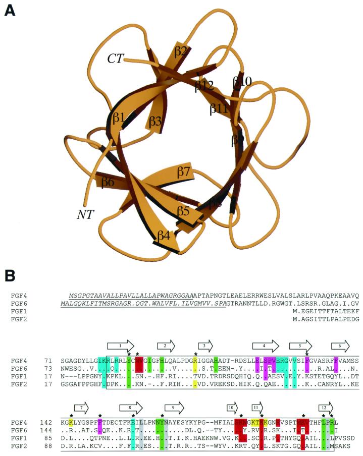
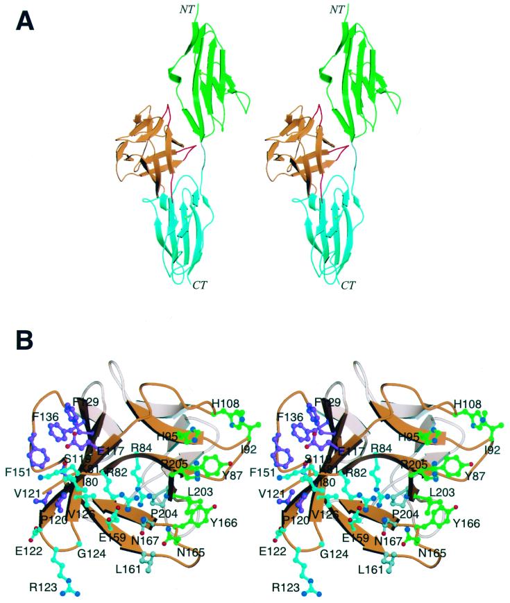
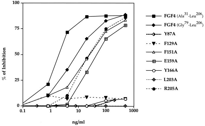
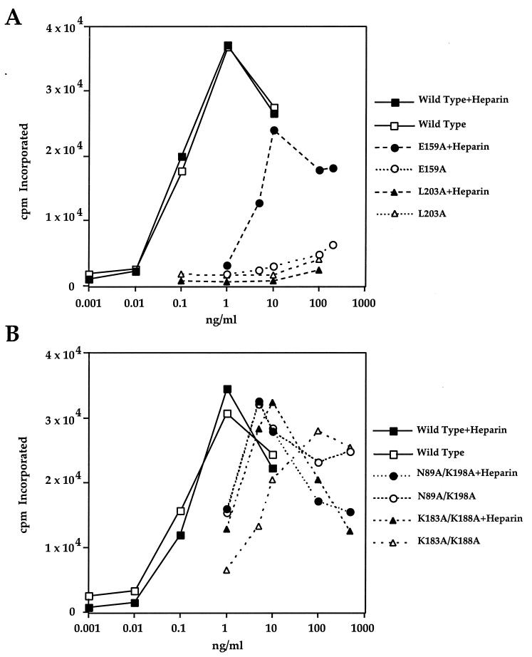
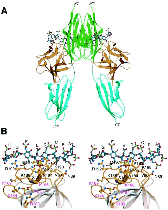
Fibroblast growth factors (FGFs) comprise a large family of multifunctional, heparin-binding polypeptides that show diverse patterns of interaction with a family of receptors (FGFR1 to -4) that are subject to alternative splicing. FGFR binding specificity is an essential mechanism in the regulation of FGF signaling and is achieved through primary sequence differences among FGFs and FGFRs and through usage of two alternative exons, IIIc and IIIb, for the second half of immunoglobulin-like domain 3 (D3) in FGFRs. While FGF4 binds and activates the IIIc splice forms of FGFR1 to -3 at comparable levels, it shows little activity towards the IIIb splice forms of FGFR1 to -3 as well as towards FGFR4. To begin to explore the structural determinants for this differential affinity, we determined the crystal structure of FGF4 at a 1.8-A resolution. FGF4 adopts a beta-trefoil fold similar to other FGFs. To identify potential receptor and heparin binding sites in FGF4, a ternary FGF4-FGFR1-heparin model was constructed by superimposing the FGF4 structure onto FGF2 in the FGF2-FGFR1-heparin structure. Mutation of several key residues in FGF4, observed to interact with FGFR1 or with heparin in the model, produced ligands with reduced receptor binding and concomitant low mitogenic potential. Based on the modeling and mutational data, we propose that FGF4, like FGF2, but unlike FGF1, engages the betaC'-betaE loop in D3 and thus can differentiate between the IIIc and IIIb splice isoforms of FGFRs for binding. Moreover, we show that FGF4 needs to interact with both the 2-O- and 6-O-sulfates in heparin to exert its optimal biological activity.


Figures

FIG. 1
FIG. 1
Structure and sequence alignment of FGF4. (A) Ribbon diagram of FGF4. Secondary structure assignments were obtained with the program PROCHECK (12). The β-strands of FGF4 are labeled according to the conventional strand nomenclature for FGF1 and FGF2 (6). NT and CT denote the amino and carboxy termini, respectively. This figure was created with the programs Molscript (11) and Raster3D (17). (B) Structure-based sequence alignment of FGFs. Sequence alignment was performed with CLUSTALW (34). All of the FGFs used in this alignment are human. The locations and lengths of the β-strands and α-helices are shown on the top. The signal sequences of FGF4 and FGF6 are indicated by italics and underlining. The box demarcates the boundaries of the β-trefoil core. A period indicates sequence identity to FGF4. A dash represents a gap introduced to optimize the alignment. FGF4 residues are colored with respect to the region on FGFR1 with which they interact: residues that interact with D2 are green, residues that interact with the linker region are gray, and residues that interact with D3 are cyan. FGF4 residues that interact with the βC′-βE loop in D3 of FGFR1 are purple. In red are FGF4 residues that constitute the conventional low- and high-affinity heparin binding sites. In addition, FGF4 residues that localize to the periphery of the high-affinity heparin-binding site and could potentially interact with heparin are yellow. A star indicates FGFR and heparin binding residues that were tested by site-directed mutagenesis.
FIG. 2
FIG. 2
Mapping of receptor binding sites in FGF4. (A) A model of the FGF4-FGFR1 structure was generated by superimposition of the Cα traces within the β-trefoil of the FGF4 structure onto the corresponding Cα traces of FGF2 in the FGF2-FGFR1 structure. Color coding is as follows: FGF4 is orange, D2 is green, D3 is cyan, and the linker region is gray. The FGF4 loop regions that clash with FGFR1 are red. NT and CT denote the amino and carboxy termini, respectively. (B) Stereo view of the receptor binding sites on FGF4. FGF4 residues are considered to be in the FGF4-FGFR1 interface if their side-chain or main-chain interatomic distance to FGFR1 is less than or equal to 3.8 Å. FGF4 residues are colored with respect to the FGFR1 regions with which they interact. FGF4 residues that interact with D2 are green, residues that interact with the linker region are gray, and residues that interact with D3 are cyan. FGF4 residues that interact with the βC′-βE loop in D3 of FGFR are purple. Oxygen and nitrogen atoms are red and blue, respectively. This figure was created by using Molscript and Raster3D.
FIG. 3
FIG. 3
Comparison of the binding affinities of various FGF4 mutants towards FGFR2. The capacity of the various FGF4 mutants to compete with binding of the N-terminally truncated wild-type FGF4 to FGFR2 binding was measured as described in Materials and Methods. The data are expressed as percent inhibition of wild-type FGF4 binding to FGFR2 by the indicated amount of unlabeled mutant FGF4. The results presented in this figure are also summarized in Table 2, where we have calculated for each mutant a 50% inhibitory concentration, or the concentration of mutant FGF4 necessary to compete off 50% of wild-type FGF4.
FIG. 4
FIG. 4
Differential effect of heparin on stimulation of DNA synthesis in NIH 3T3 cells by some FGF4 mutants. Thymidine uptake in NIH 3T3 cells in response to increasing concentrations of wild-type FGF4 and FGF4 mutants in the presence and absence of exogenous heparin was determined as described in Materials and Methods. (A) A representative experiment with the E159A and L203A mutants. (B) A representative experiment with the K183A/K188A and N89A/K198A double mutants. These results and those obtained with other mutants are summarized in Table 2, where we calculate the 50% effective dose—the concentration of mutant necessary to achieve 50% of maximum DNA synthesis produced by the wild-type FGF4.
FIG. 5
FIG. 5
Heparin binding sites in FGF4. (A) A dimeric FGF4-FGFR1-heparin model was created by superimposition of the Cα traces of two FGF4 structures onto the Cα traces of the two FGF2 molecules in the FGF2-FGFR1-heparin ternary structure. NT and CT denote the amino and carboxy termini, respectively. The coloring for FGF4 and FGFR1 is as presented in Fig. 2. The heparin oligosaccharides are rendered in the ball-and-stick format. The atom coloring for oxygens and nitrogens is as presented in Fig. 2. In addition, sulfur atoms are yellow, and the carbon atoms of oligosaccharides are gray. (B) FGF4 residues that localize to the heparin binding surface of FGF4 in the context of the ternary FGF4-FGFR1-heparin structure are mapped onto the ribbon diagram of FGF4. FGF4 residues that localize to the peripheries of the high-affinity heparin binding site and could potentially bind heparin are labeled in purple letters. A sulfate ion is bound to the conventional high-affinity heparin binding site. Another sulfate ion is bound in the additional potential heparin binding site. The atom coloring is as presented in panel A. Dotted lines represent hydrogen bonds. The sugar rings of heparin are labeled A through H, starting at the nonreducing end of the oligosaccharide. This figure was created with Molscript and Raster3D.

References

    1. Basilico C, Moscatelli D. The FGF family of growth factors and oncogenes. Adv Cancer Res. 1992;59:115–165. - PubMed
    1. Bellosta P, Talarico D, Rogers D, Basilico C. Cleavage of K-FGF produces a truncated molecule with increased biological activity and receptor binding affinity. J Cell Biol. 1993;121:705–713. - PMC - PubMed
    1. Bruenger A T, Adams P D, Clore G M, DeLano W L, Gros P, Grosse-Kunstleve R W, Jiang J S, Kuszewski J, Nigles M, Pannu N S, Read R J, Rice L M, Simonson T, Warren G L. Crystallography & NMR system: a new software suite for macromolecular structure determination. Acta Crystallogr Sect D. 1998;54:905–921. - PubMed
    1. Delli Bovi P, Curatola A M, Kern F G, Greco A, Ittmann M, Basilico C. An oncogene isolated by transfection of Kaposi's sarcoma DNA encodes a growth factor that is a member of the FGF family. Cell. 1987;50:729–737. - PubMed
    1. Eriksson A E, Cousens L S, Weaver L H, Matthews B M. Three-dimensional structure of human basic fibroblast growth factor. Proc Natl Acad Sci USA. 1991;88:3441–3445. - PMC - PubMed

Publication types

MeSH terms