. 2001 Aug;133(7):975-87.
doi: 10.1038/sj.bjp.0704152.

Agonists of proteinase-activated receptor 1 induce plasma extravasation by a neurogenic mechanism

Affiliations

Agonists of proteinase-activated receptor 1 induce plasma extravasation by a neurogenic mechanism

L de Garavilla et al. Br J Pharmacol. 2001 Aug.

Abstract

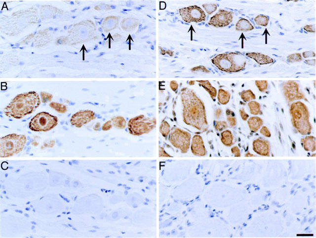
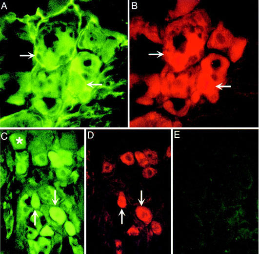
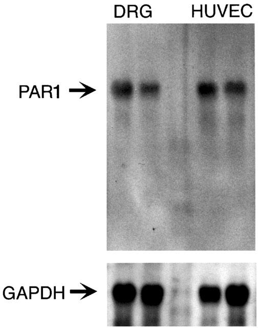
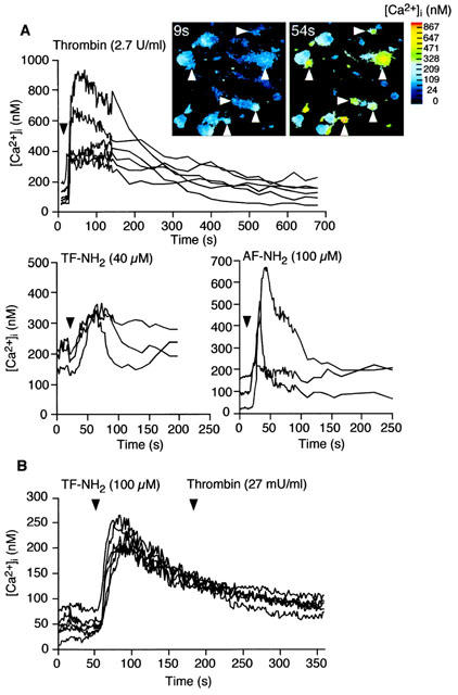
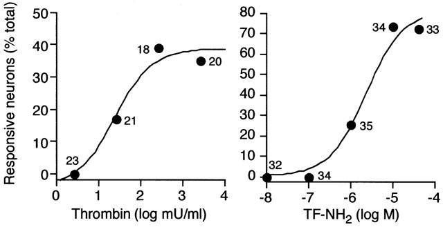
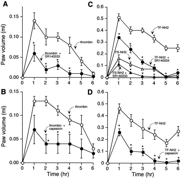
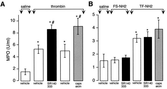
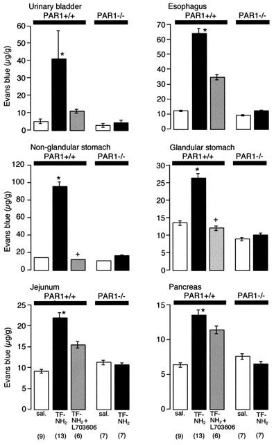
Thrombin, generated in the circulation during injury, cleaves proteinase-activated receptor 1 (PAR1) to stimulate plasma extravasation and granulocyte infiltration. However, the mechanism of thrombin-induced inflammation in intact tissues is unknown. We hypothesized that thrombin cleaves PAR1 on sensory nerves to release substance P (SP), which interacts with the neurokinin 1 receptor (NK1R) on endothelial cells to cause plasma extravasation. PAR1 was detected in small diameter neurons known to contain SP in rat dorsal root ganglia by immunohistochemistry and in situ hybridization. Thrombin and the PAR1 agonist TFLLR-NH(2) (TF-NH(2)) increased [Ca(2+)](i) >50% of cultured neurons (EC(50)s 24 mu ml(-1) and 1.9 microM, respectively), assessed using Fura-2 AM. The PAR1 agonist completely desensitized responses to thrombin, indicating that thrombin stimulates neurons through PAR1. Injection of TF-NH(2) into the rat paw stimulated a marked and sustained oedema. An NK1R antagonist and ablation of sensory nerves with capsaicin inhibited oedema by 44% at 1 h and completely by 5 h. In wild-type but not PAR1(-/-) mice, TF-NH(2) stimulated Evans blue extravasation in the bladder, oesophagus, stomach, intestine and pancreas by 2 - 8 fold. Extravasation in the bladder, oesophagus and stomach was abolished by an NK1R antagonist. Thus, thrombin cleaves PAR1 on primary spinal afferent neurons to release SP, which activates the NK1R on endothelial cells to stimulate gap formation, extravasation of plasma proteins, and oedema. In intact tissues, neurogenic mechanisms are predominantly responsible for PAR1-induced oedema.


Figures

Figure 1
Figure 1
Localization of PAR1 in rat DRG. Rat DRG (L5) were processed for immunohistochemistry (A–C) and in situ hybridization (D–F). Immunoreactive PAR1 (A) and PAR1 mRNA (D) was detected in both small and large diameter neurons (arrows). Immunoreactive NeuN (B) was detected in all neurons, and poly d(T) probe (D) identified all cells, and were used as positive controls. Non-immune serum (C) and a probe to lac Z operon (F) were used as negative controls. Scale bar=25 μm.
Figure 2
Figure 2
Localization of PAR1 in rat DRG. Rat DRG were processed for immunofluorescence to localize PAR1 (A,C), PGP9.5 (B) or CGRP (D). A, B and C, D are the same sections. (E) is a control in which the PAR1 antibody was preabsorbed with the receptor fragment that was used for immunization. Arrows indicate colocalization of PAR1, PGP9.5 and CGRP in the same neurons. *Indicates PAR1 neurons that do not contain CGRP.
Figure 3
Figure 3
Northern hybridization for PAR1 in rat DRG and HUVECs. Total RNA (10 μg/lane) was hybridized with cDNA probes to rat PAR1 or GAPDH.
Figure 4
Figure 4
PAR1 mediated Ca2+ mobilization in rat primary spinal afferent neurons in culture. (A) Effects of the PAR1 agonists thrombin (2.7 u ml−1), TF-NH2 (40 μM) and AF-NH2 (100 μM) or FS-NH2 (100 μM), which does not activate PAR1, on [Ca2+]i. Each line shows [Ca2+]i in the soma of a single neuron. The inset shows pseudocolour images of the neurons (arrows) that responded to thrombin. (B) Desensitization of PAR1 mediated Ca2+ mobilization. Exposure of neurons to TF-NH2 (40 μM for 2 min) abolished subsequent responses to thrombin (27 mu ml−1, without an intervening wash).
Figure 5
Figure 5
PAR1 mediated Ca2+ mobilization in rat primary spinal afferent neurons in culture. Concentration/response analyses for thrombin and TF-NH2. Results are expressed as the percentage of all observed neurons that responded to agonists with a detectable increase in [Ca2+]i. Numbers indicate the responsive neurons.
Figure 6
Figure 6
Effects of PAR1 agonists on oedema of the rat paw. Thrombin (5 U) (A,B) and TF-NH2 or the inactive analogue FS-NH2 (C,D) (both 100 μg) were injected into the paw and volume was measured hourly for 6 h. Results are expressed as increase in paw volume over basal. Thrombin and TF-NH2 stimulated oedema, which was inhibited by the NK1R antagonist SR140333 (A,C) and by ablation of sensory nerves with capsaicin (B,D). *P⩽0.05 compared to animals receiving TF-NH2 or FS-NH2 alone. n=6 animals per group.
Figure 7
Figure 7
Effects of PAR1 agonists on granulocyte infiltration to the rat paw. Thrombin (5 μu) (A) and TF-NH2 or the inactive analogue FS-NH2 (B) (both 100 μg) were injected into the paw and MPO activity in the paw was measured after 6 h. Thrombin and TF-NH2 but not FS-NH2 increased MPO activity. SR140333 and capsaicin exacerbated the response to thrombin but not TF-NH2. *P⩽0.05 compared to saline control; #P⩽0.05 compared to corresponding vehicle control. n=6 animals per group.
Figure 8
Figure 8
PAR1-induced extravasation of Evans blue in PAR1+/+ and PAR1−/− mice. Mice were injected with TF-NH2 (3 μmol kg−1, iv) and Evans blue extravasation was measured 10 min later. TF-NH2 stimulated extravasation in PAR1+/+ but not in PAR1−/− mice. Administration of the NK1R antagonist L-703,606 inhibited TF-NH2-induced plasma extravasation in many tissues. *Significant effect of strain and peptide treatment, ⩽0.05, two-way analysis of variance. + Significantly different from the TF-NH2 group, P⩽0.05, t-test (n).

References

    1. BOWDEN J.J., GARLAND A.M., BALUK P., LEFEVRE P., GRADY E.F., VIGNA S.R., BUNNETT N.W., MCDONALD D.M. Direct observation of substance P-induced internalization of neurokinin 1 (NK1) receptors at sites of inflammation. Proc. Natl. Acad. Sci. U.S.A. 1994;91:8964–8968. - PMC - PubMed
    1. BROKAW J.J., WHITE G.W. Differential effects of phosphoramidon and captopril on NK1 receptor-mediated plasma extravasation in the rat trachea. Agents Actions. 1994;42:34–39. - PubMed
    1. CHEN Y.H., POUYSSEGUR J., COURTNEIDGE S.A., VAN OBBERGHEN-SCHILLING E. Activation of Src family kinase activity by the G protein-coupled thrombin receptor in growth-responsive fibroblasts. J. Biol. Chem. 1994;269:27372–27377. - PubMed
    1. CHEUNG W.M., D'ANDREA M.R., ANDRADE-GORDON P., DAMIANO B.P. Altered vascular injury responses in mice deficient in protease-activated receptor-1. Arterioscler. Thromb. Vasc. Biol. 1999;19:3014–3024. - PubMed
    1. CIRINO G., BUCCI M., CICALA C., NAPOLI C. Inflammation-coagulation network: are serine protease receptors the knot. Trends Pharmacol. Sci. 2000;21:170–172. - PubMed

Publication types

MeSH terms