. 2001 Aug 15;21(16):5893-901.
doi: 10.1523/JNEUROSCI.21-16-05893.2001.

A dominant negative inhibitor of the Egr family of transcription regulatory factors suppresses cerebellar granule cell apoptosis by blocking c-Jun activation

Affiliations

A dominant negative inhibitor of the Egr family of transcription regulatory factors suppresses cerebellar granule cell apoptosis by blocking c-Jun activation

Y Levkovitz et al. J Neurosci. .

Abstract

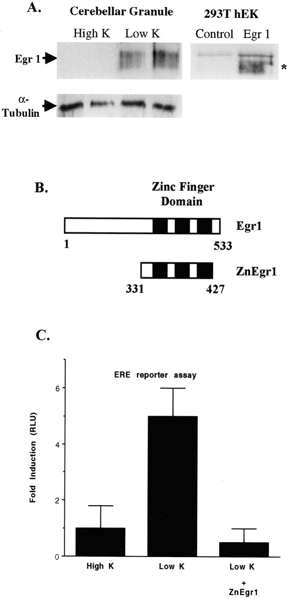
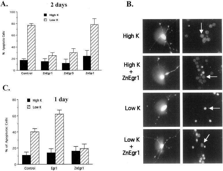
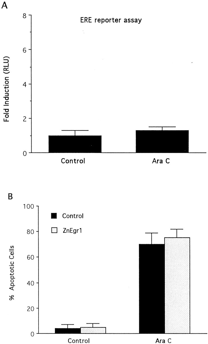
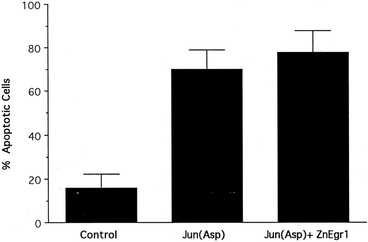
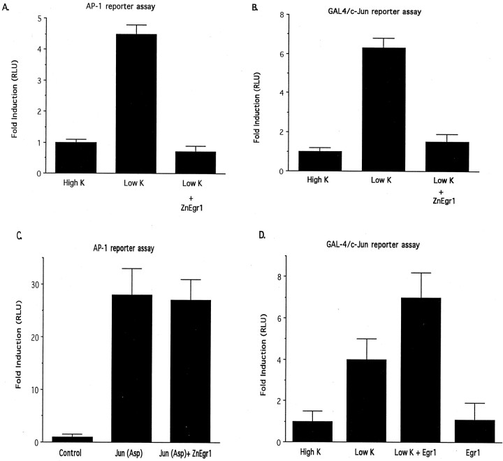
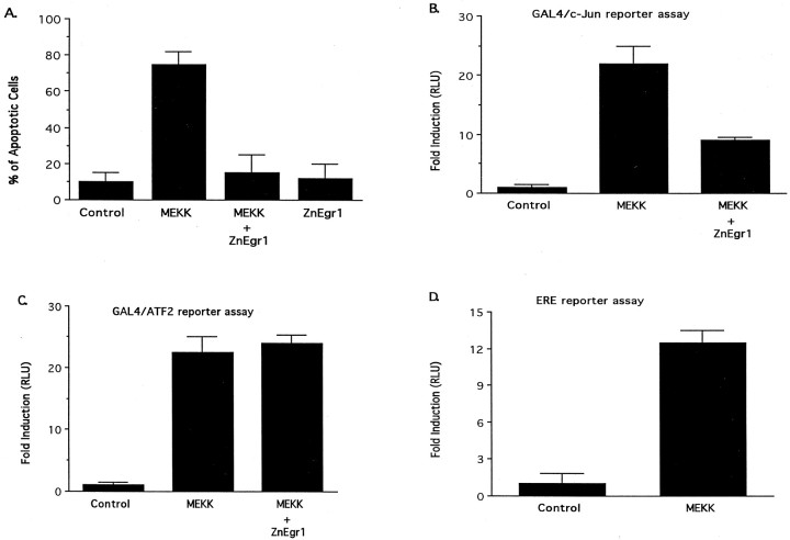
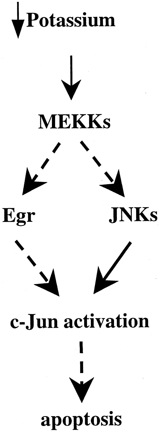
To investigate the role of the Egr family of transcription regulatory factors in neuronal apoptosis, we examined the effect of a dominant negative Egr inhibitor construct in a well characterized in vitro paradigm, cerebellar granule cell death induced by withdrawal of depolarizing concentrations of extracellular potassium. We found that this apoptotic stimulus increases the activity of a reporter gene driven by the Egr response element and that a dominant negative inhibitor of Egr-mediated transcription blocks granule cell apoptosis. In contrast, apoptosis of immature granule cells induced by cytosine arabinoside is not inhibited by the Egr inhibitor construct. Because activation of c-Jun is an essential step in granule cell death induced by potassium deprivation, but not cytosine arabinoside, we asked whether the Egr inhibitor acts by influencing c-Jun activation or its ability to induce apoptosis. We found that the Egr inhibitor does not block the ability of a constitutively active c-Jun construct to induce apoptosis in these cells but does suppress activation of c-Jun-mediated transcription induced by lowering extracellular potassium concentration. Furthermore, the Egr inhibitor blocks the ability of MEKK1 [mitogen-activated protein kinase (MAPK) kinase kinase 1], an upstream kinase capable of stimulating the JNK (c-Jun N-terminal protein kinase)-c-Jun pathway, to induce apoptosis and activate c-Jun. Together, these studies indicate that the Egr family of transcription factors plays a critical role in neuronal apoptosis and identify c-Jun activation as an important downstream target of the Egr family in this process.


Figures

Fig. 1.
Fig. 1.
Potassium deprivation increases Egr1 protein levels and ERE reporter activity. A, Immunoblot analysis of Egr1 levels. The left panel shows an immunoblot of extracts prepared from either control (High K) or potassium-deprived (Low K) granule cell cultures harvested 6 hr after switching to potassium deprivation. The broad protein band corresponding to Egr1, which migrates with a molecular weight of ∼80 kDa, is indicated by the arrow. Samples in each lane were obtained from separate culture dishes. Immunoblot analysis of rat Egr1 expressed in HEK293T cells, shown in the right panel, demonstrates that recombinant Egr1 also yields a broad protein band (indicated by asterisk), which comigrates with the band identified as Egr1 in cerebellar granule cell extracts. The Egr1 band is not detected in extracts from HEK293T cells that were transfected with an empty vector (Control). B, Schematic diagram of Egr1 and ZnEgr1. The diagram presented in this panel shows the portion of Egr1 contained in the zinc finger DNA binding domain construct, ZnEgr1, being used as an inhibitor of ERE-mediated transcription.C, Stimulation of an ERE reporter by potassium deprivation: blockade by ZnEgr1. Reducing the concentration of potassium in the medium from 25 mm (High K) to 5 mm (Low K) triggers a fivefold increase in ERE reporter activity (p < 0.01). This increase is abolished by cotransfection of the ZnEgr1 construct (Low K vs Low K + ZnEgr1,p < 0.005). Reporter activity is presented as the ratio of luciferase activities, or relative luciferase units (RLU), detected in extracts from treated and control cultures. Cells were transfected with the ERE reporter and either ZnEgr1 or an empty vector. On the second day after transfection, cell extracts were harvested for luciferase assay, 6 hr after switching the potassium concentration. Error bars shown in this and subsequent figures depict SEM values.
Fig. 2.
Fig. 2.
ZnEgr constructs block granule cell death induced by potassium deprivation. A, Quantitative analysis. In control cultures, lowering extracellular potassium concentration to 5 mm (Low K) from 25 mm(High K) greatly increases the percentage of apoptotic cells (p < 0.003). Transfection with either ZnEgr1 or ZnEgr3 blocks the increase in apoptosis elicited by potassium deprivation (Low K/control vs Low K/ZnEgr1,p < 0.008; Low K/control vs Low K/ZnEgr3,p < 0.01), whereas ZnSp1 does not. At least 200 cells were counted in each group. Scoring of GFP-positive cells was performed 2 d after initiating potassium deprivation.B, Hoechst staining of GFP-positive cells. Left column shows examples of GFP-positive granule cells. Note the presence of extensive neuronal processes studded with strongly fluorescent varicosities, typical of granule cells in culture.Right column shows the Hoechst staining of the GFP-positive cell shown in the same row along with a few surrounding GFP-negative cells. Note that potassium deprivation produces an increase in the percentage of cells with pyknotic nuclei that display stronger fluorescence, characteristic of apoptotic cells. However, the GFP-positive neuron shown from cultures transfected with ZnEgr1 and switched to 5 mm potassium (Low K+ ZnEgr1) displays dim, diffuse Hoechst staining characteristic of normal cells. Cultures shown in thefirst and third rows were transfected with an empty vector instead of the ZnEgr1 expression plasmid.C, Effects of Egr1 and ZnEgr1 at 1 d after potassium deprivation. Cells were transfected with ZnEgr1, Egr1, or empty vector (Control) and then either maintained in 25 mm KCl (High K) or switched to 5 mm potassium (Low K). One day later, GFP-positive cells were scored as apoptotic or normal. One hundred twenty cells were scored for each group. Transfection with Egr1 potentiated the apoptotic response (control/Low K vs Egr1/Low K,p < 0.03), whereas ZnEgr1 conferred protection (control/Low K vs ZnEgr1/Low K, p < 0.02).
Fig. 3.
Fig. 3.
Egr proteins do not mediate apoptosis induced in immature granule cells by cytosine arabinoside. A, AraC (500 μm) does not stimulate ERE reporter activity. Cells were transfected with the ERE reporter plasmid and then treated with either AraC (500 μm) or control medium. Eight hours later, cell extracts were harvested for luciferase assays.B, ZnEgr1 does not protect immature granule cells from apoptosis induced by AraC (500 μm). Cells were transfected with GFP and either empty vector or ZnEgr1. Two days after AraC treatment, GFP-positive cells were scored as apoptotic or normal based on Hoechst staining. Two hundred cells were scored in each group.
Fig. 4.
Fig. 4.
Effect of ZnEgr1 on apoptosis induced by c-Jun(Asp). Transfection of granule cells with ZnEgr1, which protects these cells from apoptosis induced by potassium deprivation, does not reduce the percentage of cells scored as apoptotic after transfection with a constitutively active c-Jun construct, c-Jun(Asp) [control vs Jun(Asp), p < 0.01]. Cells were scored 2.5 d after transfection with an empty vector (Control), c-Jun(Asp), or both c-Jun(Asp) and ZnEgr1 [Jun(Asp) + ZnEgr1]. Over 200 cells were scored in each group.
Fig. 5.
Fig. 5.
Effect of Egr1 and ZnEgr1 on c-Jun-mediated transcription. A, ZnEgr1 blocks stimulation of AP-1 reporter activity induced by potassium deprivation. All groups were transfected with the AP-1 reporter plasmid and either ZnEgr1 or empty vector. After transfection, cells were either maintained in 25 mm KCl (High K) or switched to 5 mm KCl (Low K). Cell extracts were harvested for luciferase assays 6 hr later. Control versus Low K,p < 0.02; Low K versus Low K + ZnEgr1,p < 0.09. B, ZnEgr1 blocks activation of c-Jun induced by potassium deprivation. All groups of cells were transfected with the GAL4/c-Jun reporter system plasmids and either ZnEgr1 or empty vector. Potassium deprivation stimulates GAL4/c-Jun reporter activity (control vs Low K, p< 0.02), and this effect is blocked by ZnEgr1 (Low K vs Low K + ZnEgr1, p < 0.04). C, ZnEgr1 does not inhibit the ability of c-Jun(Asp) to stimulate AP-1 reporter activity. Cells were transfected with the AP-1 reporter plasmid and empty vector (control), c-Jun(Asp), or both c-Jun(Asp) with ZnEgr1. Two days after transfection, cell extracts were harvested for luciferase assays. c-Jun(Asp) stimulates AP-1 reporter activity [control vs c-Jun(Asp), p < 0.02]; however, this response is not blocked by cotransfection with ZnEgr1. D, Effect of Egr1 on c-Jun activation. Cells were transfected with the Gal4/c-Jun reporter system plasmids and either Egr1 or empty vector. Two days after transfection, some cells were switched to Low K, as indicated. Cell extracts were harvested for luciferase assays 6 hr later. As shown in B, potassium deprivation stimulates GAL4/c-Jun reporter activity. Egr1 overexpression augments the response to Low K; however, the difference between these two groups does not meet the conventional criterion for statistical significance (Low K vs Low K + Egr1, p < 0.065).
Fig. 6.
Fig. 6.
Role of Egr proteins in apoptosis and c-Jun activation induced by MEKK1. A, ZnEgr1 suppresses apoptosis induced by transfection of granule cells with a constitutively active MEKK1 construct. Cells were transfected with a GFP expression vector, along with one or more of the following plasmids, as indicated below each bar: a constitutively active MEKK1 construct (MEKK), ZnEgr1, or empty vector (Control). Two days after transfection, GFP-positive cells (120 per group) were scored as normal or apoptotic. MEKK mimics the ability of potassium deprivation to induce apoptosis (control vs MEKK, p < 0.002). This effect is blocked by cotransfection with ZnEgr1 (MEKK vs MEKK + ZnEgr1,p < 0.003). ZnEgr1 does not induce apoptosis.B, ZnEgr1 inhibits the ability of MEKK1 to activate c-Jun. Cells were transfected with the GAL4/c-Jun reporter system plasmids and the plasmids indicated below each bar. Eight hours after transfection, cell extracts were harvested for luciferase assays. MEKK1 stimulates GAL4/c-Jun reporter activity (control vs MEKK, p < 0.02). This effect is blocked by ZnEgr1 (MEKK vs MEKK + ZnEgr1, p < 0.02). Coexpression of Egr1 with MEKK does not further potentiate reporter activity. C, ZnEgr1 does not inhibit MEKK1-induced activation of ATF2. Cells were transfected with the ATF2 reporter system plasmids and the plasmids indicated below eachbar. Eight hours after transfection, cells were harvested for luciferase assays. MEKK stimulates ATF2 reporter activity (control vs MEKK, p < 0.009). However, this effect is not inhibited by cotransfection with ZnEgr1. D, MEKK1 strongly stimulates ERE reporter activity. Cells were transfected with the ERE reporter plasmid and either empty vector or a constitutively active MEKK1 plasmid (MEKK). Eight hours after transfection, cells were harvested for luciferase assays. Control versus MEKK, p < 0.006.
Fig. 7.
Fig. 7.
Model of apoptotic signaling pathway linking potassium deprivation to c-Jun activation: site of the protective action of ZnEgr1. As shown in this schematic diagram, our findings indicate that ZnEgr protects granule cells from apoptosis induced by potassium deprivation by suppressing c-Jun activation. Because ZnEgr1 does not protect against the apoptotic effects of c-Jun(Asp), we infer that it acts upstream of c-Jun activation. This inference is corroborated by studies demonstrating that ZnEgr1 blocks c-Jun activation triggered by potassium deprivation. Furthermore, we have placed ZnEgr1 downstream of MEKKs because it blocks the ability of MEKK1 to stimulate c-Jun activation and induce apoptosis. Because these findings indicate that Egr1 or other Egr family members are necessary for mediating c-Jun activation by MEKK1, but Egr1 is not sufficient to stimulate c-Jun reporter activity on its own, we hypothesize that it acts in concert with JNK kinases downstream of MEKKs to activate c-Jun. Thus, according to this model, MEKK1 is able to stimulate c-Jun by triggering both induction of Egr proteins and activation of JNKs.

Similar articles

Cited by

References

    1. Ahn S, Ginty DD, Linden DJ. A late phase of cerebellar long-term depression requires activation of CaMKIV and CREB. Neuron. 1999;23:559–568. - PubMed
    1. Behrens A, Sibilia M, Wagner EF. Amino-terminal phosphorylation of c-Jun regulates stress-induced apoptosis and cellular proliferation. Nat Genet. 1999;21:326–329. - PubMed
    1. Cao X, Koski RA, Gashler A, McKiernan M, Morris CF, Gaffney R, Hay RV, Sukhatme VP. Identification and characterization of the Egr-1 gene product, a DNA-binding zinc finger protein induced by differentiation and growth signals. Mol Cell Biol. 1990;10:1931–1939. - PMC - PubMed
    1. Catania MV, Copani A, Calogero A, Ragonese GI, Condorelli DF, Nicoletti F. An enhanced expression of the immediate early gene, Egr-1, is associated with neuronal apoptosis in culture. Neuroscience. 1999;91:1529–1538. - PubMed
    1. Chapman NR, Perkins ND. Inhibition of the RelA(p65) NF-kappaB subunit by Egr-1. J Biol Chem. 2000;275:4719–4724. - PubMed

Publication types

MeSH terms