. 2001 Aug 15;21(16):6058-68.
doi: 10.1523/JNEUROSCI.21-16-06058.2001.

Activation of metabotropic glutamate receptor 1 accelerates NMDA receptor trafficking

Affiliations

Activation of metabotropic glutamate receptor 1 accelerates NMDA receptor trafficking

J Y Lan et al. J Neurosci. .

Abstract

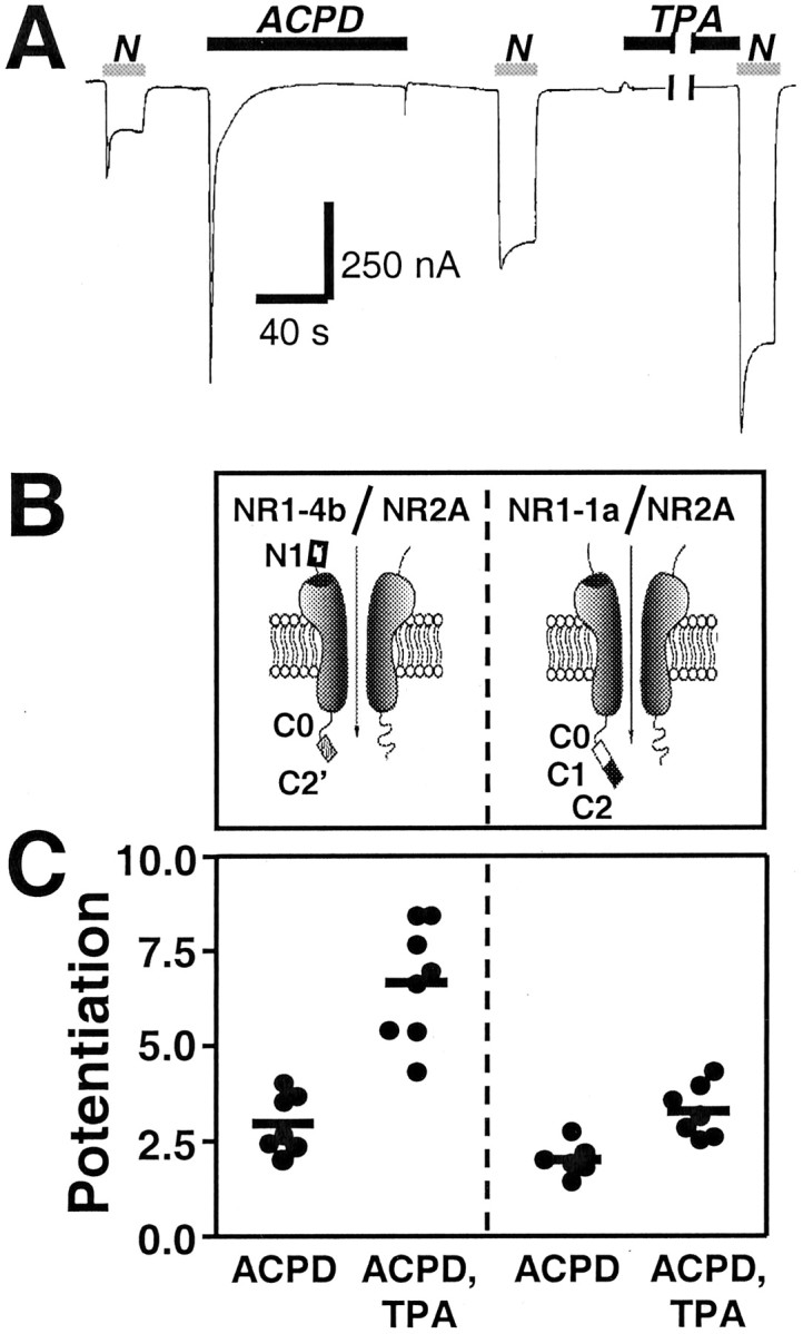
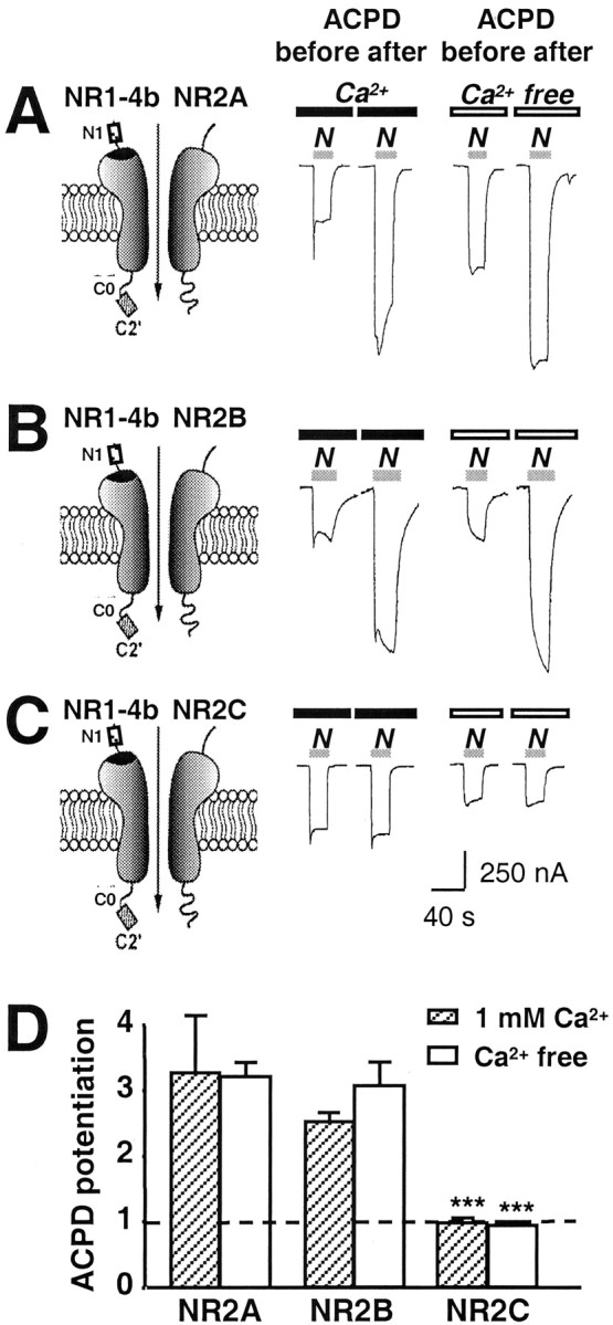
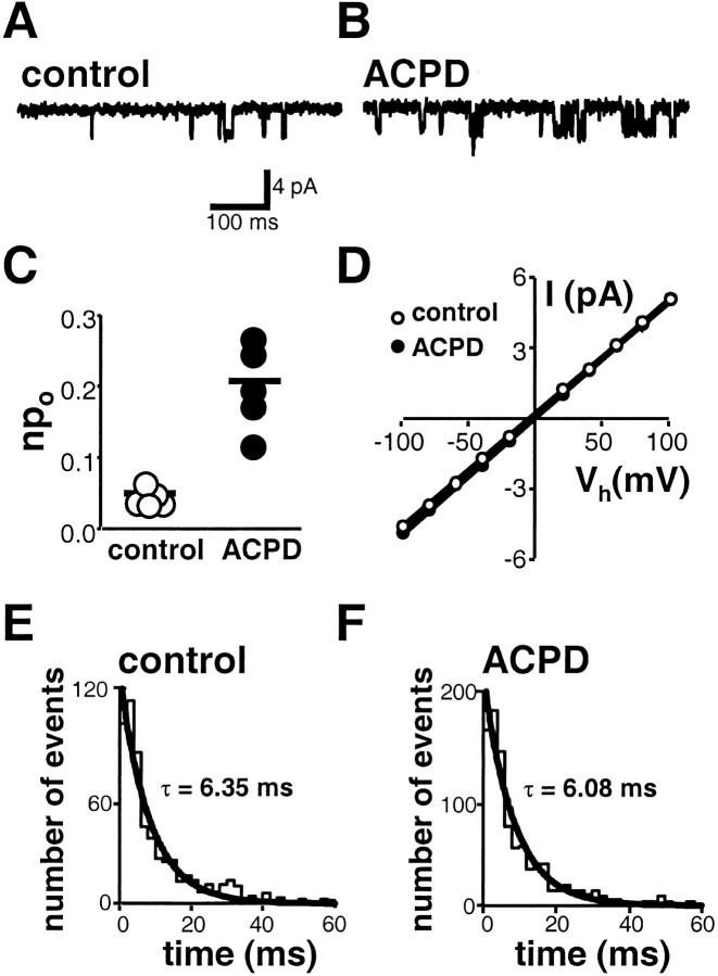
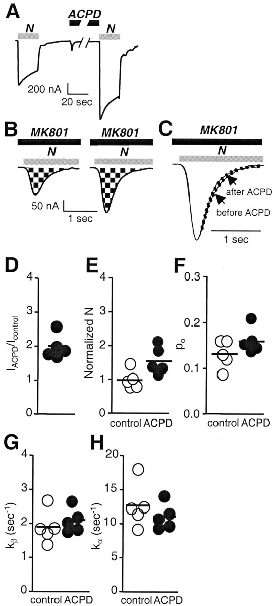
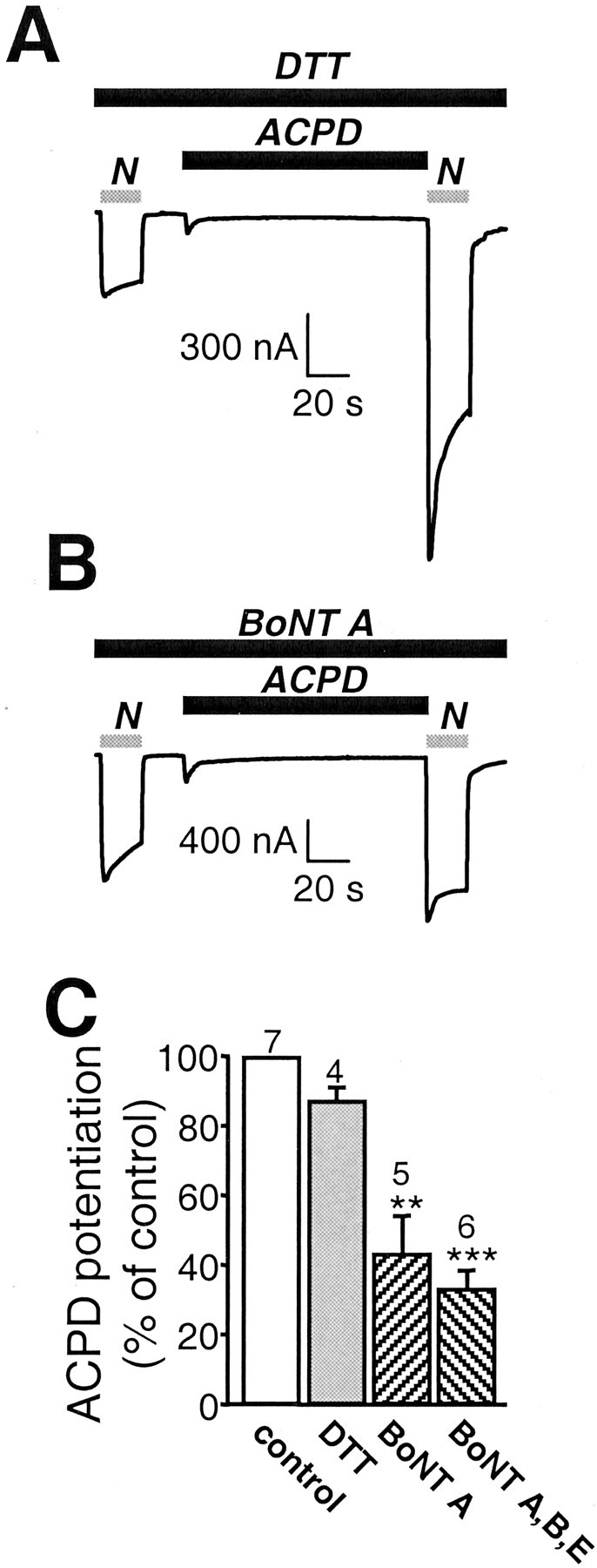
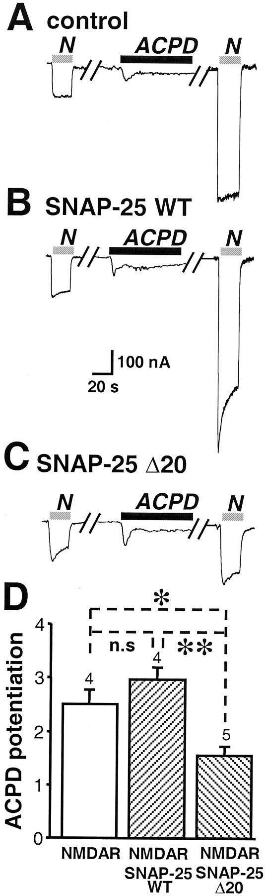
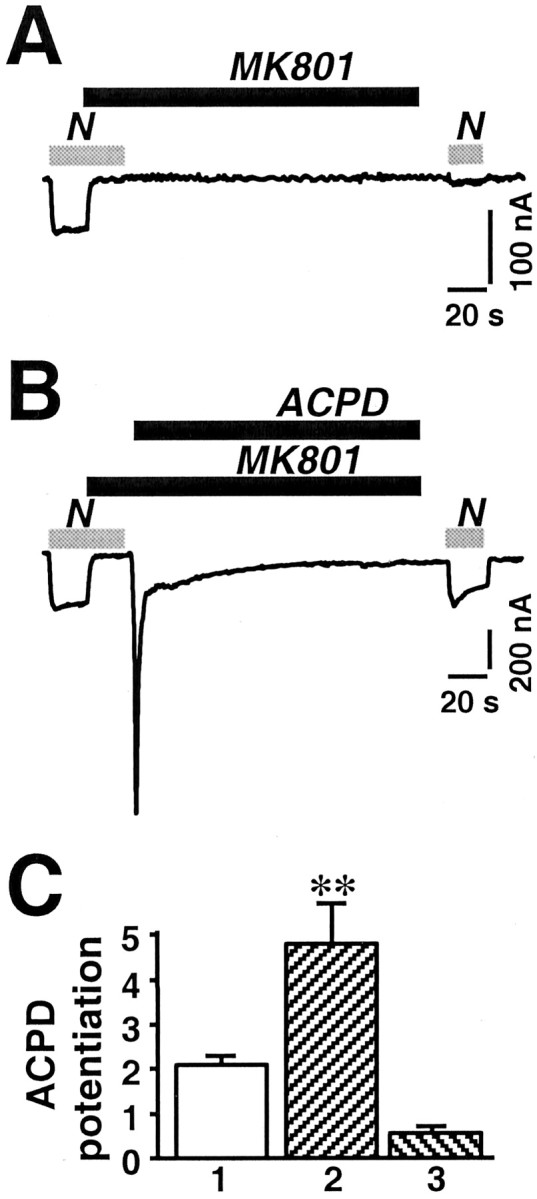
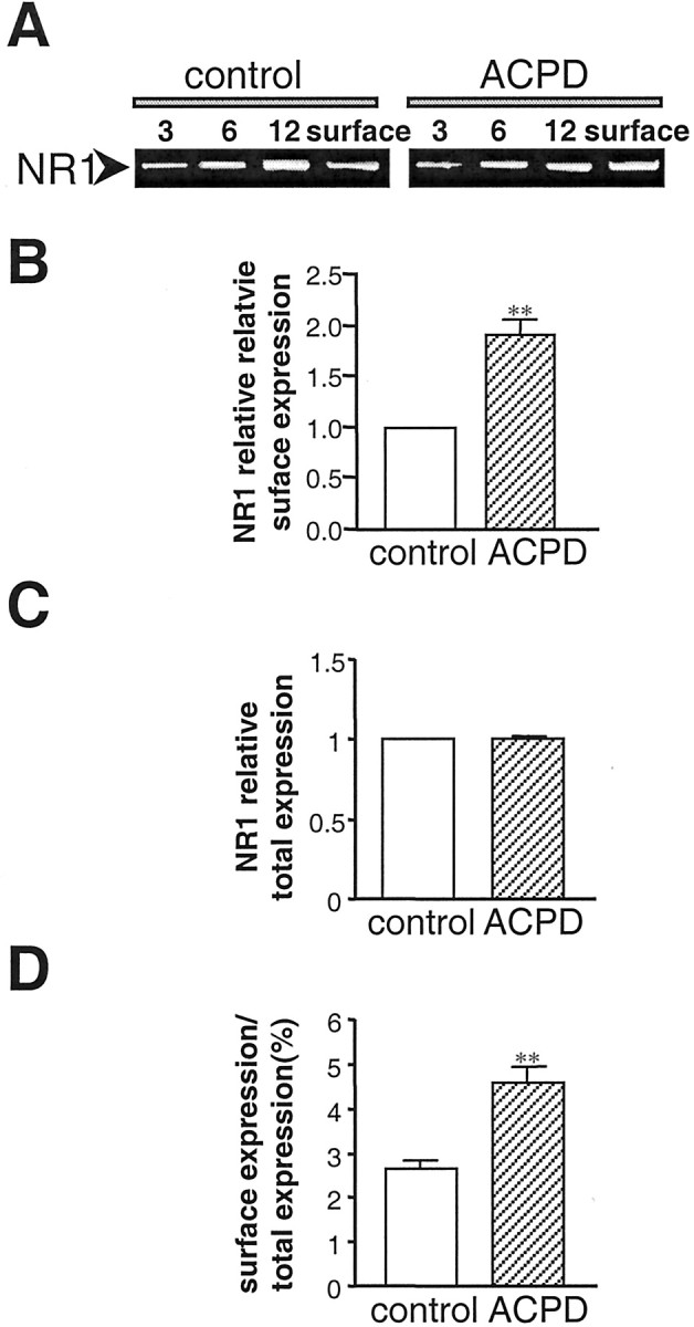
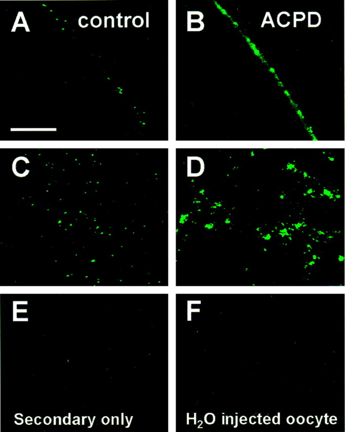
Regulation of neuronal NMDA receptors (NMDARs) by group I metabotropic glutamate receptors (mGluRs) is known to play a critical role in synaptic transmission. The molecular mechanisms underlying mGluR1-mediated potentiation of NMDARs are as yet unclear. The present study shows that in Xenopus oocytes expressing recombinant receptors, activation of mGluR1 potentiates NMDA channel activity by recruitment of new channels to the plasma membrane via regulated exocytosis. Activation of mGluR1alpha induced (1) an increase in channel number times channel open probability, with no change in mean open time, unitary conductance, or reversal potential; (2) an increase in charge transfer in the presence of NMDA and the open channel blocker MK-801, indicating an increased number of functional NMDARs in the cell membrane; and (3) increased NR1 surface expression, as indicated by cell surface Western blots and immunofluorescence. Botulinum neurotoxin A or expression of a dominant negative mutant of synaptosomal associated protein of 25 kDa molelcular mass (SNAP-25) greatly reduced mGluR1alpha-mediated potentiation, indicating that receptor trafficking occurs via a SNAP-25-mediated form of soluble N-ethylmaleimide sensitive fusion protein attachment protein receptor-dependent exocytosis. Because group I mGluRs are localized to the perisynaptic region in juxtaposition to synaptic NMDARs at glutamatergic synapses in the hippocampus, mGluR-mediated insertion of NMDARs may play a role in synaptic transmission and plasticity, including long-term potentiation.


Figures

Fig. 1.
Fig. 1.
Activation of mGluR1α potentiates NMDA whole-cell currents; alternative splicing of the NR1 subunit has little effect on ACPD potentiation. A, Typical sequence showing NMDA-activated whole-cell currents recorded in Ca2+Ringer's solution at Vh = −60 mV from oocytes expressing mGluR1α and NR1-4b/NR2A receptors before and after application of ACPD (100 μm, 2 min), followed by application of TPA (100 nm, 10 min). NMDA, 300 μm; glycine, 10 μm. ACPD elicited a large inward current that decayed rapidly to near baseline within 30 sec, ascribable to Cl efflux through Ca2+-activated Cl channels endogenous to the oocyte. ACPD significantly potentiated NMDA responses. Application of the phorbol ester TPA to oocytes after ACPD treatment further potentiated NMDA responses. B, Schematic representation of the NR1 splice variants. N1,C1, C2, and C2′ are alternatively spliced cassettes; C0 is the region between the fourth transmembrane domain and the first splice site in the C terminal (Zheng et al., 1999). C, Summary of several experiments illustrating that ACPD potentiation did not differ significantly for NR1-4b/NR2A versus NR1-1a/NR2A receptors. In contrast, potentiation by TPA (applied to oocytes after ACPD) was significantly greater for NR1-4b/NR2A receptors compared with NR1-1a/NR2A receptors (p < 0.01, one-way ANOVA followed by Bonferroni's t test). Each data point is from a single experiment; horizontal bars represent the means.
Fig. 2.
Fig. 2.
The NR2 subunit alters ACPD potentiation. NMDA-activated whole-cell currents were recorded in Ca2+ or Ca2+-free Ringer's solution from oocytes expressing mGluR1α and NMDARs.A–C, ACPD (100 μm, 2 min) potentiated NMDA currents in oocytes expressing NR1-4b/NR2A or NR1-4b/NR2B receptors; no potentiation was observed for NR1-4b/NR2C receptors.N1, C0, and C2′ are defined in the legend to Figure 1. D, Summary of three experiments illustrating ∼3.33-fold potentiation by ACPD of NR1-4b/NR2A and 2.57-fold for NR1-4b/NR2B receptors in Ca2+Ringer's solution but little or no potentiation of NR1-4b/NR2C receptors.
Fig. 3.
Fig. 3.
ACPD potentiates npo.A, B, NMDA-activated single channels recorded from outside-out patches excised from oocytes expressing recombinant NR1-4b/NR2A and mGluR1α receptors; patches were excised before (A) and after (B) application of ACPD (100 μm, 2 min). NMDA, 100 μm; glycine, 10 μm. The main unitary conductance was 44 ± 1 pS at Vh = −60 mV. C, ACPD treatment potentiated thenpo from 0.050 ± 0.006 recorded from patches excised before ACPD (n = 5) to 0.204 ± 0.034 recorded from different patches excised from the same oocytes after ACPD treatment (n = 5; p< 0.001). D, Single-channel current–voltage relationships. ACPD did not alter the main unitary conductance of NMDA-activated channels (44 ± 1 pS before ACPD;n = 5; vs 44 ± 1 pS after ACPD;n = 5). ACPD treatment did not affect the reversal potential (Erev = 0 mV before ACPD;n = 5; and 0 mV after ACPD; n = 5). E, F, Histograms of mean open time durations for NMDA-activated channels before and after application of ACPD to the oocyte. Mean open times were determined by a single exponential fit to the event lists generated by single opening and closing events. ACPD did not alter the mean open time.
Fig. 4.
Fig. 4.
mGluR1α activation increases NMDA channel number and modestly (but not significantly) increases open probability.A, ACPD (100 μm, 2 min), bath-applied, potentiated whole-cell currents elicited by application of NMDA (1 mm with 50 μm glycine). To avoid contributions by Ca2+ inactivation, Ca2+ amplification to NMDA responses, or both, recording was in Ba2+ Ringer's solution as the extracellular solution. Vh = −60 mV.B, ACPD increased the number of functional NMDA channels expressed at the cell surface (N). Currents were elicited by application of NMDA (1 mm NMDA with 50 μm glycine) in the continuous presence of the open channel blocker MK-801 (5 μm) from control (left) and ACPD-treated (right) oocytes at a holding potential of −60 mV. The NMDA inward current increased to a peak value, after which it decayed exponentially as MK-801 entered and blocked NMDA channels as they opened. The cumulative charge transfer, Q, which is the total current flow during the time interval for complete block by MK-801, was obtained by integration of the current trace over time. The larger integrated current in ACPD-treated oocytes indicated an increased number of functional channels per cell. C, Agonist-evoked currents inB were normalized to the same peak amplitude and used for kinetic analysis. The more rapid decay of the NMDA current in ACPD-treated oocytes versus control in this pair of oocytes indicates an increased rate of channel opening, kβ, but the difference was not significant in the pooled data.D–H, Quantization of data in A–C.D, Potentiation of NMDA whole-cell current,IACPD/Icontrol, was 2.2 ± 0.2 times control (p < 0.001; n = 5). E, The increase in channel number, N, was significant (p < 0.01), andNACPD/Ncontrol= 1.6 (n = 5). F, The open probabilities were not significantly different (po, control = 0.13 ± 0.01; po, ACPD = 0.16 ± 0.01;po, control/po, ACPD = 1.2; n = 5). G, Opening rates, kβ, for control (1.9 ± 0.2/sec) and ACPD-treated oocytes (2.1 ± 0.2/sec;p < 0.01). H, Closing rates,kα, for control (12.7 ± 1.5/sec) and ACPD-treated oocytes (11.1 ± 0.8/sec).
Fig. 5.
Fig. 5.
mGluR1α promotes delivery of NMDA channels to the cell membrane via exocytosis. A–C, Microinjection of the light chain of botulinum toxin type A BoNT (50 ng), an enzyme known to cleave SNAP-25, into oocytes 5 hr before recording reduced ACPD potentiation of NMDA-elicited currents by ∼50%. A, B, Representative NMDA-elicited whole-cell currents in an oocyte loaded with 50 nl of DTT (5 mm) in Aand 50 ng of BoNT A and DTT in B before and after ACPD application. BoNT A reduced potentiation but not basal NMDA-elicited currents or ACPD-elicited Cl currents.C, Quantification of A andB. A mixture of type A, B, and E BoNTs reduced ACPD potentiation by 60%.
Fig. 6.
Fig. 6.
A dominant negative mutant of SNAP-25 reduces ACPD potentiation of NMDARs. A, ACPD potentiation of NMDA-elicited currents in oocytes expressing mGluR1α and NR1-4b/NR2A receptors as in Figure 2A. B, Coexpression of wild-type SNAP-25 with NMDARs had no effect on ACPD potentiation. C, Coexpression of SNAP-25(Δ20), a dominant negative mutant of SNAP-25, with NMDARs markedly reduced ACPD potentiation. D, Quantification of the effects of wild-type and mutant SNAP-25 on ACPD potentiation.
Fig. 7.
Fig. 7.
mGluR1α potentiation increases the rate of exocytosis of NMDA channels in the cell membrane. Whole-cell recordings were obtained from Xenopus oocytes expressing NR1-4b/NR2A receptors in Ca2+-free Ringer's solution. The open channel blocker MK-801 (1 μm) was used to estimate the rate of delivery of functional channels to the cell surface in control and ACPD-treated oocytes. NMDA, 300 μm; glycine, 10 μm. A, Application of MK-801 (1 μm) in the presence of agonist completely blocked the NMDA response. Three minutes after block and washout of NMDA, MK-801 was removed; then a test application of NMDA elicited a very small response (∼10% of control), attributable to either recovery of a small number of channels from block or insertion of new channels. B, After complete block of the NMDA response by MK-801 (1 μm), ACPD (100 μm) was applied for 2 min in the continuous presence of MK-801. After washout of ACPD and MK-801, the peak of the NMDA-induced response was slightly smaller than that of the control response. The greater decay of this response is ascribable to residual MK-801. C, Quantitation of data in A and B. Test responses were normalized to initial currents. Bar 1,IACPD/Icontrol, where IACPD is the NMDA-elicited current after ACPD. In this batch of oocytes. ACPD-induced potentiation was to ∼2 times the control response. Bar 2,IACPD, MK-801/IMK-801, whereIACPD, MK-801 is the current after block of the control response by MK-801 and potentiation by ACPD, andIMK-801 is the current after block of the control response by MK-801 followed by 3 min recovery. In the presence of MK-801, ACPD potentiation was to 4.5 times the recovered response observed after block by MK-801 in A, a value that could not be accounted for by twofold potentiation of the recovered response.Bar 3, IACPD, MK-801/Icontrol. The potentiation measured as the ratio of the potentiated response after MK-801 block to the control response was somewhat smaller than the potentiated response without MK-801 minus the control response.
Fig. 8.
Fig. 8.
mGluR1α increases NMDAR surface expression. NR1 surface and total cell expression in control and ACPD-treated oocytes is shown, as assessed by Western blot analysis of surface proteins isolated by biotinylation. A, Representative Western blot of surface protein from oocytes expressing NR1-4b/NR2A with mGluR1α receptors probed with anti-NR1 antibody 54.1. 3, 6, 12, Micrograms of protein in samples of total cell extract before Neutravidin bead extraction loaded on each lane;surface, aliquot of Neutravidin bead-isolated receptors.B–D, Quantitative analysis of the effects of ACPD on surface expression (B), total cell protein (C), and fractional surface expression (D) of NMDARs. Surface expression was increased to 1.7 ± 0.2 times control (n = 4;p < 0.01). Total cell NR1 was not changed. The proportion of NR1 expressed at the cell surface increased from 2.6 to 4.5%.
Fig. 9.
Fig. 9.
mGluR1α increases NR1 surface immunofluorescence. Oocytes expressing NR1100/NR2A receptors were incubated in external recording solution in the presence or absence of ACPD (100 μm, 10 min) and subjected to immunocytochemistry. Immediately after incubation, intact oocytes were devitellinized mechanically, transferred to glass coverslips, and fixed with paraformaldehyde (4%) and sucrose (2%). To label surface NMDARs, fixed oocytes were incubated with NR1 antibody 54.1, followed by FITC-conjugated secondary antibody. NMDARs on the oocyte surface were then visualized using confocal microscopy. Surface fluorescence was expressed as the mean intensity of fluorescence per unit area. Shown are representative oocytes expressing NR1-4b/NR2A receptors from control (A, B) and ACPD (C, D) treatment groups labeled by the extracellular epitope antibody. Control oocytes labeled by a secondary antibody in the absence of primary antibody (E) and water-injected oocytes labeled as inA–D showed negligible fluorescence (F).

Similar articles

Cited by

References

    1. Aiba A, Chen C, Herrup K, Rosenmund C, Stevens CF, Tonegawa S. Reduced hippocampal long-term potentiation and context-specific deficit in associative learning in mGluR1 mutant mice. Cell. 1994a;79:365–375. - PubMed
    1. Aiba A, Kano M, Chen C, Stanton ME, Fox GD, Herrup K, Zwingman TA, Tonegawa S. Deficient cerebellar long-term depression and impaired motor learning in mGluR1 mutant mice. Cell. 1994b;79:377–388. - PubMed
    1. Akabas MH, Stauffer DA, Xu M, Karlin A. Acetylcholine receptor channel structure probed in cysteine-substitution mutants. Science. 1992;258:307–310. - PubMed
    1. Alagarsamy S, Marino MJ, Rouse ST, Gereau RW4, Heinemann SF, Conn PJ. Activation of NMDA receptors reverses desensitization of mGluR5 in native and recombinant systems. Nat Neurosci. 1999;2:234–240. - PubMed
    1. Aniksztejn L, Otani S, Ben-Ari Y. Quisqualate metabotropic receptors modulate NMDA currents and facilitate induction of long-term potentiation through protein kinase C. Eur J Neurosci. 1992;4:500–505. - PubMed

Publication types

MeSH terms

LinkOut - more resources