. 2001 Aug 15;21(16):6181-94.
doi: 10.1523/JNEUROSCI.21-16-06181.2001.

Cyclic nucleotide-mediated regulation of hippocampal mossy fiber development: a target-specific guidance

Affiliations

Cyclic nucleotide-mediated regulation of hippocampal mossy fiber development: a target-specific guidance

S Mizuhashi et al. J Neurosci. .

Abstract

The mossy fibers (MFs) arising from dentate granule cells project primarily onto a narrow segment of the proximal dendrites of hippocampal CA3 pyramidal cells. The mechanisms underlying this specific MF target selection are not fully understood. To investigate the cellular basis for development of the stereotyped MF trajectories, we have arranged the fascia dentata and hippocampal Ammon's horn tissues in diverse topographical patterns in organotypic explant coculture systems. Here we show that cyclic nucleotide signaling pathways regulate the MF pathfinding. When the dentate gyrus explants were ectopically placed facing the CA3 stratum oriens of hippocampal slices, MFs crossed the border between cocultures and reached their appropriate target area in the Ammon's horn, as assessed by membrane tracer labeling, Timm staining, electrophysiological recording of synaptic responses, and optical analyses using a voltage-sensitive dye. This lamina-specific MF innervation was disrupted by pharmacological blockade of cGMP pathway. Similar apposition of the dentate grafts near the CA1 region of host slices rarely resulted in MF ingrowth into the Ammon's horn. Under blockade of cAMP pathway, however, the MFs were capable of making allopatric synapses with CA1 neurons. These data were further supported by the pharmacological data obtained from granule cells dispersed over hippocampal slice cultures. Thus, our findings suggest that the stereotyped MF extension is mediated by at least two distinct factors, i.e., an attractant derived from the CA3 region and a repellent from the CA1 region. These factors may be regulated differently by cAMP and cGMP signaling pathways.


Figures

Fig. 1.
Fig. 1.
Extracellular signals present in hippocampal slices determine directions of MF outgrowth. A, Representative confocal image of DiI-labeled granule cell growing over the CA1 region of a hippocampal slice at 3 d in vitro (a). This neuron was reconstructed with camera lucida drawings as black and white photograph quality images (b). B, The camera lucida drawings of DiI-labeled granule cells are practically superimposed on a Nissl-stained hippocampal slice. Cells with the somata situated within 200 μm from the CA1 stratum pyramidale (red,CA1 area), in the area far from the stratum pyramidale (>400 μm) (yellow, Neutral area), or within 200 μm from the CA3 stratum pyramidale (green, CA3 area) were selected for analyzing their morphology (52.2% of total cells were suitable for the analysis). Then, their neurites were scored for length, and the cells bearing >20 μm axons were further selected for axon orientation plots (∼90% were selected). As a result, ∼53% of the neurons were excluded from this analysis. The neuron enclosed in the white dotted line is the same cell as in A. C, Axon orientation plots of granule cells plated over the CA1 area (a), the neutral area (b), and the CA3 area (c) of hippocampal slices. The longest neurites arising from cultured cells were plotted as axons in each panel. When the tip of any given neurite was placed above and below the horizontal dotted lines, the axon was considered to grow toward the CA1 stratum pyramidale and the CA3 stratum lucidum, respectively. Thus, values above and below thehorizontal dotted lines indicate the ratio of axons oriented toward the CA1 stratum pyramidale and the CA3 stratum lucidum, respectively. Granule cells growing over the CA1 area had axons oriented away from the CA1 stratum pyramidale (74.2%;n = 32; p < 0.05; χ2 test). In the CA3 area, 77.1% had axons that were directed toward the CA3 stratum lucidum (n = 35;p < 0.05; χ2 test). Granule cells in the neutral area showed an expected ratio for random orientation (∼50%) (n = 14; p > 0.1; χ2 test). The results suggest that the axon orientation is mediated by at least two distinct factors, i.e., an attractant in the CA3 region and a repellent in the CA1 region. Data were obtained from six different slices.
Fig. 2.
Fig. 2.
Explant cocultures of the fascia dentata and hippocampal Ammon's horn in various topographic arrangements. Tissues arranged as shown in schematic diagrams (left) were stained with the fluoro-Nissl method (right) at 21 d in vitro. A, An intact slice containing the fascia dentata and Ammon's horn was cultured for 21 d.B, As a control slice, the MF layer was transected by a knife cut through the slices reaching from the alvear surface of CA3 through the stratum oriens, the stratum pyramidale, and the MF layer, and the stratum radiatum into the sulcus hippocampi (Lesion). C, The DG explant was ectopically placed close to the CA3 stratum oriens of an Ammon's horn slice, in which the MFs were forced to cross the stratum oriens to reach their proper target area (DG & CA3).D, Likewise, another dislocated apposition of the DG explant to hippocampal slice at the CA1 stratum oriens was also made (DG & CA1). E, The DG slice was located immediately adjacent to two symmetrically arranged hippocampal slices (DG & Double CA3). F, The CA3 region of the Ammon's horn slice was apposed along the CA3 stratum oriens and the DG of an intact slice (DG with CA3 & CA3).G, The DG explant was placed facing the stratum oriens of the CA3 region of an intact slice (DG & CA3 with DG). The Nissl staining revealed that neurons survived in the coculture system until at least 21 d in vitro. In electrophysiological studies of Figure 3, the stratum granulosum of the DG explant (#) was stimulated, and evoked field responses were recorded from the stratum pyramidale of Ammon's horn slices (∗).
Fig. 3.
Fig. 3.
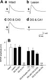
Transplanted MFs make electrophysiologically functional synapses with CA3 pyramidal cells in host hippocampal slices. Field potentials evoked by stimulation of the DG were extracellularly recorded from the CA3 or CA1 stratum pyramidale. Positions of recording and stimulating electrodes are shown in Figure2. A, Typical field responses obtained from slices cultivated according to Intact(a), Lesion(b), DG & CA3(c), and DG & CA1(d) at 14 d in vitro. Test stimulation was delivered at the time indicated by arrows. B, To estimate the number of functional synaptic contacts, the maximal amplitudes of MF-evoked fEPSP were measured from various patterns of cocultures (Fig. 2). The MFs made functional synapses with CA3 (but not CA1) pyramidal cells of the host slices, except for the CA3 pyramidal cells that received the MF inputs from the intrinsic DG. **p < 0.01 versus Intact slices; Tukey's test after ANOVA. Data represent means ± SEM of each of 15–20 slices.
Fig. 4.
Fig. 4.
MFs form Timm-positive synapses within their proper target area in host hippocampal slices. A, Representative Timm images of slices cultured for 14 d in patterns of Intact (a),Lesion (b), DG & CA3 (c), and DG & CA1(d). The MF terminals were detected predominantly in the stratum lucidum (SL), but rarely in the stratum oriens (SO) or the stratum pyramidale (SP). The DG & CA1 culture had no apparent MF terminals in any subregions of the hippocampus.B, Timm grain density was quantitatively analyzed inIntact (open columns),Lesion (solid columns), DG & CA3 (cross-hatched columns), and DG & CA1 (hatched columns) cultures. **p < 0.01 versus background density [values of the stratum radiatum (SR) of corresponding groups],## p < 0.01 versusIntact slices; Tukey's test after ANOVA. Data are means ± SEM of each of 10 slices.
Fig. 5.
Fig. 5.
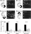
DiI-labeled MFs crossed the border between cocultures and reached their proper target area in hippocampal slices. Cocultures at 14 d in vitro were fixed with 4% paraformaldehyde, and a single crystal of DiI was placed onto the stratum granulosum of the DG explant. Using a confocal microscopy, DiI-labeled MFs were explored in the areas indicated bydotted-line boxes in the schematic drawings ofIntact (A), Lesion(B), DG & CA3(C), and DG & CA1(D) cultures. Confocal images contain the stratum radiatum (SR), the stratum lucidum (SL), the stratum pyramidale (SP), and the stratum oriens (SO) of the CA3b region (AC) or the CA1b region (D) of host hippocampal slices. The MFs extended through the stratum lucidum and made varicosity-like formations, which may represent MF giant synapses (arrowheads). InDG & CA3 slices, the varicosities were occasionally observed within the CA3 stratum oriens. In the case of DG & CA1, however, the MFs did not elongate into the CA1 region of host hippocampal slices. Similar results were obtained in every such experiment conducted (n = 12–20). Data are summarized in E. The ordinate indicates the relative incidence of distribution of the MF varicosities in each subarea of the CA3 region. Numbers in the columns show the frequency of varicosities present in the corresponding subregions. Most varicosities are observed in the stratum lucidum, but a small portion of them was found even in the stratum pyramidale and stratum oriens in the DG& CA3 slices.
Fig. 6.
Fig. 6.
Propagation of neuronal activities from DG grafts to host hippocampal slices. Activity propagation was monitored as changes in optical density of the voltage-sensitive dye RH482.Intact (A), DG & CA3 (B), and DG & Double CA3 (C) cultures displayed sequential neuron excitation along the hippocampal trisynaptic pathway, i.e., the dentate gyrus, the CA3 region, and then the CA1 region, after stimulation of the stratum granulosum of the DG explants (#). No apparent propagation from the DG explant to the host slice was observed in DG & CA1 slices (D). Experiments were repeated with 10 different slices, producing the same results.
Fig. 7.
Fig. 7.
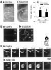
Different contributions of cAMP and cGMP signaling pathways to axon orientation of granule cells.A, Representative confocal images of a growth cone immunolabeled with antibodies to GAP-43(red) and Adenylyl cyclase(green). The immunohistochemical assessment was performed with the dentate granule cells cultured on 35 mm dishes for 3 d. Superimposition of double-labeled images (Merged) indicated that ∼80% of growth cones displayed evident immunoreactivity for adenylyl cyclase, in which case adenylyl cyclase was localized centrally in the growth cones but apparently not in the peripheral lamellipodia (n = 56). B, Axon orientation histograms of the granule cells growing over the CA1 area (a), the neutral area (b), and the CA3 area (c). The granule cells were cultured on hippocampal slices in the presence of 100 μm SQ22536 and 100 nm LY83583 for 3 d. The upward ordinates indicate the ratio of axons oriented toward the CA1 stratum pyramidale, and the downward ordinates indicate the ratio of axons oriented toward the CA3 stratum lucidum. The repulsive response of the axons of granule cells growing over the CA1 area was converted to attraction by SQ22536. The attractive responses of the axons growing over the CA3 area were disrupted by LY83583.Horizontal dotted lines indicate expected distribution for random orientation. *p < 0.05 versus the chance level of 50%; ## p < 0.01 versus Control slices; χ2 test. Data were obtained from 16–35 neurons in five to six different slices.
Fig. 8.
Fig. 8.
Involvement of the cAMP signaling pathway in MF synaptogenesis. Cocultures of DG & CA3 and DG & CA1 arrangements were incubated in the continuous presence of 100 μm SQ22536, 100 μm Rp-cAMPs, or 10 μm KT5720 for 14 d in vitro, and then synaptic responses were recorded from the CA3 stratum pyramidale inDG & CA3 cultures or from the CA1 stratum pyramidale inDG & CA1 slices after stimulation of the stratum granulosum of the DG explants. All of these inhibitors induced ectopic synaptogenesis within the CA1 pyramidal cells in the DG & CA1 cultures, whereas normal MF projections to the CA3 pyramidal cells in the DG & CA3 slices were virtually unaffected. **p < 0.01 versusControl slices; Tukey's test after ANOVA. Data are shown as means ± SEM of 20–25 slices.
Fig. 9.
Fig. 9.
Additional evidence of allopatric MF–CA1 synapse formation under low cAMP levels. DG and CA1 slices were cultured in the absence (a) or presence (b) of 100 μm SQ22536 for 14 din vitro. A, Timm staining indicated that evident MF terminals were detected in the CA1 region of SQ22536-treated cultures. Ac, Timm grain density was quantitatively analyzed in the CA1 stratum oriens (SO) and the CA1 stratum pyramidale (SP) of untreated (open columns) or SQ22536-treated slices (solid columns). **p < 0.01 versusControl slices; Tukey's test after ANOVA. Data are means ± SEM of each of 10 slices. B, In SQ22536-treated cultures, DiI-labeled MFs elongated into the CA1 region of host hippocampal slices and formed varicosity-like structures that were reminiscent of those seen in the CA3 region (Fig. 5).C, In the DG and CA1 cocultures grown in the presence of SQ22536, the optical signals of RH482 were propagated from the DG explants (#) into the host hippocampal slices. All of these experiments were repeated at least five times, leading to similar results.SR, Stratum radiatum.
Fig. 10.
Fig. 10.
Treatment with plasmin disrupted MF axon guidance. Slices arranged in DG & CA3 and DG & CA1 patterns were cultivated in the presence of 100 μm forskolin, 100 μm plasmin, or a combination of 100 μm plasmin and 100 μmforskolin for 14 d in vitro. Synaptic responses evoked by DG stimulation were recorded from CA3 stratum pyramidale inDG & CA3 cultures or from CA1 stratum pyramidale inDG & CA1 cultures. Plasmin treatment caused ectopic MF–CA1 synaptogenesis in the DG & CA1 cultures. This aberrant synapse formation was efficiently attenuated by forskolin. These agents did not affect normal MF projections to the CA3 pyramidal cells in the DG & CA3 slices. **p < 0.01 versus Control slices; Tukey's test after ANOVA. ## p < 0.01 versus plasmin. The data represent the means ± SEM of 20–25 cases.
Fig. 11.
Fig. 11.
Involvement of the cGMP signaling pathway in MF synaptogenesis. Cocultures of DG & CA3 andDG & CA1 arrangements were incubated in the presence of 100 nm LY83583 or 100 μm Rp-pCPT-cGMPs for 14 d in vitro, and then synaptic responses evoked by DG stimulation were recorded from the CA3 region in DG & CA3 cultures or the CA1 region in DG & CA1slices. These inhibitors reduced MF–CA3 synaptic responses in theDG & CA3 cultures, whereas no MF projections to the CA1 region were observed in the DG & CA1 slices. **p < 0.01 versus Control slices; Tukey's test after ANOVA. Data are shown as means ± SEM of 20–25 slices. B, Timm grain density was quantitatively analyzed in the CA3 stratum oriens (SO), the CA3 stratum pyramidale (SP), and the stratum lucidum (SL) of untreated (open columns) or LY83583-treated slices (solid columns). *p < 0.05 versus Control slices; Tukey's test after ANOVA. Data are means ± SEM of each of 10–11 slices.

Similar articles

Cited by

References

    1. Abdel-Majid RM, Leong WL, Schalkwyk LC, Smallman DS, Wong ST, Storm DR, Fine A, Dobson MJ, Guernsey DL, Neumann PE. Loss of adenylyl cyclase I activity disrupts patterning of mouse somatosensory cortex. Nat Genet. 1998;19:289–291. - PubMed
    1. Altman J, Das GD. Autoradiographic and histological evidence of postnatal hippocampal neurogenesis in rats. J Comp Neurol. 1965;124:319–335. - PubMed
    1. Amaral DG, Dent JA. Development of the mossy fibers of the dentate gyrus: I. A light and electron microscopic study of the mossy fibers and their expansions. J Comp Neurol. 1981;195:51–86. - PubMed
    1. Baranes D, Lederfein D, Huang YY, Chen M, Bailey CH, Kandel ER. Tissue plasminogen activator contributes to the late phase of LTP and to synaptic growth in the hippocampal mossy fiber pathway. Neuron. 1998;21:813–825. - PubMed
    1. Bonhoeffer F, Huf J. Recognition of cell types by axonal growth cones in vitro. Nature. 1980;288:162–164. - PubMed

Publication types

MeSH terms

LinkOut - more resources