. 2001 Aug 15;21(16):6221-32.
doi: 10.1523/JNEUROSCI.21-16-06221.2001.

Effects of progesterone synthesized de novo in the developing Purkinje cell on its dendritic growth and synaptogenesis

Affiliations

Effects of progesterone synthesized de novo in the developing Purkinje cell on its dendritic growth and synaptogenesis

H Sakamoto et al. J Neurosci. .

Abstract

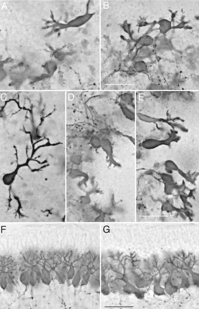
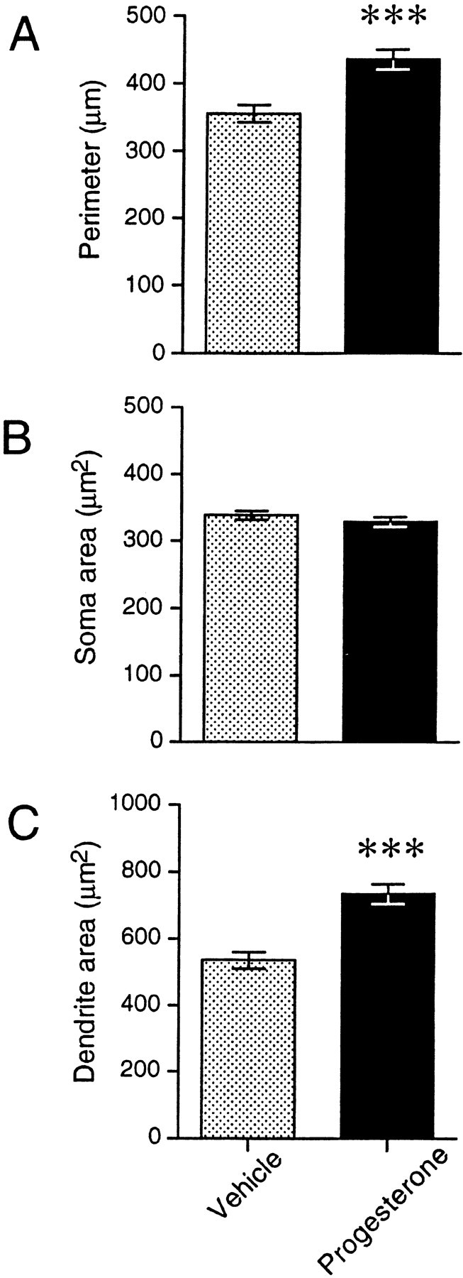
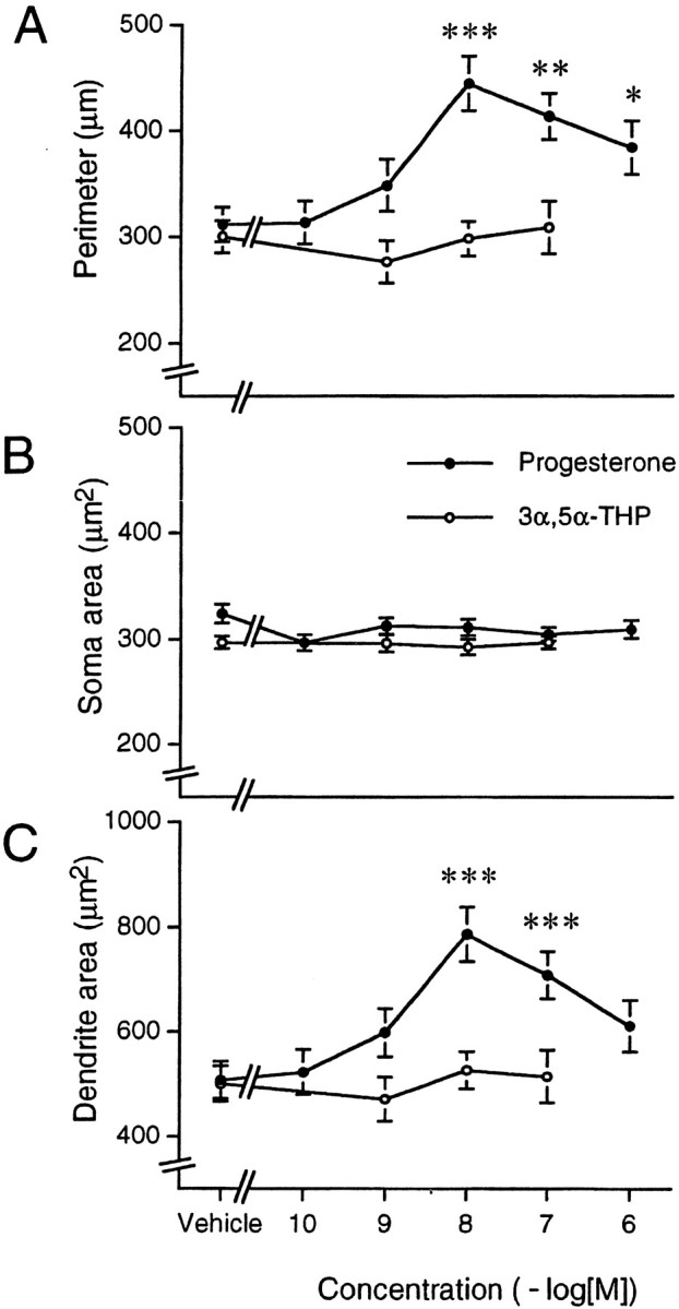
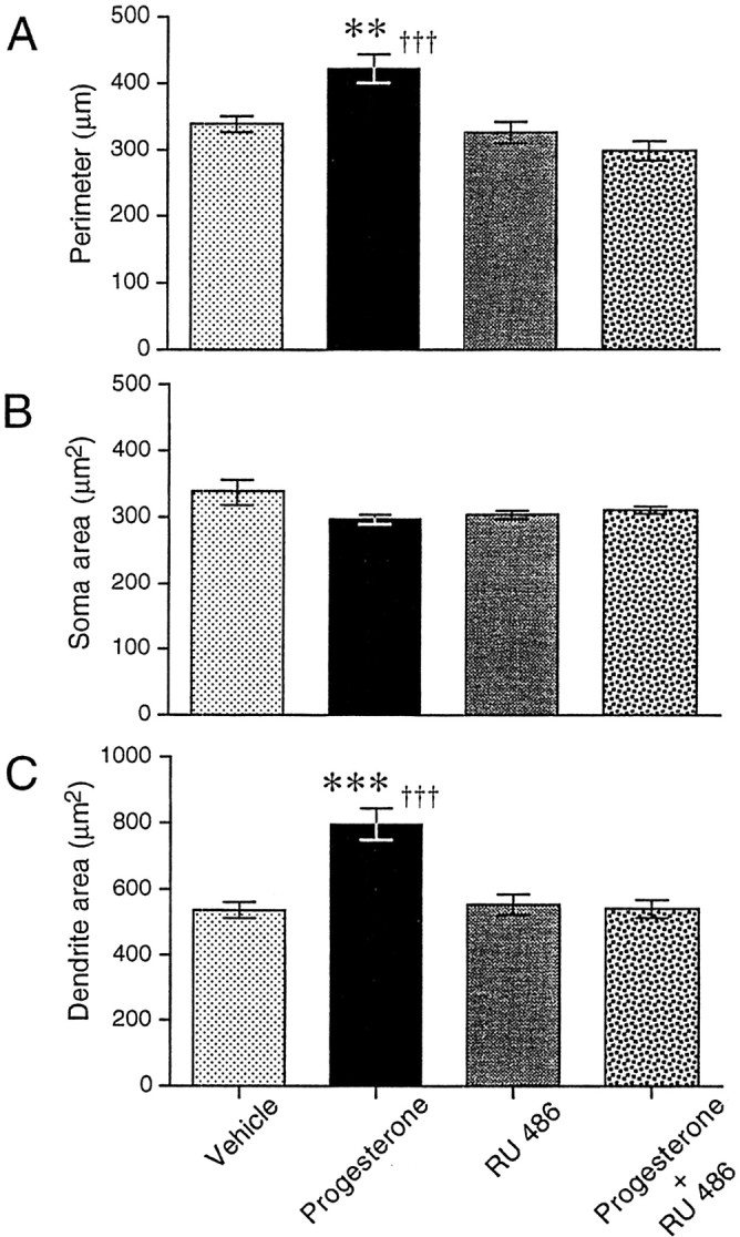
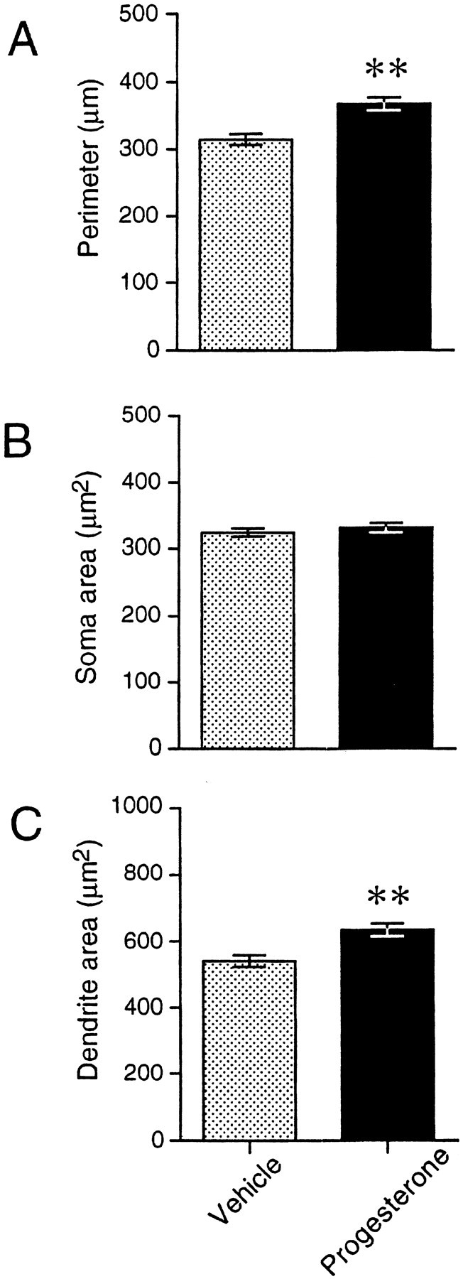
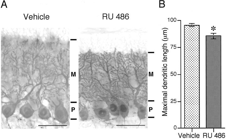
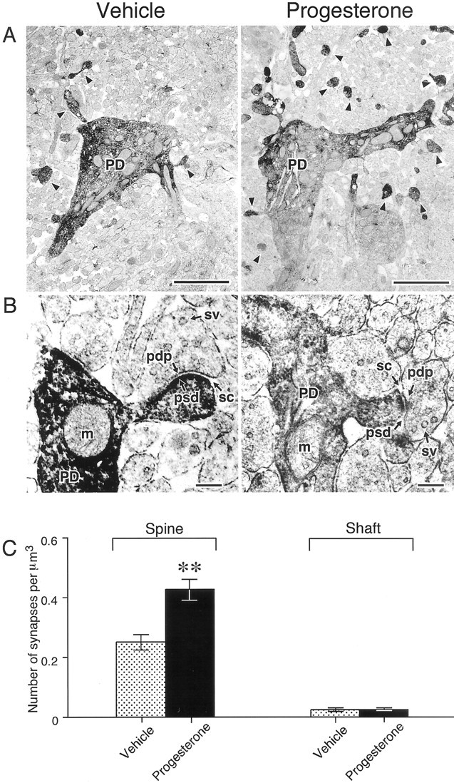
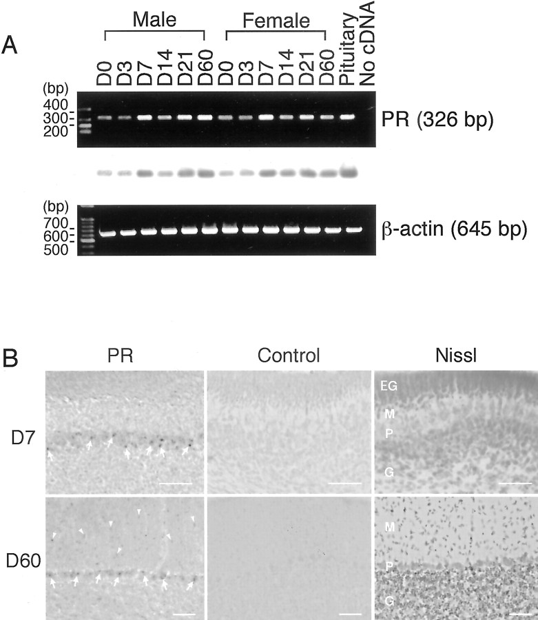
De novo steroidogenesis from cholesterol is a conserved property of vertebrate brains, and such steroids synthesized de novo in the brain are called neurosteroids. The identification of neurosteroidogenic cells is essential to the understanding of the physiological role of neurosteroids in the brain. We have demonstrated recently that neuronal neurosteroidogenesis occurs in the brain and indicated that the Purkinje cell actively synthesizes several neurosteroids de novo from cholesterol in vertebrates. Interestingly, in the rat, this neuron actively synthesizes progesterone de novo from cholesterol only during neonatal life, when cerebellar cortical formation occurs most markedly. Therefore, in this study, the possible organizing actions of progesterone during cerebellar development have been examined. In vitro studies using cerebellar slice cultures from newborn rats showed that progesterone promotes dose-dependent dendritic outgrowth of Purkinje cells but dose not affect their somata. This effect was blocked by the anti-progestin RU 486 [mifepristone; 17beta-hydroxy-11beta-(4-methylaminophenyl)-17alpha-(1-propynyl) estra-4,9-dien-3 one-6-7]. In vivo administration of progesterone to pups further revealed an increase in the density of Purkinje spine synapses electron microscopically. In contrast to progesterone, there was no significant effect of 3alpha,5alpha-tetrahydroprogesterone, a progesterone metabolite, on Purkinje cell development. Reverse transcription-PCR-Southern and immunocytochemical analyses showed that intranuclear progesterone receptors were expressed in Purkinje cells. These results suggest that progesterone promotes both dendritic outgrowth and synaptogenesis in Purkinje cells through intranuclear receptor-mediated mechanisms during cerebellar development. Such organizing actions may contribute to the formation of the cerebellar neuronal circuit.


Figures

Fig. 1.
Fig. 1.
Morphology of Purkinje cells and its modulation by progesterone treatment: in vitro and in vivo study. A–E, Cerebellar cultures from newborn male rats grown for 5 DIV and immunostained for calbindin. Cultures treated with vehicle (A), 100 nm progesterone (B), 10 nm progesterone (C), 1 μm RU 486 alone (D), and 10 nmprogesterone plus 1 μm RU 486 (E) for 3 d. Progesterone promoted dendritic outgrowth of Purkinje cells in vitro, and the effect was blocked by the anti-progestin RU 486. F, G, Parasagittal sections of neonatal cerebellum at 7 d of age were immunostained for calbindin. Male pups received daily injections of the vehicle (F) or progesterone (G) for 4 d. Purkinje cells in the progesterone group (G) demonstrated more differentiated dendrites compared with the control group (F)in vivo. All photomicrographs are of the same magnification. Scale bars, 50 μm.
Fig. 2.
Fig. 2.
Morphological comparison of Purkinje cells from vehicle- and progesterone-treated groups: in vitrostudy. Perimeter (A), soma area (B), and dendrite area (C) of Purkinje cells were measured after immunostaining for calbindin. Progesterone administration in vitro induced significant increases in the perimeter and dendrite area of Purkinje cells but did not affect their somata. Each column and error bar represent the mean ± SEM (n = 18 slices from 6 different males in each group). Data were derived from 120 Purkinje cells in each group. ***p < 0.001 (by Student's t test).
Fig. 3.
Fig. 3.
Dose–response of progesterone and its metabolite 3α,5α-THP: in vitro study. Perimeter (A), soma area (B), and dendrite area (C) of Purkinje cells were measured after immunostaining for calbindin. Progesterone administrationin vitro increased, in a dose-related manner, the perimeter and dendrite area of Purkinje cells, unlike their soma area. In contrast to progesterone, 3α,5α-THP, a progesterone metabolite, failed to alter any of the morphological parameters under a similar dose treatment. Each dot and error bar represent the mean ± SEM (n = 12 slices from 4 different males in each group). Data were derived from 80 Purkinje cells in each group. *p < 0.05, **p < 0.01, and ***p < 0.001 versus vehicle (by Student'st test).
Fig. 4.
Fig. 4.
Effect of the anti-progestin RU 486 on Purkinje cell morphology: in vitro study. Perimeter (A), soma area (B), and dendrite area (C) of Purkinje cells were measured after immunostaining for calbindin. Treatment with 10 nmprogesterone alone in vitro caused significant increases in Purkinje perimeter (A) and dendrite area (C), whereas RU 486 alone at a concentration of 1 μm did not alter any significant differences from the vehicle group. In contrast, combined treatments with progesterone and RU 486 showed that RU 486 abolished the progesterone effect in vitro. Each column and error bar represent the mean ± SEM (n = 18 slices from 6 different males in each group). Data were derived from 120 Purkinje cells in each group. **p < 0.01 and ***p < 0.001 versus vehicle; †††p < 0.001 versus progesterone plus RU 486 group (by one-way ANOVA, followed by Duncan's multiple range test).
Fig. 5.
Fig. 5.
Morphological comparison of Purkinje cells from vehicle- and progesterone-treated groups: in vivo study. Perimeter (A), soma area (B), and dendrite area (C) of Purkinje cells (lobe IX) were measured after immunostaining for calbindin. Progesterone administration in vivo induced significant increases in the perimeter and dendrite area of Purkinje cells but not soma area. Each column and error bar represent the mean ± SEM (n = 6 males in each group). Data were derived from 120 Purkinje cells in each group. **p < 0.01 (by Student's ttest).
Fig. 6.
Fig. 6.
Morphological comparison of Purkinje cells from vehicle- and RU 486-treated groups: in vivo study.A, Parasagittal sections of neonatal cerebellum at 11 d of age were immunostained for calbindin (lobe IX). Male pups received daily injections of the vehicle (left) or RU 486 (right) for 4 d from 7–10 d old during the endogenous peak of progesterone in the cerebellum. Scale bars, 50 μm.M, Molecular layer; P, Purkinje cell layer. B, Quantitative analysis of the length of molecular layer as a parameter of maximal dendritic length. Dendritic outgrowth of the Purkinje cell was significantly inhibited by RU 486 administration. Each column and error bar represent the mean ± SEM (n = 4 males in each group). *p < 0.05 (by Student's ttest).
Fig. 7.
Fig. 7.
Ultrastructural analysis of Purkinje cells:in vivo study. A, Calbindin immunoelectron micrographs of Purkinje cell dendrites in the molecular layers of vermal cerebella (lobe IX). Spine number and synapse density were increased in progesterone-administered pups.Arrowheads in A indicate presumptive spine structures. B, Higher magnification of synaptic terminals in the molecular layers of vermal cerebella (lobe IX). The morphology of synaptic boutons and neurotransmitter vesicles appeared unaltered in progesterone-administered pups. Arrowsin B indicate postsynaptic density (psd), synaptic vesicle (sv), synaptic cleft (sc), and presynaptic dense projections (pdp). C, Quantitative electron microscopic analysis using an unfolding method. Axospinous synapse density of Purkinje cells in the progesterone-treated group was significantly higher than in the control group (vehicle), unlike that of the density of dendritic shafts. Each column and error bar represent the mean ± SEM (n = 6 males in each group). Data were derived from randomly selected 24 fields (100 μm2 in each field) of vermal molecular layers in each group. **p < 0.01 (by Student'st test). PD, Purkinje cell dendrite;m, mitochondrion. Scale bars: A, 2 μm;B, 200 nm.
Fig. 8.
Fig. 8.
Expression and localization of progesterone receptor in the cerebellum: in vivo study.A, RT-PCR analysis of progesterone receptor mRNA in the male and female cerebella at 0, 3, 7, 14, 21, and 60 d of age.Top panel shows a result of gel electrophoresis of RT-PCR products for rat progesterone receptor, and middle panel shows an identification of the band by Southern hybridization using digoxigenin-labeled oligonucleotide probe for rat progesterone receptor. cDNA corresponding to 0.1 μg of total RNA extracted from each cerebellar tissue was used for a PCR reaction, and an 8 μl aliquot of each sample was applied on one lane. Pituitary tissue was used as a positive control, and a similar amount of cDNA was used in the RT-PCR. The lane labeledNo cDNA was performed without template as the negative control. Bottom panel shows a result of the RT-PCR for β-actin as the internal control, in which PCR reaction, cDNA corresponding to 0.1 μg of total RNA, was used as a template. RT-PCR studies were repeated four times using independently extracted RNA samples from different animals and produced similar results.B, Immunocytochemical analysis using progesterone receptor antiserum (left panels; PR) of the cerebellar cortex of male rats at 7 d (top panels; D7) and 60 d of age (bottom panels; D60) (adult). An intense immunoreaction for progesterone receptor was observed in both groups in large cell nuclei lying in a narrow zone between the molecular and granular layers, possibly Purkinje cells (arrows inPR). In contrast, progesterone receptor-like immunoreactivity in the cerebella of adults was also observed in relatively small cell nuclei, possibly basket and/or satellite cells (arrowheads in PR). Preadsorbing the antiserum with an excess amount of the synthetic progesterone receptor peptide used as antigen (DNA-binding domain; amino acid 533–547; 100 μg/ml) resulted in a complete absence of progesterone receptor-like immunoreactivity in all of the positively stained cells in the cerebellum (middle panels;Control). Histology of the cerebellar cortex was revealed by Nissl staining (right panels;Nissl). Immunocytochemical studies were repeated independently five times using different animals and produced similar results. EG, External granular layer; M, molecular layer; P, Purkinje cell layer;G, granular layer. Scale bars, 50 μm.

Similar articles

Cited by

References

    1. Akwa Y, Young J, Kabbadj K, Sancho MJ, Zucman D, Vourc'h C, Jung-Testas I, Hu ZY, Le Goascogne C, Jo DH, Corpéchot C, Simon P, Baulieu EE, Robel P. Neurosteroids: biosynthesis, metabolism and function of pregnenolone and dehydroepiandrosterone in the brain. J Steroid Biochem Mol Biol. 1991;40:71–81. - PubMed
    1. Altman J. Postnatal development of the cerebellar cortex in the rat. I. The external germinal layer and the transitional molecular layer. J Comp Neurol. 1972a;145:353–398. - PubMed
    1. Altman J. Postnatal development of the cerebellar cortex in the rat. II. Phases in the maturation of Purkinje cells and of the molecular layer. J Comp Neurol. 1972b;145:399–464. - PubMed
    1. Arnold AP, Gorski RA. Gonadal steroid induction of structural sex differences in the CNS. Annu Rev Neurosci. 1984;7:413–442. - PubMed
    1. Baulieu EE. Neurosteroids: a new function in the brain. Biol Cell. 1991;71:3–10. - PubMed

Publication types

MeSH terms

LinkOut - more resources