. 2001 Aug 15;21(16):6245-51.
doi: 10.1523/JNEUROSCI.21-16-06245.2001.

Remodeling of synaptic membranes after induction of long-term potentiation

Affiliations

Remodeling of synaptic membranes after induction of long-term potentiation

N Toni et al. J Neurosci. .

Abstract

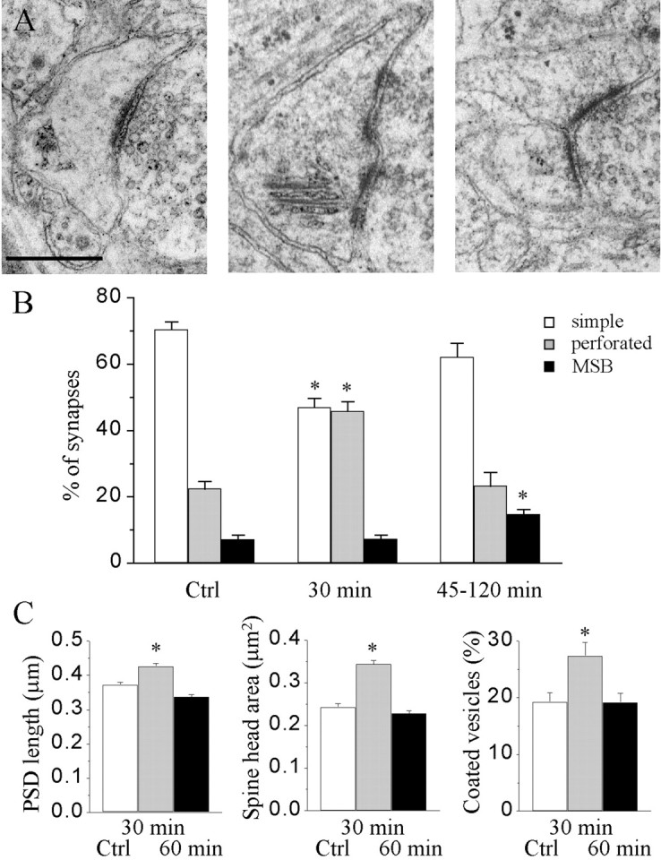
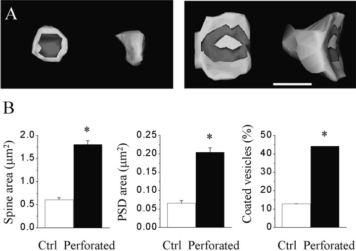
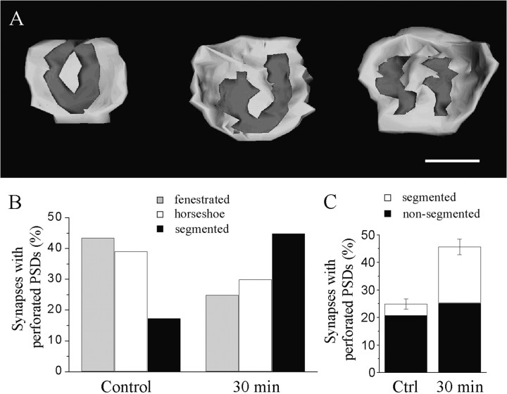
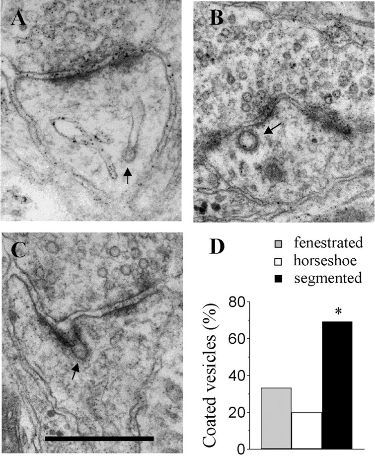
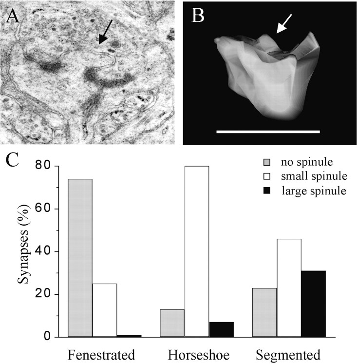
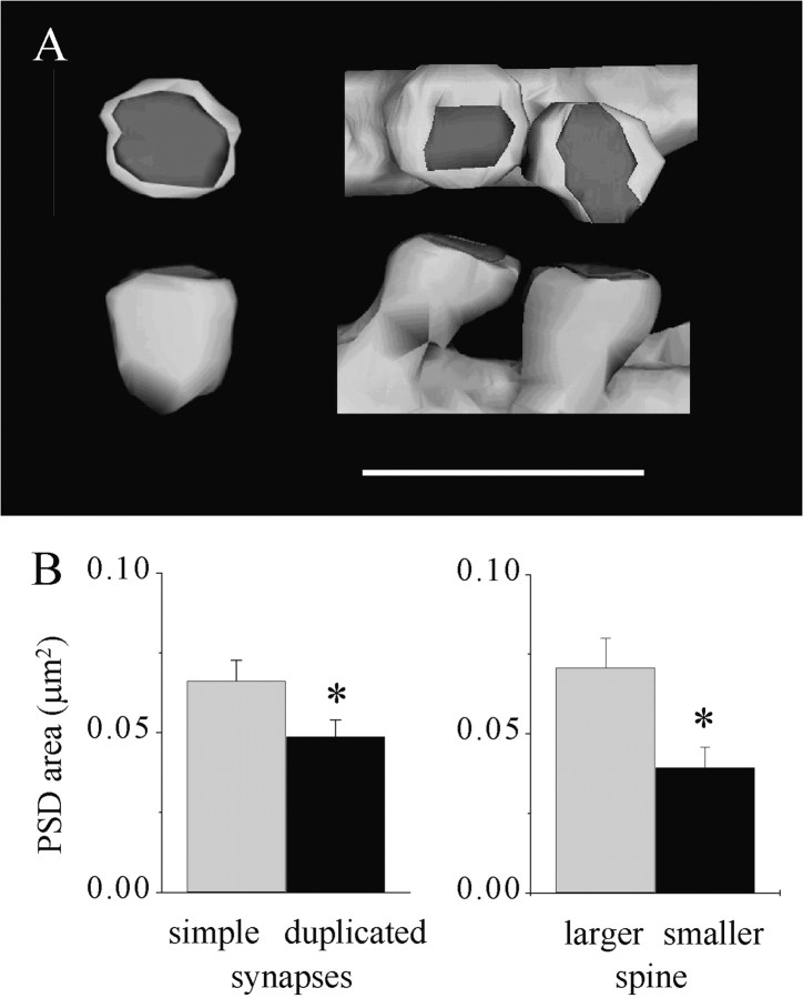
Several morphological changes of synapses have been reported to be associated with the induction of long-term potentiation (LTP) in the CA1 hippocampus, including an transient increase in the proportion of synapses with perforated postsynaptic densities (PSDs) and a later occurrence of multiple spine boutons (MSBs) in which the two spines arise from the same dendrite. To investigate the functional significance of these modifications, we analyzed single sections and reconstructed 134 synapses labeled via activity using a calcium precipitation approach. Analyses of labeled spine profiles showed changes of the spine head area, PSD length, and proportion of spine profiles containing a coated vesicle that reflected variations in the relative proportion of different types of synapses. Three-dimensional reconstruction indicated that the increase of perforated spine profiles observed 30 min after LTP induction essentially resulted from synapses exhibiting segmented, completely partitioned PSDs. These synapses had spine head and PSD areas approximately three times larger than those of simple synapses. They contained coated vesicles in a much higher proportion than that of any other type of synapse and exhibited large spinules associated with the PSD. Also the MSBs with two spines arising from the same dendrite that were observed 1-2 hr after LTP induction included a spine that was smaller and a PSD that was smaller than those of simple synapses. These results support the idea that LTP induction is associated with an enhanced recycling of synaptic membrane and that this process could underlie the formation of synapses with segmented PSDs and eventually result in the formation of a new, immature spine.


Figures

Fig. 1.
Fig. 1.
Changes in the proportion of different types of synapses and their characteristics during LTP. A, Illustration of a simple synapse with a single PSD (left), a synapse with a perforated PSD (middle), and an MSB (right). Scale bar, 0.5 μm. B, Proportion of the three types of synapses under control conditions and at 30 and 45–120 min after LTP induction (n = 4–14 hippocampal slice cultures and 358–1519 synaptic profiles analyzed; *p < 0.01).C, Changes in PSD length (left), spine head profile area (middle), and the proportion of spine profiles containing coated vesicles (right) determined via single-section analysis of the entire population of labeled synapses (n = 4–9 hippocampal slice cultures and 360–660 synaptic profiles; *p < 0.01).Ctrl, Control.
Fig. 2.
Fig. 2.
Characteristics of three-dimensionally reconstructed simple synapses and synapses with perforated PSDs observed 30 min after LTP induction. A, Illustration of the spine head of and PSD of a reconstructed simple synapse (left) and a synapse with a perforated PSD (right). Scale bar, 0.5 μm.B, Values of spine head surface area (left), PSD surface area (middle), and the proportion of spines containing coated vesicles for simple synapses (Ctrl) and synapses with perforated PSDs (Perforated). Data are the mean ± SEM of measurements made on 31 reconstructed simple synapses and 42 synapses with perforated PSDs (*p < 0.01).
Fig. 3.
Fig. 3.
Increase in the proportion of synapses with segmented PSDs 30 min after LTP induction. A, Illustration of three types of reconstructed synapses with perforated PSDs. From left to right, synapses with fenestrated, horseshoe-shaped, and segmented PSDs are shown. Scale bar, 0.5 μm. B, Proportion of the three types of synapses with perforated PSDs under control conditions and 30 min after LTP (n = 20 and 23, respectively). The two distributions are statistically significantly different (p < 0.05, χ2).C, Changes in the proportion of synapses with perforated PSDs calculated for synapses with segmented (white column) and nonsegmented (black column) PSDs according to the three-dimensional reconstruction. Data are the mean ± SEM.
Fig. 4.
Fig. 4.
Increased proportion of coated vesicles in synapses with segmented PSDs observed 30 min after LTP induction.A, Example of a coated vesicle emerging from or fusing with the spine apparatus. B, Coated vesicle within the spine head. C, Example of a coated vesicle seen fusing with the synaptic membrane at the level of the PSD. Scale bar:A–C, 0.5 μm. D, Proportion of synapses with fenestrated (gray column), horseshoe-type (white column), and segmented (black column) PSDs that contained one or several coated vesicles in a population of 15, 15, and 13 reconstructed synapses, respectively (*p < 0.05); arrows in A–Cpoint to examples of coated vesicles.
Fig. 5.
Fig. 5.
Presence of large spinules associated with the PSD of synapses with segmented PSDs. A, Illustration of a spine profile with a large spinule emerging between the two parts of the PSD. B, Illustration of a spinule on another reconstructed spine; arrows in A andB point to large spinules. Scale bar, 1 μm.C, Proportion of synapses with fenestrated, horseshoe-type, and segmented PSDs that exhibited no spinules (gray column) or spinules of small (<0.2 μm;white column) or large (>0.2 μm; black column) size in a population of 15, 15, and 13 reconstructed synapses, respectively.
Fig. 6.
Fig. 6.
MSBs with two spines arising from the same dendrite include one immature spine with a small PSD. A, Illustration of a reconstructed simple synapse (left) and an MSB with duplicated spines (right). Scale bar, 1 μm. B, Left, Size of the PSD area measured in a population of 31 reconstructed simple synapses (gray column) and 30 MSBs with duplicated spines (black column). Data are the mean ± SEM (*p < 0.05). Right, Comparison of the PSD area of the larger (gray column) and smaller (black column) spines of the reconstructed MSBs with duplicated spines. Note that the larger spine has a PSD area comparable with that of a simple synapse (B, left;gray column), whereas the PSD area of the smaller spine is almost one-half that of a simple synapse (*p < 0.01).

Similar articles

Cited by

References

    1. Buchs PA, Muller D. Induction of long-term potentiation is associated with major ultrastructural changes of activated synapses. Proc Natl Acad Sci USA. 1996;93:8040–8045. - PMC - PubMed
    1. Buchs PA, Stoppini L, Parducz A, Siklos L, Muller D. A new cytochemical method for the ultrastructural localization of calcium in the central nervous system. J Neurosci Methods. 1994;54:83–93. - PubMed
    1. Calverley RK, Jones DG. A serial-section study of perforated synapses in rat neocortex. Cell Tissue Res. 1987;247:565–572. - PubMed
    1. Calverley RK, Jones DG. Contributions of dendritic spines and perforated synapses to synaptic plasticity. Brain Res Brain Res Rev. 1990;15:215–249. - PubMed
    1. Carlin RK, Siekevitz P. Plasticity in the central nervous system: do synapses divide? Proc Natl Acad Sci USA. 1983;80:3517–3521. - PMC - PubMed

Publication types

MeSH terms

LinkOut - more resources